ORIGIN
AL RESEAR
CH
Reference values of ultrasonographic measurement
of cross-sectional area of the multiidus muscle in
children aged 6 to 9 years
Valores de referência de medidas ultrassonográicas da área de secção transversa dos músculos
multíidos em crianças de seis a nove anos
Valores de referencia de las medidas ecográicas del área de sección transversal de los músculos
multiidus en niños entre 6 y 9 años de edad
Gisela Rocha de Siqueira¹, Geisa Guimarães de Alencar², Maria Luiza Barbalho da Puriicação³
Mailing address: Geisa Guimarães de Alencar – Rua Capitão Rui Lucena, 160/904 – Boa Vista Recife-PE, Brazil – CEP:50070-080 – E-mail: geisa.guimaraes@hotmail.com Presentation: Sep. 2014 – Accepted for publication: May 2015
1PhD in Children’s and Adolescents Health from UFPE, associate professor in the Physical Therapy Department – UFPE. (PE), Brazil. 2Physical Therapist, master’s student from UFPE, member in the Human Musculoskeletal System Morpho-physiopathology Research
Group – UFPE (PE), Brazil.
3Physical Therapist – UFPE (PE), Brazil.
ABSTRACT | This study aimed to provide reference value measures from the cross-sectional area (CSA) of the multiidus muscles in children aged 6 to 9 years old. For this purpose, 41 children (20 boys and 20 girls) were interviewed about their physical exercises practice and submitted to a unilateral ultrasonographic evaluation from the CSA multiidus muscles at rest, in the level of L5, and to an anthropometric evaluation (weight, height and MMI - muscle mass index). An average of CSA from the multiidus was found from 3,11±0,84cm² with no diference between sexes (p=0,37) and between sedentary children and regular exercise practitioners (p=0,68). However, it was detected a positive correlation between the CSA and the age (r=0,75; p<0,01), the weight (r=0,83), the height (r=0,75; p<0,01) and the MMI (r=0,54; p<0,01). Thus, the CSA of the multiidus of this study showed a tendency of great dimension according to the age increase, weight, height and MMI.
Keywords | Child; Muscles; Ultrasonography.
RESUMO | Este estudo teve como objetivo fornecer valores de referência de medidas da Área de secção Transversa (AST) dos músculos multíidos em crianças de 6 a 9 anos. Para tanto, 41 crianças (20 meninos, 21 meninas) foram questionadas quanto à prática de exercícios físicos e submetidas a uma avaliação ultrassonográica unilateral da AST dos multíidos em repouso ao nível de L5 e a uma avaliação antropométrica (peso, altura e IMC). Foi encontrada uma média da AST dos multíidos de 3,11±0,84 cm², não havendo diferenças entre os sexos (p=0,37), e
176
tampouco entre as crianças sedentárias e as praticantes de exercícios físicos regulares (p=0,68). No entanto, detectou-se correlação positiva entre a AST e a idade (r=0,75; p<0,01), o peso (r=0,83; p<0,01), a altura (r=0,75; p<0,01) e o IMC (r=0,54; p<0,01). Dessa forma, a AST dos multíidos, da amostra estudada, apresentou uma tendência de maior dimensão conforme o aumento da idade, peso, altura e IMC.
Descritores | Criança; Músculos; Ultrassonograia.
RESUMEN | El propósito de este estudio es ofrecer valores de referencia de las medidas del Área de Sección Transversal (AST) de los músculos multiidus en niños con edad entre los 6 y los 9 años. Para eso a 41 niños (20 muchachos y 21 muchachas) se les preguntaron acerca de la práctica de actividades físicas y los sometieron a una evaluación ecográica unilateral de la AST de los multiidus en reposo a nivel de L5 y a una evaluación antropométrica (del peso, de la altura y del IMC). Los resultados mostraron un promedio de la AST de los multiidus con un 3,11±0,84 cm², además no hubo diferencias entre los sexos (p=0,37) ni entre los niños sedentarios y los practicantes de actividades físicas regulares (p=0,68). Sin embargo, se obtuvo una correlación positiva entre la AST y la edad (r=0,75; p<0,01), el peso (r=0,83; p<0,01), la altura (r=0,75; p<0,01) y el IMC (r=0,54; p<0,01). En este sentido, la AST de los multiidus en la muestra estudiada mostró una mayor dimensión a medida que aumenta la edad, el peso y el IMC.
INTRODUCTION
Multiidus are deep muscles which are located on both sides of the posterior and medial region of the spine, between the spinous and the transverse processes of vertebrae, and they are organized in a way to connect
one vertebra to another1. hey extend from the cervical
to the lumbar spine, the region in which they are most
developed2.
Besides that, they have a segmentary innervation, which enables those muscles to move a speciic vertebral
segment3,4. heir functions are to extend the trunk,
when activated on both sides; rotate it, when unilaterally
activated5; and promote lumbar spine stability - they are
the most important muscles regarding that last task6.
Furthermore, in recent research involving young adults, some authors have used CSA measure (Cross-Sectional Area) of the multiidus muscles as parameter to evaluate lumbar spine stability, as studies demonstrate diminished multiidus muscle CSA when lumbar spine dysfunctions occur. hey include instability and lower
back pain in young adults7,8.
Ultrasonography has stood out as a method for evaluating the multiidus muscles, as they allow to evaluate muscular contraction in a non-invasive, quick, and painless way, and it provides real-time feedback on the contraction start, also allowing the
measurement of the CSA of muscles7. hat is why it
is a resource which has been attracting the attention of physical therapists, both for clinical and scientiic
investigation purposes9.
Several studies provide CSA measurement parameters of the multiidus in adults. An important
study9 has identiied, through ultrasonographic
evaluation, an average CSA measure in men and women from 20 to 69 years of age of 7.26cm² (standard deviation = 1.37). It has demonstrated that the CSA of multiidus was found to be larger in men, and that the correlation between age and multiidus CSA was not found to be signiicant. However, no reference CSA values exist in the literature for children, nor is there an indication that factors may be related to changes in the multiidus dimension in that population.
he deinition of that reference value is essential to the early detection of diminished CSA, especially in
children who sufer from back pain, as recent studies10,11
have reported increased numbers of that pain proile in children and adolescents, with percentages ranging from 19.5% to 31.6%.
In association with that, the identiication of associated factors may allow prevention and early treatment of possible disorders which arise from diminished multiidus CSA, which may start early in the childhood and have consequences in the adult
life12.
herefore, this study aims to ill that gap in the literature and serve as a scientiic base for future research concerning this subject. hus, the aim of this study is to evaluate the ultrasonographic measures of multiidus muscle CSA at rest in children from 6 to 9 years of age.
METHODOLOGY
he subjects in this research and their legal guardians were informed about the aim, procedures, relevance, risks, and beneits from this study, and they
signed a consent form (Termo de Consentimento Livre
e Esclarecido) this project was tied to, as per resolution
no. 466/12 of the Research Ethics Committee of the Health Ministry, under CAAE no. 0347.0.172.000-10. his study was characterized as cross-sectional.
he sample was intentional, and it comprised 41 children. Children of both genders from 6 to 9 years of age were included; they were enrolled in a private elementary school. Children who complained of lower back pain, children who had musculoskeletal and neurological disorders, children who had malformations, cognitive, or behavioral deicits which prevented a data collection step from being performed, or children with scars on their backs were excluded from the study.
After the parents/guardians authorized it, the collection of data was performed through a personal information form that was developed by the researchers, and it contained questions regarding volunteers’ identiication (name, gender, birth date, residential address, and telephone) and the practice of physical exercises (type and frequency).
Following that, subjects were submitted to an ultrasonographic evaluation in order to determine the multiidus cross-sectional area and an anthropometric assessment, which included the measurement of weights and heights. All those procedures were conducted by the same researcher in a private room.
qualiied to handle the equipment and capture the best images.
he method from Stokes, Rankin, e Newham9, was
used in that evaluation, in which subjects were placed on their backs on an examination table. hey had no shirts on and their heads were positioned in the midline of said table, in its breathing hole, with arms at the sides of their bodies. One or more pillows were placed below their hips, in order to reduce the natural lumbar spine lordosis. he rater palpated the L5 spinous process, and marked the skin with a dermographic pencil. Cranially to L5, the L4 spinous process was also identiied. It was also marked with the pencil.
he device transducer was placed longitudinally at the spine midline, at the level of L4 and L5, in order that skin marks be oriented and conirmed. he transducer was rotated 90 degrees, and positioned transversely to the midline, thus producing the image of transverse processes and lumbar laminae. After that, it was moved laterally in order to obtain the image of L5 multiidus, which was measured at rest.
he image was captured from one side of the body, and CSA measures were taken through the device calipers during the exam. Multiidus CSA (cm²) was obtained by drawing, with the cursor, an ellipsis along the interior edge of the muscle, which is delimited by the subcutaneous fascia (superiorly), the lamina (inferiorly), the spinous process (medially), and by the abrupt end of the longuissimus fascia (laterally). Two images were captured and two consecutive measures were taken, and their arithmetic average was considered for analysis.
In order to determine weight (in kilograms) a digital scale from Gama brand was used. To determine height in meters, a Tonelli stadiometer, model E120P was used. In order to determine body mass index (weight divided by squared height), the following cutout points were considered: low BMI for age (< Percentile 3); proper of eutrophic BMI (> Percentile 3 and < Percentile 85); overweight (> Percentile 85 and < Percentile 97); and
obese (> Percentile 97), according to the WHO criteria13.
Data were pre-coded and processed in a microcomputer by Epi Info 6 software, in DOS mode. Classiication variables were expressed in absolute and percentage values (n; %), and quantitative variables were expressed in average values and standard deviation (average ± sd). All numeric variables were found to be normally distributed as per Shapiro-Wilk test.
Student’s t Test was used in order to compare the averages of CSA multiidus measures between genders
and among children who either practiced physical exercises or not. In order to compare CSA according to classiications regarding chronological age and BMI, ANOVA-OneWay test was employed. Following that, post-hoc Tukey test was applied in order to identify average diferences among classiications.
For measuring the extent of association between CSA (of multiidus) and anthropometric measures (weight, height, BMI) and ages, Pearson’s correlation coeicient was calculated, in which correlations were considered to be weak when coeicients were lower than 0.30, they were considered to be moderate when coeicients ranged from 0.30 to 0.7, of to be strong when coeicients were higher than 0.70. In all tests, a 5% signiicance level was considered.
he intrarater reliability of both CSA measures that were obtained by the researcher was examined by Statistical Package for the Social Sciences for Windows (SPSS) software, and determined by the intraclass correlation coeicient.
RESULTS
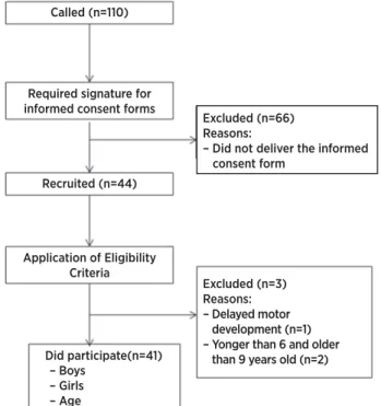
In this study, after the children and their guardians
were asked to sign consent forms, and after the
eligibility criteria were applied, 41 children of both
genders of an average age of 7.82±0.93 years, took part
in this research, as shown in the lowchart (Figure 1).
Called (n=110)
Required signature for informed consent forms
Recruited (n=44)
Application of Eligibility Criteria
Did participate(n=41) – Boys
– Girls – Age
Excluded (n=3) Reasons: – Delayed motor development (n=1) – Yonger than 6 and older than 9 years old (n=2) Excluded (n=66) Reasons:
– Did not deliver the informed consent form
Table 1 shows the sample characterization in regard to gender, chronological age, nutritional state, BMI classiication, and practice of physical exercises.
Table 1: Sample characterization
Sample characterization Gender
Masculine (n; %) Feminine (n; %)
20; 48.8 21; 51.2
Chronological age 6 years (n; %) 7 years (n; %) 8 years (n; %) 9 years (n; %)
10 (24.4) 9 (22) 16 (39) 6 (14.6) Anthropometric measures
Current Weight (Kg)* Heigh (cm)* BMI (Kg/cm2)*
30.29±8.99 1.28±0.08 17.93±3.43 BMI classiication (b)
Eutrophic (n; %) Overweight (n; %) Obese (n; %)
21 (51.2) 10 (24.4) 10 (24.4) Practices Physical exercises
Yes (n; %) 17 (41.5)
* Average ± Standard Deviation BMI: Body Mass Index
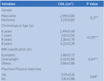
Table 2: Average CSA multiidus CSA measurement as per gender, chronological age, nutritional state, BMI classiication, and practice of physical exercises
Variables CSA (cm²) P Value
Gender Masculine Feminine
2,99±0,86
3,23±0,82 0,37* Chronological Age (a)
6 years 7 years 8 years 9 years
2,49±0,48 2,61±0,56 3,36±0,78 4,22±0,26
<0,00**
BMI classiication (b) Eutrophic
Overweight Obese
2,88±0,72 3,02±0,86 3,68±0,88
0,04**
Practices Physical exercises Yes
No
3,05±0,81
3,16±0,88 0,68*
*Student’s T Test; ** One Way – ANOVA; BMI: Body Mass Index; CSA: Cross-sectional area;
(a) Post Hoc (Tukey): 6 and 7 years (p=0.97), 6 and 8 years (p=0.07), 6 and 9 years (p=0.00), 7 and 8 years (p=0.03), 7 and 9 years (p=0.00), 8 and 9 years (p=0.03),
(b) Post Hoc (Tukey): eutrophic and overweight (p=0.89), eutrophic and obese (p=0.03), overweight and obese (p=0.17)
A multiidus CSA average of 3.11±0.84cm² was found in the studied sample. he two CSA measures that were obtained by the researcher were found to have an excellent intrarater reliability level (ICC=0.98; p=0.00).
Table 2 shows the sample characterization in regard to gender, chronological age, nutritional state, BMI classiication, and practice of physical exercises. As observed, there were a signiicant diferences for multiidus CSA according to the ages and BMI classiications. In regards to ages, diferences were observed among ages 6 and 9 years (p=0.00), 7 and 8 years (p=0.03), 7 and 9 years (p=0.00), and 8 and 9 years (p=0.03), and in regards to BMI, the diferences were between eutrophic and obese classiications (p=0.03)
Table 3 shows the correlation between the CSA measurement of multiidus muscles and chronological age, height, weight, BMI , and BMI classiications. Signiicant, strong, and direct correlation is veriied between CSA and age, height, and weight, and moderate direct correlation is observed in regards to BMI and BMI classiications.
Table 3: Correlation between the CSA measurement of multiidus muscles and chronological age, height, weight, BMI, and BMI classiications
Variables Multiidus CSA
R P
Chronological age 0.75* <0.00
Height 0.83* <0.00
Weight (Kg) 0.75* <0.00
BMI (g/cm2) 0.54* <0.00
BMI classiication
BMI: Body Mass Index; CSA: Cross-sectional area Pearson’s correlation coeicient
DISCUSSION
According to the results obtained this study, the children of 6 to 9 years of age with average height of 1.28±0.08cm, and weight of 30.29±8.99kg, ,were veriied to have average multiidus CSA of 3.11±0.84cm² at rest. he average increased with age, weight, height, and BMI, and no statistically signiicant diferences were found between genders or in regards to the practice of physical exercises.
In the surveyed literature, no multiidus CSA reference values for that age range were found to exist, which highlights the singularity of this study. However,
several studies9,14,15,16 express reference parameters for
multiidus sizes in adults. Stokes, Rankin, and Newham9
identiied an average multiidus CSA measure of L5 in men (8.91+1.68 cm²) and women (6.65+1 cm²) from
also found that average in men (7.58+1.51 cm²) and women (6.01+0.70cm²) in the same age range.
he diferences between CSA in this study, conducted with children, and the one of previously published studies with adults is regarded to the divergent age ranges of samples, as the literature that related to that subject states that there is progressive muscle mass increase with chronological age, from
childhood to adolescence17, and it is found to have more
signiicant gains during puberty18 - those values tend to
remain unaltered in the adult phase.
hat can also explain the fact that the studies by
Stokes et al.9, and Watson et al.14 have respectively
found no or weak correlations between CSA and age, unlike the results in this study, which were found to present strong correlation among variables.
Besides the age, in this study, a positive direct Relationship was found between multiidus CSA and measures of weight, height, and BMI, which is due to the fact that the osteomyoarticular system is still under development in children. hus, the progressive increase in height, weight, and consequently in BMI in childhood and adolescence is expected to cause higher overburden to the trunk, thus directly and indirectly inluencing the forces which play roles in the backbone, hence contributing, to a certain extent, to the adaptive muscular growth.
On the other hand, in studies conducted with adults, there still seems to be a lack of consensus, as Stokes
et al.9 found to signiicant correlations between the
multiidus size and general anthropometric measures,
which contradict the reports from Hides et al.15, who
found signiicant positive relationships of multiidus
CSA of L4 with height and BMI. To Stokes et al.9, it
was hard to explain those diferences; however, they highlight that their sample had an average weight above
the one from Hides et al.15.
In regards to gender, several studies state that there is a statistically signiicant relationship with multiidus
CSA. Hides et al.15, when examining that correlation
in adults, observed that men have signiicantly higher CSAs than women (6.15cm² vs 5.60cm²), which
is similar to the results obtained by Stokes et al.9,
which was expected due to the diferences in the body composition across genders.
Nonetheless, in this study that association was not found, as the volunteers were children from 6 to 9 years of age who have not reached puberty yet (a period which is linked with quick body changes, related to sexual
maturation). hat result is consistent with previous
studies19.20, which concluded that, before puberty, there
is not signiicant diference in the size of muscles across genders. hat diference is observed around 13-15 years, and it varies according to speciic muscles.
In regards to the practice of physical exercises,
Hides, Richardson et al.6 veriied signiicantly increased
multiidus CSA in adult patients with lower back pain, after speciic exercises for those muscles. However, this study has not found that correlation, as the physical exercises that were reported by the children do not cause the speciic recruitment of the multiidus muscles.
A signiicant efect from the practice of physical exercises on multiidus CSA could only be visualized if children performed exercises that were speciic for
those muscles21, such as spinal segmental stabilization.
hat technique could help answering that question in children and adolescents.
his study has limitations which relate to the fact that multiidus evaluation was only conducted at the level of L5 at rest. he study was chosen to be focused on that region, as it has been shown to be the biggest among the muscles in L2-S1, and for showing the best potential to
provide dynamic stability to its segment9,16. he lack of
measurements regarding the size of multiidus muscles during contraction is due to the fact that children have trouble voluntarily controlling those muscles, which rendered the evaluation of that measure impossible.
Besides that, the sample size was small, although
it was comparable to other similar investigations13,16.
hus, despite the sample having been varied in regards to gender, age, and anthropometric parameters, future studies must seek equally diversiied and larger samples, and include the measurement of multiidus during contraction, which is shown to be important in order to evaluate their function and to guide therapeutic
decisions23.
CONCLUSION
study allowed, in an unprecedented way, providing reference measures for the multiidus muscles in that age range, for both genders. Besides that, it reinforced the use of ultrasonography by physical therapists, as an efective and painless way to evaluate the multiidus muscles, both for research and clinical practice purposes, in order to early detect abnormalities in children.
REFERENCES
1. Patalanga N, Field D, Soames R. Anatomia do movimento humano: estrutura e função. São Paulo: Manole; 2000. 2. Moore KL, Dalley AF. Clinically oriented anatomy. Philadelphia:
Lippincott Williams and Wilkins; 2006.
3. Salmela LFT, Sakamoto ACL, Siqueira FB. Mecanismos de estabilização da coluna lombar: uma revisão de literatura. Fisioter Mov. 2004;17(4):51-4.
4. Aspden RM. Review of the functional anatomy of the spinal ligaments and the lumbar erector spinae muscles. Clin Anat. 1992;5(5):372-87.
5. Hamill J, Knutzen KM. Bases biomecânicas do movimento. São Paulo: Manole; 1999.
6. Hides JA, Richardson CA, Jull GA. Multiidus muscle recovery is not automatic following resolution of acute irst episode low back pain. Spine. 1996;21(23):2763-9.
7. Wallwork TL, Staton WR, Freke M, Hides JA. The efect of chronic low back pain on size and contraction of the lumbar multiidus muscle. Manual Ther. 2009;14(5):496-500. 8. Hides J, Gilmore C, Stanton W, Bohlscheid E. Multiidus size
and symmetry among chronic LBP and healthy asymptomatic subjects. Manual Ther. 2008;13(1):43-9.
9. Stokes M, Rankina G, Newhamb, DJ. Ultrasound imaging of lumbar multiidus muscle: normal reference ranges for measurements and practical guidance on the technique. Manual Ther. 2005;37(10):116-26.
10. Silva DAS, Gonçalves ECA, Grigollo LR, Petroski EL. Fatores associados aos baixos níveis de força lombar em adolescentes do Sul do Brasil. Rev Paul Pediatr. 2014;32(4):360-6.
11. Lemos AT, Santos FR, Moreira RB, Machado DT, Braga FCC, Gaya ACA. Ocorrência de dor lombar e fatores associados em crianças e adolescentes de uma escola privada do sul do Brasil. Cad Saúde Pública. 2013; 29(11):2177-85.
12. Balagué F, Troussier B, Salminen JJ. Non-speciic low back pain in children and adolescents: risk factors. Eur Spine J. 1999; 8(6):429-38.
13. World Health Organization. Development of a WHO growth reference for school-aged children and adolescents. Bull of the World Health Org. 2007;1(85):660-7.
14. Watson T, McPherson S, Starr K. The association of nutritional status and gender with cross-sectional area of the multiidus muscle in establishing normative data. J Man Manip Ther. 2008;16(4):E93-8.
15. Hides JA, Cooper DH, Stokes MJ. Diagnostic ultrasound imaging for measurement of the lumbar multiidus muscle in normal young adults. Physiother Theory Practice. 1992;8(1):9-26.
16. Teyhen DS, Childs JD, Stokes MJ, Wright AC, Dugan JL, George SZ. Abdominal and lumbar multiidus muscle size and symmetry at rest and during contracted States. Normative reference ranges. J Ultrasound Med. 2012; 31(7):1099-110.
17. Croix MDS. Advances in pediatric strength assessment: changing our perspective on strength development. J Sports Sci Med. 2007;6(3):292-304.
18. JR, Dante de Rose. Esporte e atividade física na infância e na adolescência. Porto Alegre: Artimed; 2002
19. Kanehisa H, Yata H, Ikegawa S, Fukunaga T. A cross-sectional study of the size and strength of the lower leg muscles during growth. Eur J Appl Physiol Occup Physiol. 1995;72(1-2):150-6. doi: 10.1007/BF00964130.
20. Kanehisa H, Ikegawa S, Tsunoda N, Fukunaga T. Strength and cross-sectional areas of reciprocal muscle groups in the upper arm and thigh during adolescence. Int J Sports Med. 1995;16(1):54-60. doi: 10.1055/s-2007-972964.
21. Danneels LA, Vanderstraeten GG, Cambier DC, Witvrouw EE, Bourgois J, Dankaerts W, De Cuyper HJ. Efects of three diferent training modalities on the cross sectional area of the lumbar multiidus muscle in patients with chronic low back pain. Braz Sports Med. 2001;35(3):186-91.
22.O’Sullivan PB. Lumbar segmental instability: clinical presentation and speciic stabilizing exercise management. Manual Ther 2000;5(1):2-12.