This work is licensed under a Creative Commons Attribution 4.0 International License.
Croat. Chem. Acta 2016, 89(1), 117–124
Published online: June 7, 2016 DOI: 10.5562/cca2797
Study of Dronino Iron Meteorite Weathering in
Clay Sand Using Mössbauer Spectroscopy
Grigoriy A. Yakovlev,1 Andrei V. Chukin,2 Victor I. Grokhovsky,1 Vladimir A. Semionkin,3 Michael I. Oshtrakh1,3,*
1 Department of Physical Techniques and Devices for Quality Control, Institute of Physics and Technology, Ural Federal University,
Ekaterinburg 620002, Russian Federation
2 Department of Theoretical Physics and Applied Mathematics, Institute of Physics and Technology, Ural Federal University,
Ekaterinburg 620002, Russian Federation
3 Department of Experimental Physics, Institute of Physics and Technology, Ural Federal University, Ekaterinburg 620002, Russian Federation
* Corresponding author’s e-mail address: [email protected]
RECEIVED: December 1, 2015 REVISED: April 29, 2016 ACCEPTED: April 29, 2016
THIS PAPER IS DEDICATED TO DR.SVETOZAR MUSIĆ ON THE OCCASION OF HIS 70TH BIRTHDAY
Abstract: Weathering products of two fragments of Dronino iron ungrouped meteorite found in the wet and drier clay sand were studied using X-ray diffraction and Mössbauer spectroscopy with a high velocity resolution. The products of metal oxidation in the internal and external surface layers were different for both fragments. The weathering products in fragment found in the wet clay sand contain magnetite (Fe3O4),
maghemite (-Fe2O3), goethite (-FeOOH) and probably ferrihydrite (5Fe2O39H2O) while those in fragment found in drier clay sand contained
ferric hydrous oxides (FeOOH) and siderite (FeCO3) mainly. Concretions found near the first fragment contain ferric hydrous oxides (FeOOH)
mainly.
Keywords: Dronino iron ungrouped meteorite, weathering, ferric oxides and hydrous oxides, siderite, Mössbauer spectroscopy.
INTRODUCTION
RONINO meteorite shower was found in July 2000 near the Dronino village, Ryazan District, Russian Fed-eration. This meteorite was classified as iron ungrouped meteorite (see Meteoritical Bulletin № 88, 2004). Terres-trial age of this meteorite can be evaluated as more than 1000 years. The first meteorite fragments demonstrated heavily weathering with a rust-covered surface of several centimeters thickness. The Meteoritical Expedition of the Ural Federal University found several fragments of Dronino meteorite in different places of the fall in the clay sand at a depth of ~0.5–~2.0 m. A photo of the place where one frag-ment was found in water-bearing soil is shown in Figure 1a. All surrounded clay sand was colored in reddish-brown due to the rust diffusion mass transfer. Several concretions formed in this place were also found. Some of these sam-ples were studied using Mössbauer spectroscopy ear-lier.[1,2] However, these first measurements were carried
out using the low velocity resolution in the Mössbauer
spectra (smaller discretization of the velocity scale in the spectra). Further, with the development of technique appli-cation, the high velocity resolution Mössbauer spectros-copy (high discretization of both the velocity reference signal and the velocity scale in the spectra) demonstrated significant advances in the study of the complicated spectra of various meteorites in order to extract more detailed in-formation.[3–7] Therefore, in the present work we discuss
the results of re-examination of the first samples of Dronino oxidation products in comparison with new sam-ples studied with the high velocity resolution Mössbauer spectroscopy.
EXPERIMENTAL
Croat. Chem. Acta 2016, 89(1), 117–124 DOI: 10.5562/cca2797 was found in the drier clay sand. The rust layer was
re-moved and fragments were cut (their slices are shown in Figure 1b,c). Oxidized surface can be conventionally divided into two layers denoted as internal (which are denoted as № 1a and № 2a for fragments № 1 and № 2, respectively) and external (which are denoted as № 1b and № 2b for fragments № 1 and № 2, respectively) surface oxidation layers which can be distinguished in their color for fragment № 2 (see Figure 1c). These layers were removed from both fragments and grounded. Two samples of concretions, found in the wet clay sand, with different size and color (small № 3 and big № 4) shown in Figure 1d,e were chosen for this study also. Two parts of each concretion were taken for analysis: the internal friable and the external solid layers from № 3 (which are denoted as № 3a and № 3b, respec-tively) and the yellow-colored internal and the dark-colored external layers from № 4 (which are denoted as № 4a and № 4b, respectively). These samples were also grounded. Additionally, the surface yellow dust (which is denoted as № 4c) was collected from concretion № 4. These powdered samples were used for X-ray diffraction (XRD) study and
then were glued on the iron free aluminum foil with a di-ameter of ~20 mm. The weight of each sample was in the range 30–60 mg that provided the thin absorbers for Möss-bauer measurements.
XRD study was carried out using PANalytical X’pert PRO diffractometer (The Netherlands) with Cu K radiation and position-sensitive X’Celerator detector with Ni filter. Measurements were done in 2 ranges between 10 and 100 with a step of 0.026 and 200 s per step.
Mössbauer spectra were measured using an auto-mated precision Mössbauer spectrometric system built on the base of the SM-2201 spectrometer with a saw-tooth shape velocity reference signal formed by the digital-ana-logue converter using quantification of 212 (discretization
with 4096 steps). This discretization of the velocity refer-ence signal (velocity resolution) produces smaller step in Doppler modulation of resonant -rays energy providing much better adjusting to resonance and significantly creases the quality of spectra measurements. This also in-creases the analytical possibilities of Mössbauer spectroscopy with extracting more reliable spectral compo-nents in the complex spectra due to larger number of spec-tral points and better measurement of the absorption line shape features. Characteristics of this system were de-scribed elsewhere.[8–10] The 1.8109 Bq 57Со(Rh) source
(Ritverc GmbH, St. Petersburg) was used at room tempera-ture. The Mössbauer spectra were measured in transmis-sion geometry with moving absorber in the cryostat at 295 K and recorded in 4096 channels. For their analysis the spectra were converted into 2048 channels to increase sig-nal-to-noise ratio for the smallest spectral components. Statistics in the Mössbauer spectra was in the range ~1.0106 and ~2.6106 counts per channel with
signal-to-noise ratio in the range 43–104. Each spectrum was meas-ured during 3–7 days. Mössbauer spectra were fitted by the least squares procedure with a Lorentzian line shape using UNIVEM-MS program. The line shape of the Möss-bauer spectrum of the standard absorber of -Fe with a thickness of 7 m was pure Lorentzian with outer lines width of 0.2360.008 mm s–1. Velocity resolution (velocity
per one channel) was ~0.010–~0.014 mm s–1 per channel
for the 2048 channels Mössbauer spectra. The spectral pa-rameters isomer shift, , quadrupole splitting (quadrupole shift for magnetically split components), EQ, magnetic hy-perfine field, Heff, line width, , relative subspectrum area,
A, and statistical quality of the fit, 2, were determined.
Cri-teria of the best fit were differential spectrum, 2, and
physical meaning of parameters. An instrumental (system-atic) error for each spectrum point was 0.5 channel (the velocity scale), the instrumental (systematic) error for the hyperfine parameters was 1 channel. The relative instru-mental error for A did not exceed 10 %. If an error
DOI: 10.5562/cca2797 Croat. Chem. Acta 2016, 89(1), 117–124 calculated during the fit (fitting error) for these parameters
exceeded the instrumental (systematic) error we used the largest error. Values of are given relative to -Fe at 295 K.
RESULTS AND DISCUSSION
XRD patterns of the studied samples are shown in Figure 2. The internal oxidation products in fragment № 1 contain mainly magnetite/maghemite Fe3O4/-Fe2O3 (it is not tooeasy to distinguish both oxides using these XRD measure-ments) and ferric hydrous oxides (FeOOH) while the exter-nal oxidation products demonstrate the presence of ferric hydrous oxides mainly (we consider iron-containing pha-ses only). In contrast, the internal and external surface oxidation products in fragment № 2 contain a large amount of siderite (FeCO3) and ferric hydrous oxides.
Both layers in concretion № 3 and the yellow internal layer in concretion № 4 contain ferric hydrous oxides only while dark external layer in concretion № 4 contains siderite additionally. An analysis of ferric hydrous oxides patterns showed that their main part was in the form of goethite (-FeOOH) while some amount of ferrihydrite (5Fe2O39H2O) and other forms of ferric hydrous oxides
could be also considered.
Mössbauer spectra of the internal and external sur-face oxidation layers in fragments № 1 and № 2 are shown in Figure 3. The spectra of the internal and external weath-ering products were different for each fragment. Moreo-ver, the spectra of both internal and external oxidation products appeared to be different for fragments № 1 and № 2, respectively. The results of the best fits of these spec-tra are shown in Figure 3 and parameters are presented in Table 1. An analysis of components 1–5 in the Mössbauer spectrum of the internal oxidation products in Dronino fragment № 1 (Figure 3a) on the basis of previously pub-lished data on iron oxides and hydroxides (see, for in-stance[11–16]) permitted us to distinguish components 1, 3
and 2, 4, 5 and assigned these components to maghemite and magnetite, respectively. Component 1 was related to the 57Fe in the octahedral sites [B] while component 3 was
related to the 57Fe in the tetrahedral sites (A) in -Fe2O3. In
contrast, component 2 was related to the (A) sites in Fe3O4
while two components 4 and 5 were related to the 57Fe in
the [B] sites in Fe3O4. Two components related to the [B]
sites may be related to the presence of ~9.3 at.% Ni in the matrix Fe-Ni-Co alloy in Dronino iron meteorite which can substitute Fe3+ in the [B] positions forming Ni2+-containing
magnetite during the weathering similar to NiFe2O4 inverse
spinel in which nickel cations can occupy [B] sites only and their different numbers in the iron local environment may be a reason of different magnetic hyperfine fields on the
57Fe nuclei (see Refs. [17,18]). Other magnetic sextets 6–8
were associated with goethite small particles with different
size and smaller values of Heff than that for bulk -FeOOH (the Néel temperature is 400 K16). Similar decrease of the
values of Heff with the decreasing of goethite nanoparticles size was observed in Ref. [19]. Three paramagnetic quadru-pole doublets demonstrate hyperfine parameters indicat-ing the presence of ferric compounds. These components can be assigned to paramagnetic goethite nanoparticles and/or ferrihydrite small particles (the latter Néel temper-ature is below 100 K). Therefore, these components were related to ferric hydrous oxides FeOOH (nanosized goethite and/or ferrihydrite). In the Mössbauer spectrum of the ex-ternal oxidation products in Dronino fragment № 1 (Figure 3b) we revealed four magnetic sextets 1–4 which can be related to -FeOOH small particles with different size and variations in the values of Heff (smaller value of for com-ponent 2 rises a question concerning the possibility to re-late this parameter to goethite). Three paramagnetic components had hyperfine parameters corresponding to ferric compounds. However, Mössbauer parameters for quadrupole doublets 5 and 6 could be related to FeOOH (nanosized goethite and/or ferrihydrite) while component 7 had larger value of than that for ferric hydrous oxides.
Quite different results were obtained for both inter-nal and exterinter-nal oxidation products in Dronino fragment № 2. The Mössbauer spectrum of the internal oxidation prod-ucts in fragment № 2 (Figure 3c) demonstrated two weak and broad magnetic sextets 1 and 2. Their hyperfine param-eters can be related to the presence of Fe-Ni-Co alloy ma-trix (due to the sample preparation) and goethite particles,
Figure 2. X-ray diffraction patterns of the internal (INT) and external (EXT) surface oxidation products in Dronino fragments № 1 (a) and № 2 (b) and the internal (INT) and external (EXT) surface layers in concretions № 3 (c) and № 4 (d). Indicated selected reflexes are magnetite/maghemite, PDF-2: 01-089-0951 (M), goethite, PDF-2: 00-029-0713 (G), ferrihydrite, PDF-2: 00-029-0712 (F) and siderite, PDF-2: 01-083-1764 (S).
8500 13500 18500 23500
20 30 40 50 60 70
IN TE N SI TY , co un ts
2,
13500 14500 15500 16500 17500 18500
20 30 40 50 60 70
IN TE N SI TY , co un ts
2, °
5000 11000 17000 23000 29000 35000 41000
20 30 40 50 60 70
IN TE N SI T Y, c ou nt s
2, °
6000 16000 26000 36000
20 30 40 50 60 70
IN TE N SI TY , co un ts
2, ° G M G M G G F F G G
M M
G G
S
F F
G F G G G G G G
G S
G S
G
G S
INT EXT INT EXT INT EXT EXT INT
a b
c d
Croat. Chem. Acta 2016, 89(1), 117–124 DOI: 10.5562/cca2797 respectively. Two quadrupole doublets 3 and 4 had
hyper-fine parameters characterized the presence of ferrous com-pounds while the other quadrupole doublets 5–8 demon-strated the presence of ferric compounds. The values of EQ
and for components 3 and 4 were similar to siderite which parameters varied in the ranges of EQ ~1.6–2.0 mm s–1
and ~1.2–1.3 mm s–1 (see Refs. [20–23]). Basing on XRD
data about the presence of FeCO3 in the weathering
products of Dronino fragment № 2 we assigned both components 3 and 4 to siderite with some possible small variations in the iron local microenvironment. Paramag-netic quadrupole doublets 5, 6 and 8 were related to FeOOH (nanosized goethite and/or ferrihydrite) with open question concerning smaller value of for component 6. Quadrupole doublet 7 had hyperfine parameters character-ized ferric compound. The Mössbauer spectrum of the
external oxidation products in Dronino fragment № 2 (Figure 3d) demonstrated more spectral components. Two magnetic sextets 1 and 2 were related to the 57Fe in the (A)
and [B] sites in magnetite while the other magnetic sextets 3–6 were assigned to goethite small particles with differ-ent size and variations in the values of Heff. Paramagnetic
quadrupole doublet 7 with Mössbauer parameters indic-ating the ferrous compound was related to FeCO3 like
similar components 3 and 4 in the Mössbauer spectrum of the internal weathered products (Figure 3c). Paramagnetic components 8 and 9 were assigned to FeOOH (nanosized goethite and/or ferrihydrite) while component 10 was related to ferric compound.
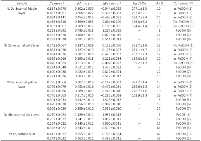
The Mössbauer spectra of concretions’ samples are shown in Figure 4. These spectra demonstrated the pres-ence of magnetic and paramagnetic (superparamagnetic)
DOI: 10.5562/cca2797 Croat. Chem. Acta 2016, 89(1), 117–124 components for the internal friable and external solid
lay-ers in concretion № 3 and for the internal yellow layer in concretion № 4. The spectra of the external dark layer and surface dust in concretion № 4 consisted of paramagnetic (superparamagnetic) components only. The results of the best fits of these spectra are shown in Figure 4 and param-eters are presented in Table 2. All magnetic sextets in these spectra demonstrated hyperfine parameters which can be related to -FeOOH small particles with different size and variations in the values of Heff. All paramagnetic quadrupole doublets demonstrating hyperfine parameters typical for ferric compounds (except component 1 in Figure 4d with parameters related to ferrous compound) were assigned to FeOOH (nanosized goethite and/or ferrihydrite). Ferrous compound observed in the Mössbauer spectrum of the ex-ternal dark layer in concretion № 4 had the values of EQ
and similar to siderite.
The results of the study of Dronino iron meteorite weathering products using XRD and Mössbauer spectros-copy demonstrated the difference in the surface oxidation of fragment № 1 found in the wet clay sand and fragment № 2 found in the drier clay sand. The internal oxidation products in fragment № 1 consisted of maghemite, mag-netite and ferric hydrous oxides, mainly goethite, while the external oxidation products contained ferric hydrous ox-ides and unidentified ferric compound. It is possible that oxidation of the Fe-Ni-Co alloy in Dronino fragment led to formation of magnetite with its further oxidation to ma-ghemite. Then maghemite interaction with water led to for-mation of ferric hydrous oxides. This process moves inside the metal while the external surface layer contains ferric hydrous oxides only. These ferric hydrous oxides are in the form of small particles and/or nanoparticles demonstrating superparamagnetic behavior at room temperature. These
Table 1. Mössbauer parameters of the surface oxidation products from Dronino iron meteorite fragments № 1 and № 2
Sample / mm s–1 / mm s–1 EQ / mm s–1 Heff / kOe A / % Component(a)
№ 1a, internal surface
oxidation products 0.3630.376±±0.073 0.036 0.449±0.047 0.314±0.081 0.664±0.068 0.776±0.070 0.776±0.026 0.776±0.026 0.776±0.026 0.233±0.026 0.284±0.026
0.397±0.015 0.332±0.013 0.277±0.017 0.425±0.023 0.578±0.018 0.380±0.021 0.316±0.019 0.286±0.025 0.444±0.013 0.389±0.013 0.390±0.013
–0.065±0.024 –0.009±0.013 0.029±0.027 0.056±0.029 –0.146±0.035 –0.456±0.040 –0.095±0.033 –0.057±0.040 1.033±0.019 0.782±0.014 0.503±0.013
507.1±2.1 494.0±0.9 483.9±1.9 472.8±1.8 449.0±1.9 326.4±1.5 253.9±1.7 189.6±1.5
– – – 5 13 10 4 8 9 10 8 16 3 14
-Fe2O3 [B] (1)
Fe3O4 (A) (2)
-Fe2O3 (A) (3)
Fe3O4 [B1] (4)
Fe3O4 [B2] (5)
-FeOOH (6) -FeOOH (7) -FeOOH (8) FeOOH (9) FeOOH (10) FeOOH (11) № 1b, external surface
oxidation products 0.7760.776±±0.026 0.026 0.776±0.154 0.776±0.026 0.423±0.034 0.300±0.026 0.767±0.106
0.425±0.040 0.104±0.048 0.349±0.040 0.442±0.051 0.322±0.013 0.379±0.013 0.604±0.032
–0.344±0.071 –0.036±0.091 –0.303±0.076 0.270±0.082 0.926±0.027 0.514±0.013 0.750±0.028
297.3±1.9 236.8±2.7 177.8±2.8 126.5±3.7
– – – 11 10 10 9 14 28 18 -FeOOH (1) ? -FeOOH (2)
-FeOOH (3) -FeOOH (4) FeOOH (5) FeOOH (6) Fe3+ compound (7)
№ 2a, internal surface
oxidation products 0.6530.776±±0.055 0.077 0.293±0.025 0.342±0.022 0.326±0.022 0.541±0.022 0.299±0.022 0.233±0.022
–0.065±0.024 0.287±0.039 1.306±0.011 1.087±0.011 0.395±0.011 0.165±0.011 0.709±0.011 0.521±0.011
–0.235±0.047 0.297±0.071 2.042±0.297 2.011±0.011 1.034±0.011 0.961±0.011 0.858±0.014 0.374±0.011
322.7±0.8 315.1±1.2
– – – – – – 7 6 4 38 13 21 6 5
? Fe-Ni-Co (1) -FeOOH (2) FeCO3 1 (3)
FeCO3 2 (4)
FeOOH (5) ? FeOOH (6) Fe3+ compound (7)
FeOOH (8) № 2b, external surface
oxidation products 0.4740.399±±0.028 0.063 0.628±0.050 0.495±0.077 0.776±0.044 0.776±0.067 0.286±0.022 0.549±0.022 0.260±0.022 0.274±0.022
0.245±0.011 0.708±0.021 0.375±0.014 0.500±0.022 0.462±0.012 0.444±0.021 1.031±0.011 0.343±0.011 0.358±0.011 0.527±0.011
0.154±0.021 –0.167±0.042 –0.592±0.038 0.030±0.043 –0.040±0.022 –0.427±0.033 2.100±0.011 1.044±0.011 0.496±0.011 0.411±0.011
487.6±0.6 459.3±1.3 331.5±0.8 329.3±1.2 274.8±1.2 192.7±1.2
– – – – 7 2 8 4 11 7 11 18 21 9
Fe3O4 (A) (1)
Fe3O4 [B] (2)
-FeOOH (3) -FeOOH (4) -FeOOH (5) -FeOOH (6) FeCO3 (7)
Croat. Chem. Acta 2016, 89(1), 117–124 DOI: 10.5562/cca2797 particles as well as individual FeOOH molecules can diffuse
or be transferred into the clay sand with changing its color and further formation of concretions. These concretions consist of ferric hydrous oxides with different sizes of FeOOH particles/nanoparticles showed different superpar-amagnetic behavior of the internal and external layers at room temperature. The oxidation process in Dronino
fragment № 2 was quite different. The internal surface oxidation products contain ferric hydrous oxides, siderite and unidentified ferric compound while the external surface weathering products consist of small amount of magnetite, ferric hydrous oxides, siderite and unidentified ferric compound. It should be noted that siderite formation in clay may be a result of bioreduction of goethite by
DOI: 10.5562/cca2797 Croat. Chem. Acta 2016, 89(1), 117–124 bacteria.[24] Therefore, bacteria can live inside the internal
oxidation layer in the drier clay sand with conversion of goethite into siderite moving inside the metal matrix with the oxidation process. Observation of small amount of siderite in the external dark layer in concretion № 4 may also be a result of goethite bioreduction, however, bacteria in this case might convert goethite into siderite at the early stage of concretion formation with further stopping their activity.
CONCLUSION
Study of Dronino iron ungrouped meteorite fragments in the clay sand was carried out using XRD and Mössbauer spectroscopy with a high velocity resolution. Surface oxida-tion products in two fragments found in the wet and drier clay sand appeared to be different. Ferric oxides and hy-drous oxides were observed in the first fragment while in the second one siderite was additionally found. It was pos-sible to distinguish the internal and external surface oxida-tion products in both fragments. In the fragment found in the wet clay sand the internal surface oxidation products contained ferric oxides and hydrous oxides while the external surface oxidation products consisted of ferric
hydrous oxides. The weathering process of the second fragment found in drier clay sand showed the possible participation of bacteria which converted goethite into siderite observed in both internal and external surface oxidation products. The transfer of ferric hydrous oxides in the wet clay sand led to concretions formation from ferric hydrous oxides with different particles size.
Acknowledgment. This work was supported in part by the basic financing from the Ministry of Education and Science of Russian Federation (basic financing for the Projects # 2085 and # 1514) and by Act 211 Government of the Russian Federation, contract № 02.A03.21.0006.
REFERENCES
[1] V. I. Grokhovsky, M. I. Oshtrakh, O. B. Milder, V. A. Semionkin, Bull. Russ. Acad. Sci., Phys. 2005, 69, 1710. [2] V. I. Grokhovsky, M. I. Oshtrakh, O. B. Milder, V. A.
Semionkin, Hyperfine Interact. 2005, 166, 671. [3] V. I. Grokhovsky, E. V. Zhiganova, M. Yu. Larionov, K.
A. Uymina, M. I. Oshtrakh, The Phys. Metals Metallogr. 2008, 105, 177.
Table 2. Mössbauer parameters of concretions matter found in Dronino fragments surrounded clay sand
Sample / mm s–1 / mm s–1 EQ / mm s–1 Heff / kOe A / % Component(a)
№ 3a, internal friable
layer 0.8340.833±±0.078 0.061 0.834±0.141 0.488±0.310 0.833±0.061 0.233±0.092 0.517±0.026 0.283±0.028
0.302±0.030 0.366±0.027 0.454±0.018 0.199±0.055 0.209±0.017 0.485±0.018 0.408±0.013 0.391±0.026
–0.044±0.055 –0.392±0.053 –0.485±0.031 0.004±0.109 –0.103±0.035 1.267±0.035 0.879±0.015 0.511±0.013
277.1±1.5 232.4±2.1 193.7±2.4 165.8±5.2 124.4±1.4
– – – 10 10 14 2 20 1 21 22 -FeOOH (1) -FeOOH (2) -FeOOH (3) ? -FeOOH (4) ? -FeOOH (5) FeOOH (6) FeOOH (7) FeOOH (8) № 3b, external solid layer 0.748±0.067
0.834±0.026 0.834±0.026 0.559±0.066 0.470±0.055 0.294±0.048 0.600±0.030 0.271±0.026
0.137±0.029 0.327±0.019 0.382±0.029 0.395±0.018 0.213±0.019 0.521±0.013 0.422±0.013 0.383±0.013
0.132±0.056 –0.173±0.027 –0.425±0.052 0.223±0.034 –0.007±0.027 1.425±0.022 0.812±0.024 0.517±0.013
321.7±1.6 281.1±1.7 233.7±2.3 184.6±1.3 135.2±1.2
– – – 13 17 12 10 7 3 22 16
? -FeOOH (1) -FeOOH (2) -FeOOH (3) -FeOOH (4) ? -FeOOH (5)
FeOOH (6) FeOOH (7) FeOOH (8) № 4a, internal yellow
layer 0.7760.776±±0.064 0.079 0.776±0.086 0.776±0.065 0.301±0.034 0.470±0.020 0.309±0.020
0.302±0.018 0.483±0.016 0.289±0.023 0.317±0.016 0.432±0.010 0.356±0.010 0.359±0.010
–0.147±0.033 –0.472±0.031 –0.245±0.040 –0.286±0.028 1.526±0.017 0.902±0.010 0.532±0.010
327.2±1.4 282.6±1.2 228.7±1.6 162.9±1.3
– – – 13 14 10 13 3 19 27 -FeOOH (1) -FeOOH (2) -FeOOH (3) -FeOOH (4) FeOOH (5) FeOOH (6) FeOOH (7) № 4b, external dark layer 0.250±0.022
0.292±0.022 0.254±0.022 0.318±0.022
1.129±0.011 0.341±0.011 0.345±0.011 0.343±0.011
1.931±0.012 1.097±0.011 0.800±0.011 0.519±0.011
– – – – 6 12 17 64 FeCO3 (1) FeOOH (2) FeOOH (3) FeOOH (4) № 4c, surface dust 0.445±0.022
0.244±0.022 0.3550.363±±0.011 0.011 0.4880.723±±0.034 0.011 – – 62 38 FeOOH (1) FeOOH (2)
Croat. Chem. Acta 2016, 89(1), 117–124 DOI: 10.5562/cca2797 [4] M. I. Oshtrakh, V. I. Grokhovsky, E. V. Petrova, M. Yu.
Larionov, K. A. Uymina, V. A. Semionkin, N. V. Abramova in Proceedings of the International Conference “Mössbauer Spectroscopy in Materials Science 2008” (Eds.: M. Mashlan, R. Zboril), AIP Conference Proceedings, Melville, New York 2008,
1070, 131.
[5] V. I. Grokhovsky, M. I. Oshtrakh, E. V. Petrova, M. Yu. Larionov, K. A. Uymina, V. A. Semionkin, Eur. J. Mineral. 2009, 21, 51.
[6] M. I. Oshtrakh, V. I. Grokhovsky, E. V. Petrova, M. Yu. Larionov, M. V. Goryunov, V. A. Semionkin, J. Mol. Struct. 2013, 1044, 268.
[7] M. I. Oshtrakh, A. A. Maksimova, M. V. Goryunov, G. A. Yakovlev, E. V. Petrova, M. Yu. Larionov, V. I. Grokhovsky, V. A. Semionkin, Mössbauer spectroscopy with a high velocity resolution: advances in the study of meteoritic iron-bearing minerals in Proceedings of the Workshop on The Modern Analytical Methods Applied to Earth and Planetary Sciences (Ed.: A. Gucsik), The MicroMatLab Kft Hungary, Sopron, 2015, pp. 43–86. [8] M. I. Oshtrakh, V. A. Semionkin, O. B. Milder, E. G.
Novikov, J. Radioanal. Nucl. Chem. 2009, 281, 63. [9] V. A. Semionkin, M. I. Oshtrakh, O. B. Milder, E. G.
Novikov, Bull. Rus. Acad. Sci.: Phys. 2010, 74, 416. [10] M. I. Oshtrakh, V. A. Semionkin, Spectrochim. Acta,
Part A2013, 100, 78.
[11] L. H. Bowen, S. B. Weed, Mössbauer Spectroscopy of Soils and Sediments in Chemical Mössbauer Spectroscopy (Ed.: R. L. Herber), Plenum Press, New York and London, 1984, pp. 217–242.
[12] E. Murad, J. H. Johnston, Iron Oxides and Oxyhydroxides in Mössbauer Spectroscopy Applied to Inorganic Chemistry, Vol. 2 (Ed.: G. J. Long),
Plenum Press, New York, 1987, pp. 507–582. [13] B. A. Goodman, Mössbauer Spectroscopy in Clay
Mineralogy: Spectroscopic and Chemical
Determinative Methods (Ed.: M. J. Wilson), Chapman & Hall, London, 1994, pp. 68–119. [14] R. Zboril, M. Mashlan, D. Petridis, Chem. Mater.
2002, 14, 969.
[15] E. Murad, J. Cashion, Mössbauer Spectroscopy of Environmental Materials and their Industrial Utilization, Kluwer Academic Publishers, New York, 2004, p. 418.
[16] E. Murad, Clay Miner. 2010, 45, 413.
[17] M. I. Oshtrakh, M. V. Ushakov, B. Senthilkumar, R. Kalai Selvan, C. Sanjeeviraja, V. A. Semionkin,
Proceedings of the International Conference “Mössbauer Spectroscopy in Materials Science 2012” (Eds.: J. Tuček, L. Machala), AIP Conference Proceedings, Melville, New York, 2012, 1489, 115. [18] M. I. Oshtrakh, M. V. Ushakov, B. Senthilkumar, R.
Kalai Selvan, C. Sanjeeviraja, I. Felner, V. A. Semionkin, Hyperfine Interact. 2013, 219, 7. [19] I. Mitov, D. Paneva, B. Kunev, Thermochim. Acta
2002, 386, 179.
[20] S. Musić, S. Popović, J. Radioanal. Nucl. Chem. 1987,
111, 27.
[21] B. B. Ellwood, B. Burkart, K. Rajeshwar, R. L. Darwin, R. A. Neeley, A. McCall, G. J. Long, M. L. Buhl, C. W. Hickcox, J. Geophys. Res.1989, 94, 7321.
[22] S. Ram, K. R. Patel, S. K. Sharma, R. P. Tripathi, Fuel 1998, 77, 1507.
[23] G. Medina, J. A. Tabares, G. A. Pérez Alcazar, J. M. Barraza, Fuel 2006, 85, 871.