ÓRgÃO OfICIAL dA SOCIEdAdE PORTUgUESA dE REUMATOLOgIA
78 CASO CLÍNICO
1. Serviço de Reumatologia, Centro Hospitalar e Universitário de Coimbra
stages of the CD19+B cell maturation process. An accu -rate estimate of the incidence or prevalence of XLA is difficult to obtain because the disease is uncommon. However, a minimal estimate of approximately 1 in 379,000 live births was provided from a national regis -try1. Typically, XLA patients present lymphoid hypo -plasia, almost complete absence of CD19+B cells in the peripheral blood, severe hypogammaglobulinemia with severe antibody deficiency and increased susceptibili-ty to infection. The most common clinical manifesta-tions of XLA are recurrent and chronic infecmanifesta-tions (bacte rial - encapsulated pyogenic bacteria, viral, fun-gal or parasitic). More rarely, some malignancies, sen-sorineural hearing loss and auto-immune manifesta-tions have been associated with XLA. In 1956, Janeway et al. reported a syndrome with cutaneous and muscu-lar manifestations resembling dermatomyositis in pa-tients with XLA2. Since then this condition has been termed dermatomyositis-like syndrome (DLS).
This rare association of conditions is usually pro-gressive and fatal and is characterized by erythema, ede-ma and induration of the skin, muscle weakness, and flexion contractures of the extremities2-4.
This syndrome is typically accompanied by central nervous system manifestations associated with persis-tent infection by an Echovirus. The involvement of Echovirus at the onset of the cutaneous and muscular manifestations has also been suggested5.
cAse report
We report the case of a 27-years old male with XLA, whose medical history included xerophthalmia, de-creased visual acuity, repeated conjunctivitis, severe dental caries, chronic obstructive pulmonary disease (with a forced expiratory volume in 1 second (FEV1)=38,4% and a spirometry/FEV1 relationship of of 51,3), cylindrical bronchiectasis with Haemophilus
Dermatomyositis-like syndrome
in x-linked agammaglobulinemia
Carvalho PD1, Costa C1, Rodrigues M1, Salvador MJ1, Pereira da Silva JA1, Malcata A1
AbstrAct
Primary immunodeficiencies (PIDs) encompass more than 250 different pathological conditions. X-linked agammaglobulinemia (XLA) has been occasionally as-sociated with cutaneous and muscular manifestations resembling dermatomyositis, often termed dermato-myositis-like syndrome (DLS). This syndrome has been associated with cutaneous, muscular and central ner-vous system manifestations, accompanying a persistent infection by an Echovirus. According to sixteen pre -viously reported cases, this syndrome has a poor progno sis. We report the case of a 27-years old male, with XLA and DLS, successfully treated with 6 cycles of human immunoglobulin and methotrexate. Clinical symptoms improved dramatically with a complete re -solution of the musculoskeletal manifestations. Despite this clinical response, prognosis should remain re-served. The evolution of this syndrome remains un-predictable and therapeutic options are limited. To the best of our knowledge, there are only a few reports of similar cases which have survived so many months af-ter the diagnosis.
Keywords: Dermatomyositis-like syndrome; X-linked agammaglobulinemia; Immunoglobulin; Primary im-munodeficiencies
IntroductIon
Primary immunodeficiencies (PIDs) encompass more than 250 different pathological conditions. X-linked agammaglobulinemia (XLA) is a primary humoral de-ficiency due to defects in a signal transduction molecule – Bruton tyrosine kinase – which is expressed in every
ÓRgÃO OfICIAL dA SOCIEdAdE PORTUgUESA dE REUMATOLOgIA
79 Carvalho PD et al
influenzacolonization, pansinusitis, peptic esophagitis, Giardia lambliagastrointestinal infection and low height and weight. He also described progressive sen-sorineural hearing loss requiring placement of audito-ry prostheses. XLA had been confirmed several years before by demonstration of 16 and 18 exons deletion of Bruton Tyrosine Kinase (BTK) gene.
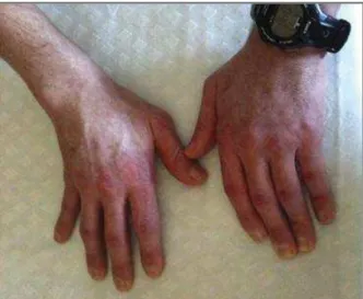
The patient presented to the Rheumatology De-partment due to persistent symmetric polyarthritis evolving over the last 5 years, associated with proximal muscle weakness and paresthesia of both feet over the past 2 years. The patient was receiving naproxen, anal-gesics, 19 mg of prednisolone-equivalent and “re-placement” doses of human immunoglobulin (ac-cording to serum levels) for his XLA. At first observa-tion, he was 166cm tall, he weighted 49 Kg and the cardiopulmonary sounds were normal. We observed a pronounced proximal muscular atrophy of the lower limbs (Figure 1) and the presence of erythematous cu-taneous lesions bila terally on the 2nd, 3th and 4th metacarpophalangeal joints (Figure 2). Limited range of motion was present in the elbows, wrists, hips, knees and ankles. Swelling and tenderness of several metacarpophalangeal and proximal interphalangeal joints was documented. Distal strength of both upper and lower limbs was notably normal, but reduced muscle strength of the pelvic girdle was observed (3+ in a five grade scale).
Laboratory results showed no significant abnor-malities, namely in the inflammatory parameters and muscle enzymes. The electromyogram revealed signs of muscle fibers lesion in the right iliopsoas muscle. Following electromyography the patient started com-plaining of involuntary muscle contractions (mainly in the right lower limb, where the exam had been per-formed). Muscle biopsy was performed in the right deltoid muscle and demonstrated abundant inflam-matory infiltrate in muscle fibres (mainly CD3+, CD4+ and CD8+ T cells) with evident perifascicular atrophy suggesting dermatomyositis.
We started treatment with methotrexate in gradual increase (10 mg per week until 15 mg per week), as-sociated with human immunoglobulin (400 mg/Kg/ /day during 5 days, each month, for 6 months). The clinical symptoms improved dramatically, including a complete resolution of the articular manifestations, paraesthesia and muscle involuntary contractions. Muscle weakness was gradually improved over a few months and the grading scale for assessment of func-tion in patients with myositis6evolved from 19/30 to
28/30. Currently, 9 months after starting the im-munoglobulin, the patient remains well under this treatment of methotrexate, 15 mg per week, orally, prednisolone, 10 mg per day, orally, and monthly re-FIGure 1. Pronounced proximal muscular atrophy of the lower limbs
FIGure 2. Erythematous cutaneous lesions overlying the 2nd,
ÓRgÃO OfICIAL dA SOCIEdAdE PORTUgUESA dE REUMATOLOgIA
80
Dermatomyositis-like synDrome in x-linkeD agammaglobulinemia
placement do ses of human immunoglobulin (doses ac-cording to serum levels), to control XLA.
dIscussIon
The hypothesis usually proposed for the pathogenesis of this syndrome is persistent viral infection, namely by an Echovirus. In some cases presented on literature, viruses were isolated usually from the cerebrospinal fluid but also occasionally from muscle. Typically a sin-gle type of Echovirus is found, although an Echovirus--Adenovirus combination or an association of two Echoviruses have been described7. It should be men-tioned that neurologic signs are often usually mild, par-ticularly at the onset, and can even be completely
ab-sent8. The Echovirus infection had been associated with meningoencephalitis and with DLS. However, it is not clear if the persistent viral infection has to be present in the central nervous system or in other tissue, namely muscle. In our case report, there were no neurologi -cal signs that could justify a lumbar puncture to search for the presence of an Echovirus. We did not search for viruses elsewhere, because, based on available evidence, their identification would not change the thera -peutic approach.
In 1985, Lederman et al9, performed an analysis of 96 patients with XLA. DLS was observed in 6 patients, all of which had meningitis/encephalitis. Five of these patients also had arthritis. Five of the six patients had disseminated Adenovirus infection. All six patients died. One patient had DLS at diagnosis of XLA and the
tAble I. XlA And dls (16 lIterAture cAses As oF 1990)6
Nº Clinical manifestations Neurologic manifestations Viral cultures Evolution
1 Erythema, edema, Meningoencephalitis Echovirus, adenovirus Died
muscle infiltration
2 Erythema, edema, Meningoencephalitis Echovirus Died
muscle infiltration
3 Erythema, edema, Seizures, headache, Not performed Died
muscle infiltration altered mental status
4 Erythema, edema, polymyositis Confusion, loss of memory Echovirus Died
5 Cutaneous atrophy, edema, Headaches, Echovirus Died (10 weeks)
polymyositis nuchal rigidity
6 Polymyositis Deafness Echovirus Died (2,5 years)
7 Erythema, polymyositis No signs Echovirus Died (7 months)
8 Erythema, edema, polymyositis Headaches, seizure Echovirus Died (29 months)
9 Erythema, edema, polymyositis Seizures, confusion Echovirus Died (3 years)
10 Polymyositis No signs Echovirus Alive (4 years)
11 Cutaneous atrophy, Nerve VI + VII, paresis, Echovirus Died (4 years)
polymyositis ataxia, deafness
12 Polymyositis, edema Lethargy Echovirus Alive (5,5 years)
13 Polymyositis, edema Seizures, deafness Echovirus Died (2 years)
14 Erythema, edema, polymyositis Seizures, confusion Echovirus Died (1 year)
15 Cutaneous atrophy, Cranioencephalic computed Echovirus Died (1 month)
edema, polymyositis tomography (CE-CT) alterations
16 Erythema, polymyositis Seizures Negative Died (2 years)
tAble II. XlA And dls (17thcAse reported)
Nº Clinical manifestations Neurologic manifestations Viral cultures Evolution
ÓRgÃO OfICIAL dA SOCIEdAdE PORTUgUESA dE REUMATOLOgIA
81 Carvalho PD et al
others developed it while on gamaglobulin prophyla -xis9. A case report of a successful treatment of this syn-drome with intravenous immunoglobulin therapy in a patient with XLA was described10.
In 1990, Thyss et al7, made a review of the literature and found 16 reported cases of DLS associated with XLA. The most interesting results of that review are shown in the Table I. The majority of patients have a conjugation of the following symptoms: erythema, ede-ma, muscle infiltration, cutaneous atrophy and polymyositis. Most of them had neurological manifes-tations, the Echovirus was found in 14 (viral cultures were performed only in 15) and the prognosis was dra-matic (14 of 16 died within 5.5 years). In Table II we perform a similar classification of the case we report herein. Since 1990, several cases of a dermatomyositis-like syndrome were described, but none was reported in association to XLA.
The patient reported in this paper had a good res -ponse to high-dose immunoglobulin treatment. Wag-ner et al11, described a patient with XLA and growth hormone deficiency who developed an Echovirus-as-sociated meningoencephalitis and DLS while being treated with intramuscular gammaglobulin (replace-ment dose) and human growth hormone. Initiation of highdose intravenous gammaglobulin resulted in reso -lution of the clinical symptoms and the patient re-mained asymptomatic over 55 months.
Another reported case of treatment with gamma-globulin, methotrexate and corticosteroids (case 14, Table I), who had a poor prognosis, was found in the literature12. In our case, an off-label immunomodulator treatment was added to the immunoglobulin the -rapy as well. Our patient had a clinical relevant pe-ripheral polyarthritis and a monthly intense treatment with immunoglobulin was interpreted has a transitory situation. Hence, regarding the existence of no recent reports of infections on this patient, and the expectan-cy that a sustained reduction on the immunoglobulin therapy frequency could be done without clinical flares of the myositis, methotrexate was added. On the o ther hand, the prednisolone dose was gradually tapered un-til a smaller dose could be reached, regarding the in-fectious risks concerns.
We describe an additional case of excellent response to high-dose intravenous immunoglobulin, at least in the short term. Besides the viral cultures for Echovirus were not performed, it was possible to identify and successfully treat this rare syndrome. Although the pre sence of an Echovirus was observed in most of the clini
-cal cases described in literature, there is no evidence to associate the presence of this virus with clinical mani-festations, recommended treatment or outcome. Gi ven the rarity of these conditions, no clinical trials are ex-pected in the near future, thus underlining the impor-tance of sharing clinical experience.
correspondence to Pedro Carvalho
Serviço de Reumatologia, Centro Hospitalar e Universitário de Coimbra, Portugal
E-mail: pedrodcsc@gmail.com
reFerences
1. Winkelstein JA, Marino MC, Lederman HM, Jones SM, Sullivan K, Burks AW, et al. X-linked agammaglobulinemia: report on a United States registry of 201 patients. Medicine (Baltimore). 85. United States2006. p. 193-202.
2. Craig JM, Gitlin D, Grice DS, Janeway CA. Collagen disease in patients with congenital agammaglobulinemia. Trans Assoc Am Physicians 1956;69:93-97.
3. Page AR, Hansen AE, Good RA. Occurrence of leukemia and lymphoma in patients with agammaglobulinemia. Blood 1963;21:197-206.
4. Gotoff SP, Smith RD, Sugar O. Dermatomyositis with cerebral vasculitis in a patient with agammaglobulinemia. Am J Dis Child 1972;123(1):53-56.
5. Bodensteiner JB, Morris HH, Howell JT, Schochet SS. Chronic ECHO type 5 virus meningoencephalitis in X-linked hypo-gammaglobulinemia: treatment with immune plasma. Neuro-logy 1979;29(6):815-819.
6. John K, Paul D. Rheumatology. Second Edition ed2008. 7. Thyss A, el Baze P, Lefebvre JC, Schneider M, Ortonne JP.
Der-matomyositis-like syndrome in X-linked hypogammaglobuli-nemia. Case-report and review of the literature. Acta Derm Ve-nereol 1990;70(4):309-313.
8. Wilfert CM, Buckley RH, Mohanakumar T, Griffith JF, Katz SL, Whisnant JK, et al. Persistent and fatal central-nervous-system ECHOvirus infections in patients with agammaglobulinemia. N Engl J Med 1977;296(26):1485-1499.
9. Lederman HM, Winkelstein JA. X-linked agammaglobulinemia: an analysis of 96 patients. Medicine (Baltimore) 1985;64(3): 145-156.