Article
A 5-Year Prospective Follow-Up Study of Lipid-Rich
Adrenal Incidentalomas: No Tumor Growth or
Development of Hormonal Hypersecretion
Camilla Schalin-Jäntti1, Merja Raade2, Esa Hämäläinen3, Timo Sane1
1Department of Endocrinology, Abdominal Center, 2Department of Radiology, HUS Medical Imaging Center, Helsinki
University Central Hospital, University of Helsinki; 3
HUSLAB, Department of Clinical Chemistry, University of Helsinki, Helsinki, Finland
Background: Current guidelines for follow-up of adrenal incidentalomas are extensive and hampered by lack of follow-up stud-ies. We tested the hypothesis that small lipid-rich adrenal incidentalomas, initially characterized by tumor size <40 mm and <10
Hounsfield units (HUs) on unenhanced computed tomography (CT) may not demonstrate excessive growth/hormonal hyperse-cretion on follow-up.
Methods: Sixty-nine incidentalomas in 56 patients were restudied with unenhanced CT and screening for hypercortisolism (dexamethasone suppression test [DST], plasma adrenocorticotropic hormone) and pheochromocytoma (24-hour urinary meta-nephrines and normetameta-nephrines) 5 years later. Primary hyperaldosteronism was excluded at base-line.
Results: Tumor (n=69) size was similar before and after 5 years follow-up (19±6 mm vs. 20±7 mm). Mean tumor growth was
1±2 mm. Largest increase in tumor size was 8 mm, this tumor was surgically removed and histopathology confirmed cortical
ad-enoma. DST was normal in 54 patients and two patients (3.6%) were still characterized by subclinical hypercortisolism. Initial tumor size was >20 mm for the patient with largest tumor growth and those with subclinical hypercortisolism. All patients had
normal 24-hour urinary metanephrines and normetanephrines. Low attenuation (<10 HU) was demonstrated in 97% of 67
mass-es re-evaluated with unenhanced CT.
Conclusion: None of the patients developed clinically relevant tumor growth or new subclinical hypercortisolism. Biochemical screening for pheochromocytoma in incidentalomas demonstrating <10 HU on unenhanced CT is not needed. For such
inciden-talomas <40 mm, it seems sufficient to perform control CT and screen for hypercortisolism after 5 years.
Keywords: Adrenal incidentaloma; Follow-up; Computed tomography; Pheochromocytoma; Metanephrine; Normetanephrine
INTRODUCTION
Adrenal incidentalomas are common with a prevalence ranging from 1% to 8.7% [1-6]. Most authors recommend hormonal
screening for pheochromocytoma and excessive cortisol secre-tion for all and screening for primary aldosteronism in hyper-tensive patients [6-9]. Surgical resection is recommended for functional tumors and tumors greater than 40 mm in diameter.
Received: 21 January 2015, Revised: 24 February 2015, Accepted: 14 August 2015
Corresponding author: Camilla Schalin-Jäntti
Department of Endocrinology, Abdominal Center, Helsinki University Central Hospital, P.O. Box 340, Helsinki 00029, Finland
Tel: +358-9-47172589, Fax: +358-9-4715798, E-mail: camilla.schalin-jantti@hus.fi
Copyright © 2015 Korean Endocrine Society
For smaller masses that are compatible with benign adenomas (<10 Hounsfield units, HU) on unenhanced computed
tomog-raphy (CT), imaging and biochemical re-evaluation at 1 to 2 years is generally recommended [6-9]. However, current guide-lines on follow-up of adrenal incidentalomas are hampered by the lack of prospective studies.
We previously reported that, in the initial work-up of adrenal incidentalomas, pheochromocytoma can be ruled out based on an unenhanced attenuation value <10 HU on CT [10] and that it
therefore is not necessary to also perform biochemical screening for pheochromocytoma for such lipid-rich incidentalomas. However, the long-term natural history of adrenal incidentalo-mas is not well characterized and there are open questions, such as whether imaging findings may predict who should or should not undergo further testing, what percentage of incidentalomas will increase in size to ≥40 mm over time and when hypercorti-solism or subclinical hypercortihypercorti-solism will develop. Morelli et al. [11] recently published a large retrospective study on 206 pa-tients with adrenal incidentalomas and concluded that initial ad-enoma size >24 mm associates with future development of
subclinical hypercortisolism. However, the study did not include data on unenhanced CT, and did not evaluate the subgroup of lipid-rich adrenal incidentalomas. Furthermore, the retrospec-tive design of the study does not allow for a standardized study protocol [11].
The present study was undertaken to evaluate the hypothesis that lipid-rich adrenal incidentalomas, a hallmark of benign ad-renal adenomas, may not show excess growth and/or develop excess hormonal secretion during short-term follow-up and that it might be possible to re-evaluate them after 5-year fol-low-up instead of at 1 to 2 years intervals. If this would be the case, it would allow for a less extensive and more cost-effec-tive follow-up scheme for such adrenal incidentalomas in the future. We prospectively evaluated our cohort of adrenal inci-dentalomas initially characterized by a low unenhanced CT value (<10 HU) and tumor size <40 mm [10] for rate and
ex-tent of tumor growth, evaluated possible excess cortisol secre-tion (serum cortisol >100 nmol/L after a 1 mg dexamethasone
suppression test [DST] and measured plasma adrenocortico-tropic hormone [ACTH; <10 ng/L]), screened for
pheochro-mocytoma by measuring 24-hour urinary metanephrines and normetanephrines and re-evaluated the HU units of these inci-dentalomas with unenhanced CT 5 years later. We invited all patients (n=78) of our original cohort [10] who did not
under-go surgery or, who after unilateral adrenalectomy had a lipid-rich adrenal incidentaloma in their remaining adrenal gland.
Primary hyperaldosteronism had been ruled out in all of these patients at baseline and was not re-studied. Fifty-six patients with altogether 69 adrenal masses agreed to participate. All pa-tients underwent repeat CT, biochemical screening and clinical examination. The results of the follow-up study were compared to those at baseline.
METHODS
Material
The study material consists of 56 patients, who all belong to the original cohort of 115 patients who were referred to the outpa-tient Department of Endocrinology at the Helsinki University Central Hospital between 1 January 2007 and 31 December 2009 because of an adrenal mass. Of the 115 patients, 15 un-derwent surgery after initial work-up. The patients participating in the present study all had a follow-up of at least 5 years. The adrenal masses had been detected incidentally on abdominal or chest CT in all cases. Only adrenal masses of 10 mm or more were included in the study. Patients diagnosed with or treated for any cancer within 5 years were excluded from the primary study [10]. The patients underwent clinical examination (CSJ and TS), repeat non-contrast CT imaging as well as measure-ments of 24-hour urinary metanephrines and normetanephrines, a 1 mg DST test and measurements of plasma ACTH.
Imaging studies
CT imaging data was evaluated by an experienced adrenal ra-diologist (MR). All study subjects underwent repeat adrenal CT imaging at the Department of Radiology at the Helsinki University Central Hospital using a 64-slice CT scanner and a 3 mm reconstructed slice thickness in the axial and coronal plane. Maximal diameter of the adrenal mass was determined with a distance cursor in the axial plane of the CT scan. The density of the adrenal mass expressed as HU values was mea-sured from unenhanced CT scans by placing a circular region-of-interest (ROI) cursor on each mass. The diameter of ROI was dependent of the size of the mass and a ROI as large as possible still avoiding the edges of the mass was always used. Measurement of each mass was done mostly thrice and the mean value was used in analysis. None of the tumors had be-come cystic or necrotic during follow-up.
Biochemical analysis
bio-chemical evaluation of pheochromocytoma, 24-hour urine col-lections were performed in all study subjects and fractionated urinary metanephrine (reference value <1.7 µmol) and
normeta-nephrine (reference value <4.0 µmol) were measured by high
pressure liquid chromotography (HPLC) as previously described [10]. For evaluation of autonomous cortisol production, an over-night DST was performed with a dose of 1 mg taken at 11:00 PM, blood samples for determination of serum cortisol were drawn at 8:00 AM the following morning. Serum cortisol was measured using a chemiluminescent microparticle immunoassay (Abbott Architect i2000SR analyser, Abbott Diagnostics, Lake Forest, IL, USA) and plasma ACTH with a chemiluminescent immunometric assay (Immulite 2000 Xpi, Siemens Healthcare Diagnostics, Munich, Germany). The cut-off criteria for autono-mous cortisol production were impaired suppression of serum cortisol >100 nmol/L after an overnight 1 mg DST and a low
plasma ACTH concentration of <10 ng/L (reference value <46).
Primary hyperaldosteronism
At baseline aldosterone overproduction was excluded in all hy-pertensive patients by measuring plasma renin activity and se-rum aldosterone with RIA and using increased aldosterone to re-nin ratio (>800) and an increased diurnal aldosterone excretion
(>40 nmol) as criteria for aldosterone overproduction. These
data have been presented in a previous study of the cohort [10].
Ethics
The study was approved by the Ethics Committee of the Hel-sinki University Central Hospital and the Board of the Depart-ment of Internal Medicine. All results are given as mean±SD.
The statistical difference between the subgroups was analyzed with the Student t test or Mann-Whitney U test when
appropri-ate and with chi-square for cappropri-ategorical data. Paired t test were
used to analyse the differences between baseline and follow-up. Statistical analyses were performed with the SPSS version 19.0 (IBM Co., Armonk, NY, USA). A P<0.05 was considered
statistically significant.
RESULTS
Characteristics of the study cohort
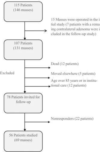
A flowsheet depicting the original cohort and the 56 patients comprising the present study cohort is given in Fig. 1. The pa-tients participating in the follow-up study had altogether 69 ad-renal masses. Characteristics of patients are given in Table 1. Of all study patients, 44 (78%) had one, 11 (20%) had two, and
one patient (2%) three separate adrenal incidentalomas. Of all 69 adrenal masses, 23 (33%) were located in the right adrenal gland and 46 (67%) in the left adrenal gland.
Change in tumor size after 5 years follow-up
Mean change in tumor size after follow-up for the whole cohort was 1±2 mm (P<0.001) (Table 1). Change in tumor size ranged
from –4 to 8 mm (Fig. 2). Five masses (7.2%) demonstrated a decrease in tumor size of 1 to 4 mm. When the incidentalomas were divided into two groups based on tumor size at baseline (<20 or ≥20 mm), the mean change in size was 0.3 and 1.9 mm, respectively (Table 1). The tumor disposing the largest tumor growth of 8 mm during follow-up was also the largest one (39 mm) after follow-up. It was non-functional and characterized by a density of <10 HU at follow-up. The patient, a female aged 67
years, underwent laparoscopic adrenalectomy and histopatho-logical examination demonstrated a cortical adenoma. For all other masses, size at 5 years follow-up remained <37 mm.
Fig. 1. The original study cohort with adrenal incidentaloma and
number of patients and adrenal masses eligible for the 5-year fol-low-up study.
115 Patients (146 masses)
15 Masses were operated in the ini-tial study (7 patients with a remaing contralateral adenoma were in-cluded in the follow-up study)
Dead (12 patients)
Excluded Moved elsewhere (5 patients)
Age over 85 years or in institu-tional care (12 patients)
Nonresponders (22 patients) 107 Patients
(131 masses)
78 Patients invited for follow-up
Re-evaluation of Hounsfield units
At baseline, all masses were characterized by <10 HU
attenua-tion on CT. After 5 years follow-up (Table 1), 66 of 69 masses (96 %), for which evaluation of HU on nonenhanced CT was available were characterized by <10 HU. Three incidentalomas
classified as lipid-rich adenomas at baseline had 5-year follow-up HU values of 10, 10, and 22, respectively. At baseline, the mean diameter of these masses was 19±6 mm (range, 10 to
34) and the corresponding diameter after follow-up was 20±7
mm (range, 10 to 39). Fig. 3 demonstrates the typical CT find-ings of one of the adrenal incidentalomas of the present series after follow-up.
Biochemical screening for excess cortisol secretion
In the whole cohort, at the end of follow-up, mean serum
corti-sol concentration after 1 mg DST was 44±22 nmol/L and
mean basal plasma ACTH 15±8 ng/L. Morning serum cortisol
after 1 mg DST was <50, 50 to 100, and >100 nmol/L in 33,
19, and 2 patients, respectively. Corresponding plasma ACTH concentrations in patients with normal (<50 nmol/L) or
sub-normal (50 to 100 nmol/L) serum cortisol after the 1 mg DST were 16.2±8.1 ng/L versus 14.5±8.5 ng/L, respectively (not
significant).
Two females aged 62 and 70 years had non-suppressed
se-Fig. 2. Distribution of tumor size changes after 5 years follow-up.
40 35 30
25 20 15 10 5
0
No. of pati
ents
Distribution of change in size
–4 –3 –2 –1 0 1 2 3 4 5 6 7 8 Change of diameter (mm)
Fig. 3. Unenhanced computed tomography of a typical adrenal
in-cidentaloma (arrow) on the right. Placement of region of interest (ROI) cursor for measurement of Hounsfield units (HU) is also shown. HU was negative.
Table 1. Characteristics of 56 Patients with 69 Lipid-Rich Adrenal Incidentalomas (<10 HU)
Characteristic All patients Size <2 cm Size ≥2 cm
No. of patients 56 26 30
Age, yr 64±8 65±9 63±8
Sex, female:male 32:24 16:10 16:14
Body mass index, kg/m2 29±6 29±5 29±5
No. of tumors 69 35 34
HU <10 after follow-up, n (%) 63/69 (96) 31/33 (94) 32/33 (97)
Size at baseline, mm 19±6 14±2 24±4a
Size after follow-up, mm 20±7 14±3 26±4a
Growth during follow-up, mm 1.0±2.0 0.3±1.1 1.9±2.6b
Increased 24-hour urine metanephrines/normetanephrines after follow-up, % 0 0 0
Serum cortisol (DST) at baseline, nmol/L 38±23 36±18 40±29
New subclinical hypercortisolism after follow-upc, n (%) 0/54 (0) 0/26 (0) 0/28 (0)
Values are expressed as mean±SD unless other indicated. Data for incidentalomas <2 and ≥2 cm in diameter is given separately. HU, Hounsfield unit; DST, dexamethasone suppression test.
rum cortisol concentrations of 110 and 167 nmol/L after 1 mg DST, respectively and both of them also had low plasma ACTH concentrations (6 and <5 ng/L, respectively). These
pa-tients had had biochemical findings indicating subclinical hy-percortisolism also at baseline, but had rejected operation at that time. Neither of them had any clinical signs of hypercorti-solism. Their body mass index was 30.8 and 28.7 kg/m2
, re-spectively. They both had normal blood pressure and they did not have type 2 diabetes. The 62-year-old female underwent surgery, and a cortical adenoma was confirmed. The 70-year-old female did not want surgery and she was scheduled for fur-ther follow-up. None of the ofur-ther patients fulfilled the bio-chemical criteria for subclinical hypercortisolism. Thus, the conversion rate to subclinical hypercortisolism after 5 years follow-up was 0%.
Biochemical screening for pheochromocytoma
As shown in Table 1, none of the patients had increased 24-hour fractionated urinary metanephrine or normetanephrine concen-trations after 5 years of follow-up. Urinary metanephrine con-centrations ranged from 0.2 to 1.1 µmol/L (reference value
<1.7) and normetanephrines from 0.1 to 2.2 µmol/L (reference
value <4).
DISCUSSION
This is the first prospective study on lipid-rich adrenal inciden-talomas. The main findings of the present study are that there was no relevant tumor growth after 5 years of follow-up and that the conversion rate to subclinical or clinical hypercorti-solism was zero. In addition, we could confirm that pheochro-mocytoma is outruled in such adrenal incidentalomas also after medium-term follow-up, as measurements of 24-hour fraction-ated metanephrines and normetanephrines were normal. The recommendations for initial diagnostic work-up of adre-nal incidentalomas are fairly uniform [7-9,12-14]. The different algorithms currently proposed for follow-up of adrenal inci-dentalomas are rather extensive and hampered by the lack of prospective follow-up studies [7-9,12-14]. There are several open questions regarding what percentage of incidentalomas will increase in size to ≥40 mm over time, when hypercorti-solism or subclinical hypercortihypercorti-solism will occur, as well as whether imaging findings could predict who should or should not undergo further testing.
Most authors recommend continuous annual screening for hyperfunction up to 4 to 5 years and one to three interval CT
scans to evaluate potential tumor growth. The initial work-up should adequately identify all potentially malignant and hor-monally active adrenal tumors in order to refer these patients for surgery. The follow-up scheme should confirm the benign nature of the adrenal mass and, alternatively, identify malignant transformation and ensure that the adrenal mass does not be-come hormonally active. The follow-up strategy should not only be safe but also cost-effective, avoiding unnecessary and expensive investigations of patients who do not need such in-vestigations.
In our 5-year prospective follow-up study of altogether 69 incidentally found lipid-rich adrenal masses in 56 patients, we demonstrate that mean growth of all lipid-rich adrenal masses was only 1±2 mm, i.e., not significant. Some of these adrenal
masses even decreased in size over time. A limitation of the present study is the fairly small number of patients (n=56) and
adrenal masses (n=69). Therefore, future prospective studies
including larger numbers of lipid-rich adrenal tumors are war-ranted. However, in line with the present study, we did not ob-serve tumor growth in our larger initial cohort including 174 patients and 214 lipid-rich adrenal masses during 15.8 months of follow-up [10].
In the present study, the tumor demonstrating the largest in-crease in diameter, 8 mm, was also the largest one (39 mm) at the end of follow-up. The patient harbouring this tumor under-went laparoscopic surgery and histopathology confirmed a be-nign cortical adenoma. When the patients were divided into two groups based on initial tumor size <20 or ≥20 mm, we found that none of the tumors (35 tumors, 26 patients) charac-terized by initial tumor size <20 mm demonstrated clinically
significant tumor growth during follow-up and none had or de-veloped subclinical hypercortisolism.
Another important finding of the present study is that the conversion rate to subclinical hypercortisolism in such lipid-rich adrenal incidentalomas was 0%. Two patients (3.6%) who were biochemically characterized by subclinical hypercorti-solism at baseline had subclinical hypercortihypercorti-solism also after 5 years of follow-up. None of the patients developed overt hy-percortisolism. While one of these females was operated on and histopathology demonstrated a benign adenoma, the other female did not want surgery. These females did not show clini-cal signs of hypercortisolism and had not developed type 2 dia-betes or hypertension during follow-up. In the future, further studies including larger sample size are needed to confirm these results.
that pheochromocytoma does not underlie homogenous tumors originally characterized by a HU <10 on non-contrast CT. It is
not necessary to perform biochemical screening for pheochro-mocytoma in such tumors, neither at baseline nor at follow-up. The NIH consensus statement from 2002 [7] suggest initial endocrine testing with 1 mg DST, plasma free metanephrines and measurement of potassium and screening for aldosterone overproduction in hypertensive patients. Annual biochemical screening for 4 years is recommended in masses <4 cm, with
two repeat CTs at least 6 months apart. The algorithm proposed be Young [3] in 2007 is very similar, recommending initial test-ing with 1 mg DST, urinary metanephrines and catechol-amines, potassium and screening for aldosterone overproduc-tion in hypertensive patients. According to this algorithm, bio-chemical screening should also be performed yearly for 4 years and masses <4 cm should be monitored by CT at 6, 12, and 24
months. The French Society of Endocrinology recommend similar initial endocrine testing in their consensus statement of 2008 [12], with repeat 1 mg DST and plasma and urinary meta-nephrines at 6 months, thereafter repeat 1 mg DST at 2 and 5 years, while CT imaging of masses <4 cm is recommended at
6 months, 2 and 5 years.
The American Association of Clinical Endocrinologists/ American Association of Endocrine Surgeons Medical Guide-lines from 2009 [13] is rather extensive, recommending screen-ing for aldosterone overproduction in hypertensive patients and otherwise endocrine testing annually for 5 years and imaging re-evaluation of masses <4 cm at 3 to 6 months and then
annu-ally for 1 to 2 years. Nieman [9] recommends annual endocrine testing (except for aldosteronism if excluded at baseline) for 4 years in masses <3 cm, non-functional and benign as
charac-terized by imaging at 1 to 2 years. She suggests imaging moni-toring of masses <4 cm at 1 to 2 years and the use of additional
imaging criteria in addition to size. Imaging is recommended at 1 to 2 years or more and when needed at 3 to 6 months. In the Italian Association of Clinical Endocrinologists’ (AME) position paper from 2011 [6], frequency and duration of repeat endocrine testing is recommended to be judged indi-vidually, after clinical monitoring. Imaging characteristics oth-er than size should be included and masses 2 to 4 cm in size should be monitored. CT or magnetic resonance imaging (MRI) is recommended at 3 to 6 months, thereafter, no repeat imaging is recommended in masses <2 cm with benign
fea-tures, while imaging of masses >2 cm is recommended on an
individual base.
Lastly, in their review from 2012, Arnaldi and Boscaro [14]
recommend annual biochemical screening for 5 years (except for aldosterone overproduction), monitoring of masses <4 cm
with CT or MRI and the use of additional imaging criteria in addition to size. Repeat imaging is recommended at 6 months. In conclusion, this is the first 5-year follow-up study on the natural course of lipid-rich adrenal incidentalomas <40 mm,
not characterized by overt hypercortisolism or aldosteronism at baseline. The results indicate that it is not necessary to bio-chemically screen for pheochromocytoma in patients with such incidentalomas and that their next follow-up can be scheduled 5 years ahead and should include an unenhanced CT and screening for hypercortisolism. It is debatable whether further screening is needed at all in elderly subjects with lipid-rich in-cidentalomas <2 cm in size. The results of the present study
should, however, be confirmed in future studies including larg-er patient numblarg-ers.
CONFLICTS OF INTEREST
No potential conflict of interest relevant to this article was re-ported.
ACKNOWLEDGMENTS
The technical help of Ms. Marketta Halinen is greatfully ac-knowledged. This study was supported by grants from Re-search Funding of Helsinki University Hospital (erityisvaltion-osuus).
REFERENCES
1. Bulow B, Ahren B; Swedish Research Council Study Group
of Endocrine Abdominal Tumours. Adrenal incidentaloma: experience of a standardized diagnostic programme in the Swedish prospective study. J Intern Med 2002;252:239-46.
2. Barzon L, Sonino N, Fallo F, Palu G, Boscaro M.
Preva-lence and natural history of adrenal incidentalomas. Eur J Endocrinol 2003;149:273-85.
3. Young WF Jr. Clinical practice: the incidentally discovered
adrenal mass. N Engl J Med 2007;356:601-10.
4. Terzolo M, Bovio S, Pia A, Reimondo G, Angeli A.
Man-agement of adrenal incidentaloma. Best Pract Res Clin En-docrinol Metab 2009;23:233-43.
5. Zeiger MA, Siegelman SS, Hamrahian AH. Medical and
6. Terzolo M, Stigliano A, Chiodini I, Loli P, Furlani L,
Arnal-di G, et al. AME position statement on adrenal incidentalo-ma. Eur J Endocrinol 2011;164:851-70.
7. Grumbach MM, Biller BM, Braunstein GD, Campbell KK,
Carney JA, Godley PA, et al. Management of the clinically inapparent adrenal mass (“incidentaloma”). Ann Intern Med 2003;138:424-9.
8. Thompson GB, Young WF Jr. Adrenal incidentaloma. Curr
Opin Oncol 2003;15:84-90.
9. Nieman LK. Approach to the patient with an adrenal
inci-dentaloma. J Clin Endocrinol Metab 2010;95:4106-13.
10. Sane T, Schalin-Jantti C, Raade M. Is biochemical
screen-ing for pheochromocytoma in adrenal incidentalomas ex-pressing low unenhanced attenuation on computed tomog-raphy necessary? J Clin Endocrinol Metab 2012;97:2077-83.
11. Morelli V, Reimondo G, Giordano R, Della Casa S,
Polico-la C, Palmieri S, et al. Long-term follow-up in adrenal inci-dentalomas: an Italian multicenter study. J Clin Endocrinol Metab 2014;99:827-34.
12. Tabarin A, Bardet S, Bertherat J, Dupas B, Chabre O,
Ham-oir E, et al. Exploration and management of adrenal inci-dentalomas. French Society of Endocrinology Consensus. Ann Endocrinol (Paris) 2008;69:487-500.
13. Zeiger MA, Thompson GB, Duh QY, Hamrahian AH,
An-gelos P, Elaraj D, et al. The American Association of Clini-cal Endocrinologists and American Association of Endo-crine Surgeons medical guidelines for the management of adrenal incidentalomas. Endocr Pract 2009;15 Suppl 1:1-20.
14. Arnaldi G, Boscaro M. Adrenal incidentaloma. Best Pract