Jebmh.com
Original Article
J. Evid. Based Med. Healthc., pISSN- 2349-2562, eISSN- 2349-2570/ Vol. 3/Issue 71/Sept. 05, 2016 Page 3860
PEDICLE TONGUE FLAP SURGERY IN ORAL SUBMUCOUS FIBROSIS
Muthubabu K1, Srinivasan M. K2, Jeeva G3, Metali Rai4, Gayathri S5, Assadi Rekha6
1Assistant Professor, Department of ENT & Head and Neck Surgery, Meenakshi Medical College & Research Institute,
Kanchipuram.
2Professor, Department of ENT & Head and Neck Surgery, Meenakshi Medical College & Research Institute, Kanchipuram. 3Junior Resident, Department of ENT & Head and Neck Surgery, Meenakshi Medical College & Research Institute, Kanchipuram. 4Junior Resident, Department of ENT & Head and Neck Surgery, Meenakshi Medical College & Research Institute, Kanchipuram. 5Junior Resident, Department of ENT & Head and Neck Surgery, Meenakshi Medical College & Research Institute, Kanchipuram. 6Junior Resident, Department of ENT & Head and Neck Surgery, Meenakshi Medical College & Research Institute, Kanchipuram.
ABSTRACT
BACKGROUND
Oral submucous fibrosis is a disease of unknown aetiology and is a legacy of Indians. It has been variously treated both medically and surgically but neither has been found to be rewarding. Various groups have been studying the therapy schedules and aetiological association, but the conclusions have remained unclear.
AIM
The study aims to focus on newer surgical therapy stressing on the mechanics and use of pedicle tongue flap in the management of this condition.
METHODS AND MATERIALS
The study comprised of 40 patients from our outpatient department suffering from oral submucous fibrosis in the age group of 11 to 70 years. The contributory factors of oral submucous fibrosis and the symptoms of the disease were evaluated and the role of pedicle tongue flap surgery in the management of this disease which is a premalignant condition is discussed.
RESULTS AND CONCLUSION
Pedicle tongue flap surgery has given promising results in the treatment of trismus due to oral submucous fibrosis. After the surgery, none of our patients developed any malignant change.
KEYWORDS
Oral Submucous Fibrosis (OSF), Trismus, Leucoplakia, Tongue Flap.
HOW TO CITE THIS ARTICLE: Muthubabu K, Srinivasan MK, Jeeva G, et al. Pedicle tongue flap surgery in oral submucous fibrosis. J. Evid. Based Med. Healthc. 2016; 3(71), 3860-3863. DOI: 10.18410/jebmh/2016/825
INTRODUCTION: Oral submucous fibrosis is a premalignant condition.(1) which has evoked the interest of
different workers over the last few decades. Information regarding aetiology, pathology and management of this disease has been sketchy and inconclusive. There have been differing pointers to this unusual syndrome and the OSF complex seems to involve all areas of upper gastrointestinal tract. With our present knowledge, we may define submucous fibrosis as an insidious chronic disease affecting any part of the oral cavity, the pharynx and the oesophagus.,(2) alone or in combination. Clinical
presentation varies from glossitis, stomatitis, vesicles, ulcers resulting in pale fibrotic board like oral submucous membrane in the retromolar, soft palate, tonsillar, pharyngeal and rarely glossal and vallecular region.
Fibrosis of masseter and retromolar area causes trismus and odynophagia. Pedicle tongue flap surgery plays a promising role in the treatment of trismus due to OSF. The mean age of presentation is around 43 years and male female ratio is 2:1.(3)
METHODS: This study comprised of 40 patients from our outpatient department. There were 24 males and 16 females and age distribution varied from 11 years to 70 years. There was preponderance of patients in the 3rd to 4th decade. The
contributory factors for OSF are areca nut, tobacco, betel leaves and nuts (Table. 1). The dominant symptoms seen were inability to open the mouth fully, in addition, the complaints included both sensory alteration in the oral cavity and mucosal ulceration (Table. 2). Clinical examination of the oral cavity in this group revealed
1. Pale buccal mucosa in all patients. 2. Thick band like pterygomandibular raphe.
3. Interincisor distance ranged between 1.5 to 2.5 cm. 4. Poor dental hygiene.
Patients were thoroughly investigated. 3 patients had associated microcytic hypochromic anaemia with low haemoglobin levels.
Financial or Other, Competing Interest: None. Submission 02-08-2016, Peer Review 12-08-2016, Acceptance 24-08-2016, Published 03-09-2016. Corresponding Author:
Dr. Muthubabu K, Assistant Professor,
Department of ENT & Head and Neck Surgery,
Meenakshi Medical College & Research Institute, Kanchipuram. E-mail: muthubabu67@gmail.com
Jebmh.com
Original Article
J. Evid. Based Med. Healthc., pISSN- 2349-2562, eISSN- 2349-2570/ Vol. 3/Issue 71/Sept. 05, 2016 Page 3861
The patients were screened routinely for associated systemic illness and were taken up for surgical management. The operative procedure required release of the pterygomandibular raphe and the mucosal area around this ensuring adequate blood supply using a pedicle flap.
No. Contributory Factors No. of Patients
1. Only Areca Nut 2
2. Only Tobacco 7
3. Areca Nut, Lime and Betel
Nuts 10
4. Pan Masala 12
5. Combination of Betel Leaves
and Tobacco 9
Table 1: Showing the Incidence of Contributory Factors
No. Symptoms No. of Patients
Complained
1. Inability to Open Mouth
Fully 40
2. Burning Sensation in the
Mouth 35
3.
Burning Sensation in the Mouth on Taking Spicy
Food
36
4. Alteration in the Taste
Sensation 30
5. Aphthous Ulceration 6
Table 2: Showing the Symptoms
AIM OF THE SURGERY:
1. To Relieve Trismus.
2. To Prevent intermolar Fibrosis.
3. To Provide Neovascularisation to the Fibrosed Palate.
Criteria of Patient Selection:
1. Mouth Opening of Less than 2.5 cm (Normal 4 to 5 cm).
2. Failure of Conservative Line of Treatment to Relieve Trismus.
3. Absence of any other pathology viz. Infections, ulceration.
4. Absence of malignancy in the oral cavity.
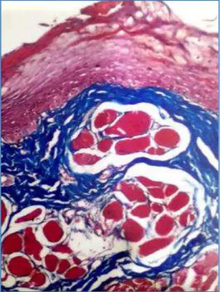
5. Biopsy was done to confirm the diagnosis (Fig. 1).
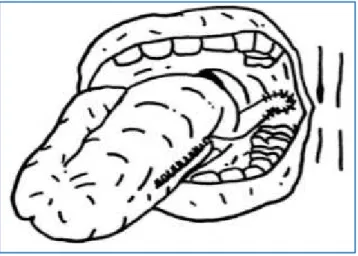
PROCEDURE: Endotracheal tube was placed under GA. The endotracheal tube was positioned by railroading along a flexible fibreoptic laryngoscopy through the nose. The last molar teeth in both jaws on either side were extracted. Mouth was opened using Doyens mouth gag. A raw area was created in the buccal mucosa starting from the anterior pillar through the retromolar area and the pterygomandibular raphe forward for about 3 to 4 cm. In some cases, a part of buccal mucosa was excised to house the elliptical tongue flap.
The cheek incision goes up to the level of the masseter taking care not to allow the buccal pad of fat to herniate into the wound. An elliptical myomucosal tongue flap (Dorsolateral), posterior based tongue flap from the ipsilateral anterior 2/3rd of the tongue was rotated behind
the retromolar trigone (Fig. 2 and 3). It is then approximated to the raw area in the cheek. 2 or 3 stay sutures were applied to the under surface of the tongue and the raw area of cheek. The tongue defect was closed with interrupted Vicryl stitches. The flap is approximated to the edges of the area using interrupted Vicryl sutures. A nasogastric tube was inserted prior to extubation. Patient was maintained on nasogastric feeds for 3 days and slowly switched to semisolids. The procedure can be done unilaterally or bilaterally in the same sitting.
Fig. 1: Showing Masson’s Trichrome 10x Buccal
Mucosal Biopsy Section Showing Collagen Infiltration into Skeletal Muscle
Jebmh.com
Original Article
J. Evid. Based Med. Healthc., pISSN- 2349-2562, eISSN- 2349-2570/ Vol. 3/Issue 71/Sept. 05, 2016 Page 3862
Fig. 3: Showing Flap Suturing
Post-operative Care: Patient was put on, 1. Antibiotics.
2. Collosol iodine - orally 1 teaspoon 3 times a day for 20 days and later 1 teaspoon once a day for another 20 days.
3. Antiseptic mouth gargles.
4. Mouth opening encouraged by asking the patient to progressively bite 2 cc, 5 cc, and finally 10 cc syringes.
5. Post-operative beta carotenoids for 6 months.
Followup: There was an appreciable increase in the interincisor distance. About 1 cm increase in the interincisor distance was noted (Fig. 4 and 5).
Pre-Operative Picture of a Patient:
Fig. 4: Showing Interincisor Distance Less Than 2 cm
Post-Operative Picture of a Patient:
Fig. 5: Showing Increase in Inter Incisor Distance
RESULT: Pedicle tongue flap surgery facilitated better articulation, mastication and swallowing. Appreciable increase in the interincisor distance was noted. Colour of soft palate and cheek were noted to turn pink from the pale colour after surgery due to revascularisation of the areas. None of the patients complained of any impairment of taste sensation after surgery.
DISCUSSION: Oral submucous fibrosis is a crippling disease characterised by abnormal accumulation of collagen fibre in the oral submucosa. It is regarded as precancerous lesion.(1) The exact aetiology is not known but possible
aetiological factors like betel nut, areca nut(4) are attributed.
Prolonged irritation due to tobacco and chillies can cause OSF.(5) Vitamin deficiency is also attributed among the
aetiological factors.(6) Iron deficiency anaemia has also been
noted in patients with OSF.(7) In OSF it is likely that there is
a genetic predisposition to OSF and genes situated in the HLA region are important determinants in genetic susceptibility in OSF.(8) Arecoline.(9) in areca nut is a major
alkaloids in the collagen synthesis and causes OSF.(10) The
presenting symptoms are mainly burning sensation in the mouth, pain, dryness of mouth, stomatitis, ulceration and vesicles. Increased salivation and alteration of taste sensation is seen.(11) The symptoms varies according to the
stage of the disease,
1. Stage 1 - stage of stomatitis and vesiculation. 2. Stage 2 - stage of fibrosis - patient has stiffness in
opening the mouth.
3. Stage 3 - stage of sequelae and complication - leucoplakia changes develop and may change into malignancy.
There are various treatment modalities like local injection of placentrex.(12) injection of hydrocortisone in the oral
mucosa.(13) surgical treatment like incision of retromolar
band, condylectomy, excision of fibrosis and free skin graft has been tried. Posterior based pedicle tongue flap is a rewarding procedure to treat OSF because it is a versatile flap with excellent blood supply.(14) This procedure was used
Jebmh.com
Original Article
J. Evid. Based Med. Healthc., pISSN- 2349-2562, eISSN- 2349-2570/ Vol. 3/Issue 71/Sept. 05, 2016 Page 3863
CONCLUSION: Oral submucous fibrosis is a crippling disease of unknown aetiology and is a legacy of India not reported elsewhere. Though there are various modalities of treatment, pedicle tongue flap surgery has given promising results. Patients who underwent medical line management without any relief of symptoms came to us with trismus. Pedicle tongue flap surgery performed on this patient proved to be a success to the patients and changed the life style. Patients were periodically followed up and found to be normal and symptom free. Though OSF is regarded as premalignant condition after pedicle tongue flap surgery, none of our patients developed any malignant changes.
REFERENCES
1. Pillai R, Balaram P, Reddiar KS. Pathogenesis of oral submucous fibrosis. Relationship to risk factors associated with oral cancer. Cancer 1992;69(8):2011-2020.
2. Maher R, Ahmed W, Qureshi H, et al. Esophageal changes in oral submucous fibrosis using fiberoptic endoscopy-a pilot study. JPMA J Pak Assoc 1991;11(12):312-313.
3. Sankaranarayanan R. oral cancer in India. An epidemiological and clinical review. Oral Surg Oral Med Oral Pathol 1990:69(3):325-330.
4. Murti PR, Gupta PC, Bhonsle RB, et al. Effect of incidence of oral submucous fibrosis of intervention in the areca nut chewing habit. Journal of oral pathological medicine 1990;19(2):99-100.
5. Pindborg JJ, Bhatt M, Devanath KR, et al. Oral
submucous fibrosis. Indian J Med Sci
1966;20(5):349-350.
6. Wahi PN, Kapur VL, Luthura VK, et al. Oral submucous fibrosis. Bull WHO 1966;35:793.
7. Rajendran K, Vaudevan DN, Vijayakumar T. Serum iron levels and proteins in oral submucous fibrosis. Ann of Dentistry 1990;49(2):23-25.
8. Canniff JP, Batchelor JR, Dodi IA, et al. HLA typing in
oral submucous fibrosis. Tissue Antigens 1985:26(2):138-142.
9. Sinor PN, Moorthy PR, Gupta PC, et al. A case control study of oral submucous fibrosis special reference to etiological role of areca nut. Journal of oral pathological medicine 1990;19(2):94-98.
10. Harvey W, Scutt A, Maghji S, et al. Stimulation of human buccal mucosa fibroblasts in vitro by betel nut alkaloids. Arch Oral Biology 1986;31(1):45-49. 11. Chaturvedi VN, Maratha NG. Electrogustometry in
oral submucous fibrosis -A study in 50 cases. Indian J Dent Res 1992;3(3):90-93.
12. Anil S, Beena VT. Oral submucous fibrosis in a 12 year old case report. Pediatric Dentistry 1993;15(2):120-122.