UNIVERSIDADE DE LISBOA
FACULDADE DE CIÊNCIAS
DEPARTAMENTO DE BIOLOGIA VEGETAL
NOVEL APPLICATIONS OF A FLOW CYTOMETRIC
SENSITIVITY ASSAY FOR PLASMODIUM SPP.:
COMPOUND SCREENING AND GAMETOCYTE
DETECTION
Carolina Isabel Glória Tempera
DISSERTAÇÃO
MESTRADO EM MICROBIOLOGIA APLICADA
UNIVERSIDADE DE LISBOA
FACULDADE DE CIÊNCIAS
DEPARTAMENTO DE BIOLOGIA VEGETAL
NOVEL APPLICATIONS OF A FLOW CYTOMETRIC
SENSITIVITY ASSAY FOR PLASMODIUM SPP.:
COMPOUND SCREENING AND GAMETOCYTE
DETECTION
Carolina Isabel Glória Tempera
DISSERTAÇÃO ORIENTADA POR PROF. DR. THOMAS HÄNSCHEID (FMUL)
E PROF.ª DR.ª ANA TENREIRO (FCUL)
MESTRADO EM MICROBIOLOGIA APLICADA
2013
NOVEL APPLICATIONS OF A FLOW CYTOMETRIC
SENSITIVITY ASSAY FOR PLASMODIUM SPP.:
COMPOUND SCREENING AND GAMETOCYTE
DETECTION
Carolina Isabel Glória Tempera
M
ASTER
T
HESIS
2013
This thesis was fully performed at the Institute of Microbiology in
Faculty of Medicine of the University of Lisbon under the direct
supervision of Prof. Dr. Thomas Hänscheid.
Prof. Dr. Ana Tenreiro was the internal designated supervisor in the
scope of the Master in Applied Microbiology of the Faculty of
Sciences of the University of Lisbon.
i
Acknowledgments
I would like to acknowledge Prof. Dr. Thomas Hänscheid for supervising me, for the time lost to advise me and for the opportunity to be constantly learning. I thank Prof. Dr. Ana Tenreiro for guidance and availability. To Prof. Dr. José Melo Cristino, director of Institute of Microbiology in the Faculty of Medicine of the University of Lisbon and to Prof. Dr. Mário Ramirez, director of the Molecular Microbiology and Infection Unit in the Instituto de Medicina Molecular for providing facilities and equipment.
I would like to thank to Prof. Dr. Rui Moreira and Dr. Ana Ressurreição for kindly providing compounds for me to investigate them.
I would like to thank Dr. Rui Gardner and his group, Cláudia Bispo and Cláudia Andrade, from Instituto Gulbenkian de Ciência for all gametocyte sorting experiments conducted in their laboratory.
Thank to Maria Rebelo for her advises and time lost. To Rosangela Frita, Ana Góis and Márcia Boura for the help and support.
Thank to my mother and my sister for always giving me motivation, confidence and encouragement, and to the rest of my family. Thank to my friends. Thank you.
ii
Abstract
Malaria is caused by a parasite of the genus Plasmodium and remains the most important parasitic disease. The emergence of Plasmodium falciparum parasites resistant to all known antimalarial drugs is of major concern. New antimalarial drugs are needed, not only a drug that overcome the undesirable side effects of current antimalarial drugs but new highly active ones against the asexual stages, as well as, drugs that could also eliminate the transmissible sexual form of the parasite to the mosquito.
To test the susceptibility of the parasite to drugs a variety of sensitivity assays can be used to screen new compounds, such as: hypoxanthine incorporation, ELISA based assays like pLDH and HRP2, fluorometric and flow cytometric assays. Recently, a novel flow cytometric sensitivity assay based on hemozoin detection was described. Using this novel sensitivity assay new antimalarial compounds were screened at 1 and 3 µM. The SYBR green DNA staining assay and the HRP2 were also perfomed as mean of comparison. Results showed that none of the tested compounds presented inhibitory activity against P. falciparum strains 3D7 and Dd2 at 1 µM, independently of the method used. Only one of the compounds showed more than 50% inhibition at 3µM.
The flow cytometric hemozoin detection method was also assessed for its potential to detect gametocytes. Gametocyte may have a different depolarizing profile, based on the underlying hemozoin distribution. Thus, we further investigated if they could be distinguished from other parasitie forms based on their higher degree of depolarization. A culture enriched in gametocytes was FACS sorted by selecting the higher depolarizing population. Results showed that gametocytes were selectively present only in the high depolarizing population and not in the middle and non-depolarizing events.
Therefore, this recently described sensitivity assay based on hemozoin detection can be used as a novel approach to screen for new antimalarial drugs. This approach has as major advantages the fact that results can be obtained in only 24 h of incubation and no additional reagents or additional incubation times are required. Another important characteristic of this method is that it might be able to detect gametocytes based on the particular hemozoin distribution in these forms, which can lead to the use of this method to test antimalarial transmission blocking drugs.
Key-words: Malaria, antimalarial drugs, antimalarial drug resistance, in vitro sensitivity assays, hemozoina, flow cytometry.
iii
Resumo
A malária é uma doença que segundo os mais recentes dados da Organização Mundial de Saúde (OMS) foi responsável por cerca de 660 000 mortes e 219 milhões de casos em 2010. É a doença parasitária que mais mortes causa sendo a região mais afetada por esta doença a África subsariana, e com uma maior incidência em crianças até aos 5 anos de idade.
A malária é causada pelo parasita do género Plasmodium. As cinco espécies que podem causar doença no humano são: P. falciparum, P. vivax, P. ovale, P. malariae e P. knowlesi. No entanto P. falciparum é a espécie responsável pelo maior número de casos e mortes por malária sendo também o que pode levar a uma maior severidade da doença. A malária manifesta-se por períodos de febres altas e calafrios, mal-estar generalizado, torpor e dor de cabeça, naúsea e dor abdominal, por vezes até vómitos e diarreia. No caso de doença severa esta implica anemia severa, malária cerebral, síndrome de dificuldade respiratória aguda, insuficiência renal e nos casos mais graves a morte.
Segundo estes factos, a maária é uma doença que levanta preocupação e tem impacto a nível mundial. Isto porque, por várias circunstâncias se pensou que estaríamos no caminho da erradicação da doença e no entanto, apesar de o número de casos e mortes ter diminuído na última década, esta continua a afetar milhões de indivíduos. Hoje em dia, um dos maiores problemas face a esta doença prende-se com o facto de P. falciparum já ter apresentado resistência a todos os fármacos utilizados no tratamento da doença. Entre os antimaláricos já usados encontram-se a quinina, a cloroquina, a primaquina, a pirimetamina conjugada com a sulfadoxina, a mefloquina entre outros. No entanto, as atuais directrizes para o tratamento da malária correspondem a terapias de combinação com recurso a derivados da artemisinina (“Artemisinin-based combination therapies” – ACTs). Alguns exemplos destas combinações são: artesunato combinado com amodiaquina, artesunato com mefloquina, arteméter e lumefantrina, entre outros.
O principal objectivo detas terapias é eliminar as formas do parasita que circulam no sangue, pois é este estadio que conduz à manifestação da doença. Plasmodium é um parasita que apresenta um ciclo de vida complexo. É transmitido ao humano através da picada de um mosquito infectado femea do género Anopheles. Durante a picada, o parasita que existe nas glândulas salivares do mosquito é injetado na corrente sanguínea do humano, e dirige-se até ao fígado. No fígado vários parasitas vão invadir os hepatócitos, dentro dos quais maturam e replicam antes de serem libertados novamente na corrente sanguínea. Esta fase hepática é assintomática. Uma segunda vez na corrente sanguínea, os parasitas vão invadir eritrócitos dentro dos quais vão maturar e replicar antes de lisarem o eritrócito, libertando novos parasitas para invadir novos eritrócitos. Uma vez dentro de um eritrócito, um parasita segue um ciclo de maturação que compreende as seguintas formas: forma de anel, trofozoíto, e esquizonte. No final, irá libertar vários merozoítos. No entanto, ocasionalmente alguns destes merozoitos, quanto invadem um eritrócito vão originar formas sexuadas do parasita, os gametócitos. Os gametócitos apresentam-se sob a forma de percursores masculinos e femininos, e são estas formas que quando em
iv
circulação podem ser ingeridas por um mosquito durante uma nova picada. Após ingeridas pelo mosquito, estas formas vão recombinar genéticamente, dando origem a uma nova descendência que será novamente transmitida ao humano, recomeçando este ciclo.Colocam-se assim dois grandes objectivos à erradicação da malária: (i) um passa pelo desenvolvimento de novos agentes antimaláricos, devido à evidênca de resistência de P.
falciparum a todos os actuais antimaláricos, incluindo os derivados da artemisinina que
compreendem as atuais diretrizes de tratemento da doença; (ii) o outro prende-se com a eliminação das formas sexuadas do parasita em circulação no hospedeiro, pois eliminando estas formas, quebra-se o ciclo de transmissão hospedeiro-vector.
Para a contínua pesquisa de novos compostos antimaláricos é necessário ensaios que detectem a sensibilidade dos parasitas aos diferentes fármacos. Os ensaios de sensibilidade actualmente existentes são: (i) o teste de microscópia da OMS; (ii) o teste por incorporação de hipoxantina; os testes baseados na detecção de anticorpos (Enzyme-Linked Immunosorbent Assay – ELISA) para quantificação de proteinas do parasita como a (iii) lactase desidrogenase (Parasite lactate dehydrogenase – pLDH e a (iv) proteina rica em histidina 2 (Histidine Rich Protein - HRP2); testes baseados na deteção do DNA do parasita por (v) ensaios fluorométricos e por (vi) citometria de fluxo.
No entanto, como até agora não existe o teste de sensibilidade ideal, e devido a várias limitações de cada ensaio, novos ensaios são desenvolvidos. Um dos mais recentes baseia-se na deteção de hemozoina por citometria de fluxo. A hemozoína, também denominada pigmento malárico, é um bioproduto que resulta da destoxificação de heme livre produzido após a metabolização da hemoglobolina pelo parasita, e acumula-se no interior do eritrócito durante a maturação do mesmo. A hemozoína é um cristal com propriedades birefringentes que levam à despolarização da luz. Devido a esta propriedade, foi desenvolvido um ensaio de citometria de fluxo que detecta a hemozoína presente no interior de eritrócitos infectados. Assim, com o acumular de hemozoina ao longo do tempo, a deteção dos parasitas vai aumentando, havendo um maior número de eventos a despolarizar entre as 24h-30h de um primeiro ciclo de maturação, altura em que a maioria dos parasitas se apresenta como esquizonte, o estadio que também tem maior quantidade de hemozoína. Este ensaio permitiu detectar a sensibilidade de P.falciparum a vários antimaláricos e foi agora utilizado para testar a sensibilidade do parasita a novos antimaláricos cedidos pelo grupo do Professor Doutor Rui Moreira da Faculdade de Farmácia.
A pesquisa da actividade dos novos compostos (“screening”) a 1 µM e a 3 µM foi realizada em duas estirpes de laboratório de P. flaciparum, a 3D7 (sensível à cloroquina) e a Dd2 (resintente à cloroquina). Após incubar a estirpe Dd2 em presença dos vários compostos, a sua inibição foi avaliada por citometria de fluxo, utilizando o ensaio da deteção de hemozoína e pelo ensasio da deteção de parasitas cujo DNA foi corado com SYBR green. Os resultados de ambos os ensaios demonstraram que nenhum dos 18 compostos testados a uma concentração de 1 µM levou à inibição do parasita. Com a estirpe 3D7 foram testados 24 novos compostos, não só utilizando os dois médotos de citometria de fluxo já referidos como também através de HRP2-ELISA. Os resultados demonstaram que independentemente do ensaio utilizado nenhum dos novos
v
compostos apresentou actividade inibitória a 1 µM. Porém os compostos foram testados a uma concentração mais elevada, e a 3 µM um dos compostos demonstrou actividade inibitória (o composto 321). Determinou-se a concentração à qual este composto inibe 50 % do crescimento do parasita (CI50) de acordo com os três ensaios para detectar a sensibilidade do parasita obtendo-se um valor à cerca de 2 µM. No entanto, esta concentração é demasiado alta para o composto poder ser posteriormente testado como agente antimalárico na fase sanguínea do parasita, pois os actuais antimaláricos actuam na ordem nos nanomolar (nM).Para erradiacar a malária, a eliminação de gametócitos é um dos principais objectivos, de modo a quebrar o ciclo de transmissão da doença. Porém, existem muito poucos tratamentos que inibam estas formas sexuadas, e por isso é necessário continuar a investigar novos fármacos que actuem com mais impacto neste estadio do parasita. Para tal, uma vez mais são necessários ensaios que possam averiguar a sensibilidade dos gametócitos a diversos fármacos. No entanto, não exitem tantos ensaios para tal, como os que existem para testar a sensibilidade dos estadios assexuados. Uma vez que os gametócitos também apresentam hemozoína no seu interior, pressupôs-se que o ensaio de sensibilidade baseado na deteção de hemozoína permitisse também detectar gametócitos, retirando partido do facto de a hemozoína acumulada nos gametócitos apresentar-se de forma diferente nos esquizontes, pois ocorre sob a forma de vários e pequenos fragmentos ao contrário de um único e grande aglumerado. Assim, colocou-se a hipótese de se detectar os gametócitos por citometria de fluxo, e que estes corresponderiam a uma diferente população de acordo com a despolarização da sua hemozoína.
Para corroborar esta ideia, estabeleceu-se a diferenciação de gametócitos a partir de culturas contínuas e posteriormente, através de análise por citometria de fluxo, os eventos da cultura de gametócitos foram separados (“sorting”) com base na sua depolarização. Partindo do princípio que os gametócitos têm mais cristais, estes podem levar a uma maior quantidade de luz a depolarizar, logo iriam localizar-se num nível de despolarização mais elevado do que os esquizontes. Assim, três populações foram separadas do seguinte modo: (i) população que não despolariza; (ii) população que despolariza a um nível médio (semelhante ao nivel de despolarização de esquizontes) e (iii) população a um nível mais elevado de despolarização. Após observação de cada uma das populações por microscopia, constatou-se que dos eventos adquiridos, os gametócitos só se encontravam na população cuja despolarização era a mais elevada.
Com este recente ensaio baseado na deteção de hemozoína foi então possivel testar a actividade inibitória de novos compostos antimaláricos, obtendo as mesmas conclusões que outros dois métodos utilizados em paralelo. Este ensaio poderá assim ser utilizado no rastreio da actividade de novos compostos, como alternativa aos existentes actualmente, aos quais, há resistências desenvolvidas pelo parasita assim como o facto de conduzirem a efeitos secundários indesejáveis. Em relação aos outros ensaios de sensibilidade, este método apresenta-se como um método rápido de obter resultados (24h) e em tempo real, sem a necessidade de adição de reagentes ou tempos adicionais de incubação com reagentes. Este método permitiu ainda detetar uma população específica do parasita, os gametócitos, os quais representam um dos principais
vi
alvos para a eliminação da malária. Assim, este método poderá também ser investigado como ensaio de sensibilidade dirigido a gametócitos.Palavras-chave: malária, Plasmodium spp, resistência a antimaláricos, testes de sensibilidade in
vii
Index
Pag. 1. Introduction 1 1.1 Introduction to Malaria 1 1.2 Treatment of Malaria 11.2.1 History and overview of antimalarial drugs 2
1.2.2 Current guidelines for malaria treatment 3
1.2.3 Antimalarial drug resistance 5
1.2.4 Artemisinin drug resistance 5
1.3 Antimalarial drug research 6
1.4 Flow Cytometry and Malaria 8
1.5 Hemozoin detection by Flow Cytometry 9
1.6 Specific Gametocyte detection for drug testing 11
2. Objectives 12
3. Materials and Methods 13
3.1 Culture media, solutions and reagents 13
3.2 P. falciparum in vitro cultures 13
3.2.1 P. falciparum continuous cultures maintenance 13
3.2.2 Giemsa staining of blood smears 14
3.2.3 Culture synchronization 14
3.2.4 Frozen stocks of P. falciparum 14
3.3 Antimalarial drugs sensitivity assays 15
3.3.1 Inhibition of Chloroquine (IC50 determination) – Influence of Oxygen 15
3.3.2 Flow Cytometric Assays – General protocol 15
3.3.3 Screening novel compounds from Faculdade de Farmácia 16
3.3.4 Flow cytometer 18
3.3.5 Flow cytometric assays - results analysis 18
3.3.6 Histidine-rich protein-2 sensitivity assay 19
3.3.7 HRP2-ELISA Sample analysis 20
viii
3.4 Gametocyte cultures 20
3.4.1 Culture Method A 20
3.4.2 Culture Method B 21
3.4.3 Erythrocytes fixation using paraformaldehyde 21
3.4.4 Sorting Gametocyte cultures 21
4. Results 23
4.1. Baseline Chloroquine IC50 determination for screening 23
4.2 Compounds screening at 1 µM against P. falciparum strain Dd2 24
4.3 Compounds screening at 1 µM against P. falciparum strain 3D7 24
4.4 Compounds screening at 3 µM against P. falciparum strain 3D7 25
4.5 Inhibitory concentration (IC50) values of compounds: 256, 291 and 321 26
4.6 P. falciparum 3D7 Gametocytes detection 27
4.6.1 Results Culture Method A 27
4.6.2 Results culture Method B 32
5. Discussion 35
5.1 Drug sensitivity assays 35
5.1.1 P. falciparum growth in two different atmospheres (low and high oxygen) 36
5.1.2 Screening results 36
5.1.3 P. falciparum: use of strains Dd2 and 3D7 37
5.1.4 Sensitivity assays: analysis of depolarization and SYBR green detection
and HRP2-ELISA 37
5.2 Gametocytes detection 40
5.2.1 Gametocytes cultures 41
5.2.3 Detection of gametocytes using side scatter depolarization 42
6. Conclusion 43
1
1. Introduction
1.1 Introduction to Malaria
Malaria is a parasitic blood disease that according to World Health Organization latest report caused an estimated 219 million cases of malaria and 660 000 deaths in 2010 [1]. It affects mostly the sub-Saharan Africa territory, and has a higher incidence in children under 5 years old [1].
Malaria is caused by one of the five known Plasmodium spp in humans: P. falciparum, P.
vivax, P. ovale, P. malariae and P. knowlesi [2]. In general, symptoms include periodic chills and fevers, malaise, lethargy, headache, nausea, abdominal pain and sometimes vomiting and diarrhea. P. falciparum is the major strain that can cause severe disease such as severe anemia, cerebral malaria, pulmonary edema, acute respiratory distress syndrome and renal failure, and thus is the strain causing most deaths [3].
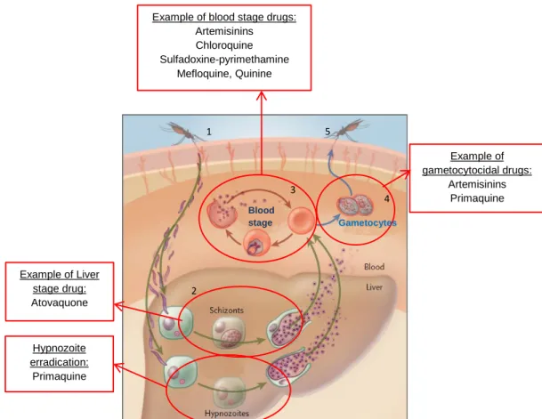
Plasmodium has a complex life cycle [4] which comprises an asexual reproduction cycle in the human host and a sexual reproduction cycle in the mosquito vector (Figure 1). The cycle starts when an infected female Anopheles mosquito bites an individual, and injects sporozoites, present in mosquito’s salivary glands, into the host blood stream during its blood meal. These sporozoites migrate to the liver where they mature and multiply within hepatocytes. These forms are known as schizonts (Figure 1, point 2). This extra-erythrocytic stage is asymptomatic and usually lasts 6 days to 14 days, although sometimes it can last up to several months or even years in the case of P.
vivax or P. ovale. These two human Plasmodium species can produce hypnozoites in the liver [5]
(Figure 1, point 2). Hypnozoites are a dormant form of the parasite, also called cryptic form, which can stay in the liver for long periods of time and are the cause of the disease relapse.
After the liver stage, tens of thousands of merozoites will be released into the blood, where they will invade and develop within erythrocytes. The blood stage of infection includes asexual forms of the parasite that undergo repeated cycles of multiplication in erythrocytes (Figure 1 point 3), causing parasite numbers to rise rapidly. This stage is responsible for the symptoms of malaria. Within the erythrocyte, the asexual forms of the parasite passes through different sequential maturation stages: ring, trophozoite and schizont forms (Figure 2). In the end, the erythrocyte ruptures and new merozoits are released and ready to infect new erythrocytes (Figure 1, point 3). Some parasites will develop into the sexual forms, responsible for transmission, known as gametocytes Figure 1, point 4). The female and male gametocytes (macro-gametocyte and micro-gametocyte, respectively) will be ingested by the mosquito vector during its feeding, and sexual reproduction occurs inside the mosquito midgut before the parasite is transmitted to another human host and the whole cycle starts again [6].
1.2 Treatment of Malaria
Most available antimalarial drugs were designed to target the symptomatic blood stages and thus act only against the sexual blood forms [7]. Treatment of an individual diagnosed with P.
falciparum malaria is of great concern because contrary to the other species, it can be rapidly fatal
[3]
2
transmission and drugs which eliminate the asymptomatic cryptic hepatic forms of P. vivax andovale, are also needed [8].
1.2.1 History and overview of antimalarial drugs
Quinine is one of the oldest known antimalarial drugs, and occurs naturally in the bark of cinchona trees in South America. It is an alkaloid and was first isolated in 1820 and used for many decades [9]. During the First World War, due to quinine stocks declining, the development of the first synthetic antimalarial was conducted. Work with synthetic dyes led to the development of the acridines and the 8-aminoquinolines, such as pamaquine (and subsequently primaquine) [10].
In the 1940s chloroquine, a 4-aminoquinoline, was introduced as an antimalarial chemotherapy after having been synthesized in Germany. Chloroquine was not only highly effective and well tolerated as treatment but was also the main drug of choice in the WHO Global Eradication Programme of the 1950s and 1960s [9].
During World War II, chloroquine was only one of many antimalarials that resulted from scientific advances. Others were developed, and some of them focused on derivatives of pyrimidine. Research in this direction resulted in the development of the antifolates like proguanil. Then, pyrimethamine was developed shortly afterwards. Proguanil and pyrimethamine were used as prophylactic and therapeutic agents, despite their slow schizontocidal action. The antifolates are dihydrofolate reductase (DHRF) and dihydropteroate synthase (DHPS) inhibitors that disrupt folate synthesis in the parasite [10]. Later it was discovered that in combination with pyrimethamine, sulfa drugs like sulfadoxine, were more effective against Plasmodium infection. However, after the introduction of sulfa drug-DHRF inhibitor combinations, the U.S. army developed further aryl-amino alcohol derivatives from quinine, such as mefloquine and halofantrine. More recently, atovaquone was introduced as an antimalarial drug, and is used also as prophylactic agent since inhibits not only the blood stages forms of the parasite but the liver stages as well [11]. It is administered in synergy with proguanil [12].
Most of antimalarial drugs therapeutics acts upon the asexual blood stages of parasite, like quinine, chloroquine, mefloquine and the antifolates (Table 1). Primaquine is the only that acts against liver stages parasites, more specifically only against hypnozoites, that occur in P. vivax and
P. ovale. Tough, primaquine also inhibits gametocyte forms.
Overall, the blood stages antimalarial drugs interfere with parasite hemoglobin degradation and heme detoxification, or with parasite folate biosynthesis. Others drugs such as tetracyclines and clindamycin inhibit protein synthesis in the apicoplast [13], [14].
3
Figure 1 – Plasmodium life cycle and antimalarial drugs interference. Plasmodium life cycle frommosquito (1), through liver stage (2), blood stage (3), gametocytes (4) and these are passed again to the mosquito vector (5). Adapted from Baird, J.K. 2005 [7]. The antimalarial drugs can act upon liver forms, blood forms and/or gametocytes.
Figure 2 - P. falciparum intra-erythrocytic maturation cycle. Representative images of infected erythrocyte with P. falciparum parasite at ring stage (1), trophozoite stage (2) and schizont (3). Then the erythrocyte ruptures, freeing new merozoites as well as hemozoin (4). The new merozoites invade uninfected erythrocytes (5).
1.2.2 Current guidelines for malaria treatment
Although the guidelines may vary somewhat, especially between affluent non-endemic countries and endemic countries, the first-line treatment of uncomplicated falciparum malaria relies on artemisinin-based combination therapies (ACTs) [15]. Artemisinin comes from Artemisia annua
Hypnozoite erradication: Primaquine
Example of blood stage drugs: Artemisinins Chloroquine Sulfadoxine-pyrimethamine Mefloquine, Quinine Example of gametocytocidal drugs: Artemisinins Primaquine Blood stage Gametocytes 1 2 3 4 5 Example of Liver stage drug: Atovaquone 1 2 3 4 5
4
also known as qinghao, and it was long used in Chinese traditional medicine [16]. However, it was only in the early 1970s that its potent antimalarial activity was discovered. Artemisinin and derivates such as arthemeter, artesunate and dihydroartemisinin, are associated with a high rate of recrudescence if used as monotherapy [17]. This is probably related with pharmacodynamic properties of these agents which include fast acting but short half-lives. Therefore, they are usually combined with longer acting antimalarials. Artemether plus lumefantrine, artesunate plus amodiaquine, artesunate plus mefloquine or/and artesunate plus sulfadoxine-pyrimethamine are examples of ACTs. Although still not clear, artemisinins seem to affect the hemoglobin catabolism during parasite maturation within the erythrocyte [18], [19].Artemisinin and artemisinin combination therapies also have some effectiveness at reducing gametocyte carriage [20], although well inferior to primaquine [21]. Primaquine remains the principal available drug for radical treatment to eliminate the cryptic liver stage forms of P. vivax or ovale (Figure 1, point 2). However, because of possible severe side effects like hemolytic anemia it is difficult to use in individuals with glucose-6-phosphate dehydrogenase deficiency (G6PDd) [22], [23] (Table 1).
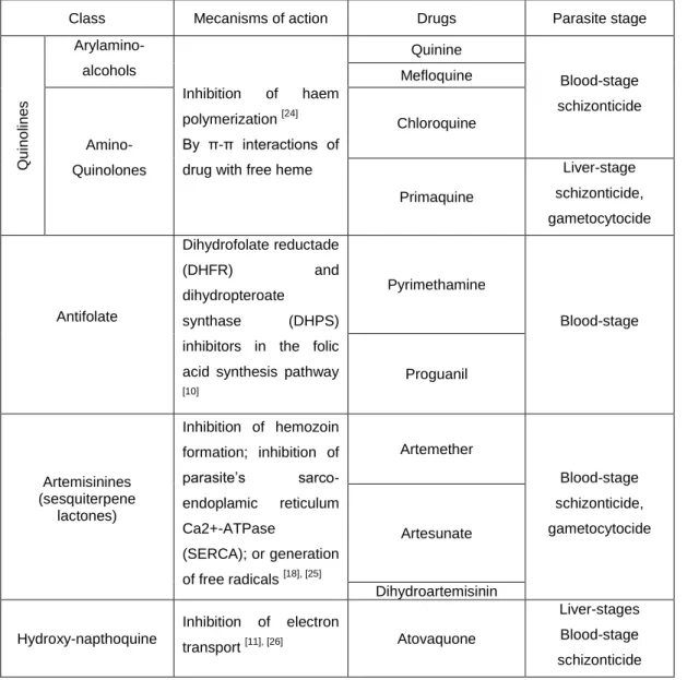
Table 1 – Major antimalarial drugs actions
Class Mecanisms of action Drugs Parasite stage
Q u in o lin e s Arylamino-alcohols Inhibition of haem polymerization [24] By π-π interactions of drug with free heme
Quinine Blood-stage schizonticide Mefloquine Amino-Quinolones Chloroquine Primaquine Liver-stage schizonticide, gametocytocide Antifolate Dihydrofolate reductade (DHFR) and dihydropteroate synthase (DHPS)
inhibitors in the folic acid synthesis pathway [10] Pyrimethamine Blood-stage Proguanil Artemisinines (sesquiterpene lactones) Inhibition of hemozoin formation; inhibition of parasite’s sarco-endoplamic reticulum Ca2+-ATPase (SERCA); or generation of free radicals [18], [25] Artemether Blood-stage schizonticide, gametocytocide Artesunate Dihydroartemisinin Hydroxy-napthoquine Inhibition of electron transport [11], [26] Atovaquone Liver-stages Blood-stage schizonticide
5
Overall, there is a need for novel antimalarial drugs that overcome the limitations of the currently available antimalarials. The side effects of antimalarial drugs and their lack of activity against liver parasites and sexual stages has presented an important concern in malaria eradication.1.2.3 Antimalarial drug resistance
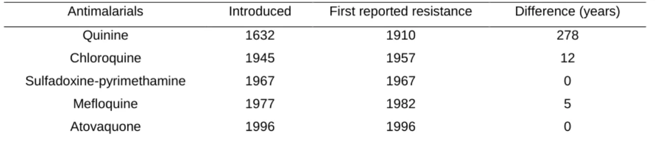
Since malarial drug treatment exists, Plasmodium parasites have developed resistance to the most of the drugs that have been used to treat malaria [24]. The advent of chloroquine resistance was probably the most relevant, because chloroquine was thought to lead to malaria erradication yet, resistance appeared and spread globally [27]. In the late 1950s, resistance to chloroquine was noted on the Thai-Cambodian border in Colombia, than in 1980 all endemic areas in South America were affected, and by 1989 almost all Asia and Oceania. In Africa, chloroquine resistance was first documented in the east in 1978 [28]. Mefloquine, sulfadoxine-pyrimethamine, and atovaquone-proguanil, are antimalarial drugs to which P. falciparum also developed resistance shortly after their introduction (Table 2).However, the use of proguanil and pryrimethamine alone as prophilaxis had a major impact in the insurgent of parasite resistance against these drugs [10].
Table 2 – Dates of introduction and first reports of antimalarial drug resistance.
Antimalarials Introduced First reported resistance Difference (years)
Quinine 1632 1910 278
Chloroquine 1945 1957 12
Sulfadoxine-pyrimethamine 1967 1967 0
Mefloquine 1977 1982 5
Atovaquone 1996 1996 0
(Adapted from Wongsrichanalai, C., et al. 2002 [28])
1.2.4 Artemisinin drug resistance
One of the greater concerns in malaria control is associated with the development of drug resistance, mainly because resistance of P. falciparum to almost all antimalarial drugs has been described, including to the first line treatment with artemisinin [29], [30] .
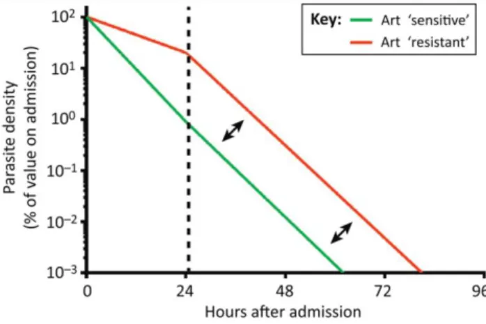
Resistance to artemisinin is characterized by slow parasite clearance in vivo[31] (Figure 3) with no corresponding reductions of susceptibility detected in in vitro [32].
Commonly, for other antimicrobial agents, resistance is defined by clinical failures and decreased susceptibility in vitro [32]. However, for the current definition of “artemisinin resistance”, based on delayed clearance of parasites, neither criterion is fulfilled. However, it is still an issue of big concern [33].
In this scenario, it is of great importance to continue the search for new antimalarial drugs, as well as the continuous surveillance of P. falciparum resistance, especially in endemic countries.
6
Figure 3 - Parasite clearance in artemisinin sensitive and resistant parasite populations. Parasiteclearance profile of artimisinin resistant and sensitive parasite populations. A broken line at 24h shows a shift in clearance between populations, and arrows represent an identical slope thereafter. Abbreviations: Art = artemisinin (From: Ferreira, P. E., et al. 2013) [32].
1.3 Antimalarial drug research
In the antimalarial drug research area, different approaches to study P. falciparum drug sensitivity have been developed. There are in vivo tests, originally defined by the World Health Organization (WHO) in terms of parasite clearance, which comprise the follow classification: sensitive [S] or one of the three degrees of resistance [RI, RII, RIII] [34]. Later a modified protocol was introduced based on the clinical outcome with the following classifications: adequate clinical response, early treatment failure, and late treatment failure (Table 3). There are also phenotypic in
vitro tests that take advantage of the possibility of continuous culture of the parasite (developed in
the 1970s) to perform the assays [35]. Although molecular in vitro tests also exist, the correlation between genetic changes and resistance is not well known yet for most antimalarial drugs.
All in vitro tests share the same principle: comparison of cultures incubated with different concentrations of the tested compound to a drug free control, yet, they differ considerably in the methods used to assess parasite growth or viability.
The main phenotypic in vitro drug sensitivity assays include [36] (Table 4):
Light microscopy methods like the WHO microtest to assess parasite maturation. It requires little technical equipment, can be used for samples with low parasite densities, and usually requires only 24 hours of incubation. On the other hand, its biggest disadvantage is the fact that it is labour-intensive, subjective (inter-operator variation) and requires highly trained personal.
Isotope incorporation assays, such as the [3H]-hypoxanthine incorporation (tritium-labeled hypoxanthine) [37]. It allows a high degree of automatization and therefore is faster to perform than the tests based on the morphological assessment of parasite growth. On the negative side, it only measures metabolic activity over the second half of the parasite life cycle and implies work with isotopic/radioactive material. It also requires high purchase cost for infrastructure, equipment and thus is very badly suited for field studies.
7
(adapted from Wongsrichanalai et al. 2002 [28])
Antigen detection of parasite lactate dehydrogenase (pLDH) or histidine-rich protein (HRP2), produced by malaria parasites during their growth and multiplication, using an Enzyme-Linked Immunosorbent Assay (ELISA).
Parasite lactate dehydrogenase (pLDH) is a terminal enzyme in the Embden-Meyerhof pathway (glycolysis) of the malaria parasite [38]. Its production and accumulation can be used both for malaria diagnosis ex vivo and in in vitro P. falciparum cultures as indices of the presence and viability of the parasite. As a drawback, this assay requires monoclonal antibodies (mAbs) specific for pLDH, which are expensive, have limited supplies and lack an optimal specificity.
Histidine- and alanine-rich protein is produced by P. falciparum in the course of its growth and multiplication [39]. Histidine-rich protein 2 (HRP2) levels are closely associated with parasite density and development, and studies have implicated HRP2 as an important factor in the detoxification of heme [40]. This protein stability can be considered an advantage for in vitro drug susceptibility assays. The assessment of this protein production can be simply measured in a commercial available, double-site sandwich, Enzyme-Linked Immunosorbent Assay (ELISA) test kit. This assay, based on the HRP2 produced by P. falciparum and measured using the ELISA has the advantage of requiring low parasitemia (0.05%). However is an assay that requires a time period of drug incubation of 72 h, followed by a series of steps to detect the protein through ELISA. Table 3 – Classifications of in vivo antimalarial sensitivity test outcomes according to the original and modified protocol
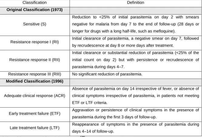
Classification Definition
Original Classification (1973)
Sensitive (S)
Reduction to <25% of initial parasitemia on day 2 with smears negative for malaria from day 7 to the end of follow-up (28 days or longer for drugs with a long half-life, such as mefloquine).
Resistance response I (RI) Initial clearance of parasitemia, a negative smear on day 7, followed by recrudescence at day 8 or more days after treatment.
Resistance response II (RII)
Initial clearance or substantial reduction of parasitemia (<25% of the initial count on day 2) but with persistence or recrudescence of parasitemia during days 4–7.
Resistance response III (RIII) No significant reduction of parasitemia.
Modified Classification (1996)
Adequate clinical response (ACR)
Absence of parasitemia on day 14 irrespective of fever, or absence of clinical symptoms irrespective of parasitemia, in patients not meeting ETF or LTF criteria.
Early treatment failure (ETF) Aggravation or persistence of clinical symptoms in the presence of parasitemia during the first 3 days of follow-up.
Late treatment failure (LTF) Reappearance of symptoms in the presence of parasitemia during days 4–14 of follow-up.
8
Parasitemia measuring through fluorescent DNA dyes, such as SYBR green I [41], YOYO, PicoGreen [42], DAPI [43] and Hoechst with either spectrophotometric or cytometric readout. These assays take advantage of the lack of DNA in the mature RBCs, detecting the stained DNA from the parasite, thus assessing parasitemia in blood samples. With spectrophotometric readouts, the fluorescence intensity, which is proportional to the amount of DNA in individual samples, is measured with a mini fluorometer, fluorescence spectrophotometer or fluorescence activated microplate reader. However, this method is not as sensitive as other methods and it requires long incubation period (48 - 96 h).
With the cytometric readout using a flow cytometer, which enables to assess the parasitemia percentage and preform double stain for a better evaluation of the parasite as mentioned below.
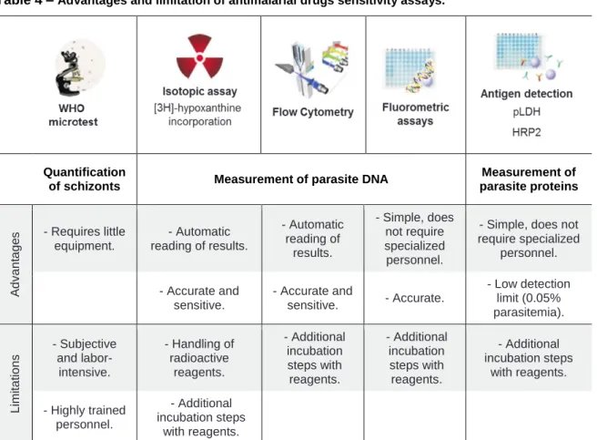
Table 4 – Advantages and limitation of antimalarial drugs sensitivity assays.
Quantification
of schizonts Measurement of parasite DNA
Measurement of parasite proteins Ad v a n ta g e s - Requires little equipment. - Automatic reading of results. - Automatic reading of results. - Simple, does not require specialized personnel.
- Simple, does not require specialized personnel. - Accurate and sensitive. - Accurate and sensitive. - Accurate. - Low detection limit (0.05% parasitemia). L im ita ti o n s - Subjective and labor-intensive. - Handling of radioactive reagents. - Additional incubation steps with reagents. - Additional incubation steps with reagents. - Additional incubation steps with reagents. - Highly trained personnel. - Additional incubation steps with reagents.
Adapted from: Rebelo, M. et al. 2013 [49] – Supplement info
1.4 Flow Cytometry and Malaria
Flow cytometry is becoming particularly important for the study of malaria parasite growth and invasion due to the speed and amount of information it provides and also because it overcomes some of the limitation of existing non-cytometric methods. Flow cytometers have also been progressively more cost affordable and portable, conferring important features for field research.
9
Using flow cytometry it is possible to assess blood parasitemia after specifically staining the nucleic acid (DNA) of the parasites, taking advantage of the fact that normal circulating red blood cells (RBCs) lack nucleic acids, while reticulocytes contain only RNA. Parasitemia in blood samples can therefore be determined by counting and compering the ratio of RBCs which stain positive for DNA to the total number of RBCs analysed. As mentioned, there is an exception that can potentially confound cytometric analysis of malaria, which is the presence of reticulocytes [44]. These are erythrocytes which have been recently released from the bone marrow and still contain small amounts of RNA and they only present at levels less than 1.5% in healthy adults. The presence of reticulocytes could be confused as a parasite if one was not using DNA specific stains. For example, acridine orange [44] and SYTO 9 [45] are not specific, thus staining both DNA and RNA. However, the remnant RNA in the circulating erythrocytes degrades quickly and within a few days in in vitro cultures, leading to a small presence of this type of cell in blood samples [46].Flow cytometers with more than one laser also enable the use of combinations of stains [45], [47]
to study more aspects of the malaria parasite. The variety of different nucleic acid stains as well as the membrane potential stains has been combined together with additional stains to learn more about malaria parasite biology. Shapiro [48] described the use of a double staining to differentiate nucleic acids by combining Hoechst 33342 with a RNA selective stain and used this to increase sensitivity for cell cycle detection using flow cytometry. Pyronin Y, a homolog of acridine orange, was a better choice because it is more selective for double stranded RNA, does not fluoresce when attached to DNA, and can detect the phases of the cell growth cycle.
With flow cytometry assays it is possible to set up different approaches for the study of
Plasmodium parasites thus obtaining information about growth and development, allowing the
evaluation of current drugs, tracking levels of resistance and screening for new and effective drugs through determination of the concentration of drugs which inhibit more than 50% (IC50) of the parasite growth. In these assays the goal is to determine the number of DNA positive cells in the presence of different concentrations of drugs, all of which is compared to a drug-free control.
1.5 Hemozoin detection by Flow Cytometry
To overcome some of the drawbacks of the available in vitro tests referred above, a novel flow cytometric sensitivity test for P. falciparum has been proposed recently [49]. This test is based on the detection of hemozoin (Hz).
Hz crystals are formed during the intra-erythrocytic stage (Figure 2), when the parasite digests hemoglobin to obtain amino acids, iron and space to grow [50]. As a result of this digestion free toxic heme is produced and detoxified by the parasite that converts it into Hz [51].
Hz, also known as malaria pigment, is a paramagnetic, birefringent pigment that depolarizes light and therefore it can be detected using optical methods such as flow cytometry [52]. In P. falciparum Hz is detectable at developmental erythrocyte stages beyond the ring form, which include trophozoites (Figure 4 - A and D), schizonts (Figure 4 - B and E), and also gametocytes (Figure 4 - C and F). However, in mature schizonts, Hz crystals aggregate as they start to appear and form a big clump. On the other hand, in the gametocytes, Hz pigment does not
10
form an agglomerate and instead it seems to be more disperse (Figure 4), and also present in Garnham bodies [53].A)
B) C)
D)
E) F)
Figure 4 – Hemozoin within P. falciparum. In A) and D) the thin arrow point to a ring form and the thick
arrows points to young trophozoites. B) and E) are schizont forms. In C) and F) a gametocyte is presented. From A)-C) light microscopy images. From D)-F) depolarization microscopy images where it is possible to distinguish bright white spots of depolarizing Hz crystals. Smears were stained with 10x Giemsa, amplification (1000x).
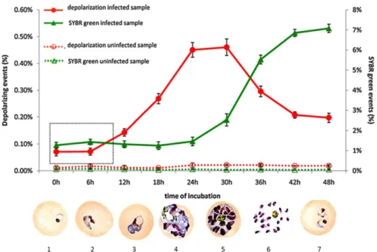
Consequently Hz can be considered an optimal parasite maturation indicator since its amount increases as the parasite matures (Figure 5).
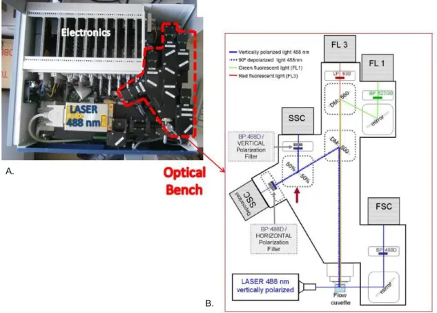
Due to the fact that Hz is birefringent it rotates the plane of polarized light, a process called depolarization, which is possible to detect by flow cytometry. LASER light, commonly used as a light source in flow cytometers, has polarized light. Thus, by placing a polarization filter orthogonally (90º rotated) to the plane of the LASER light in front of a second Side-Scatter (SSC) detector, allows to detect depolarized light and consequently Hz (Figure 6) [54].
With a small modification of a CyFlow® Blue (Partec, Münster, Germany), creating two SSC detectors, using a 50%/50% beam splitter (Figure 6) it was possible to develop a drug sensitivity assay based on the detection of Hz depolarization. It allows the detection of cells with Hz relatively to a total number of cells, thus assessing the percentage of parasites that maturate. Because the majority of the available antimalarial drugs act on the intra-erythrocytic stage of the malaria infection and, moreover, some of them act directly upon Hz formation it appeared possible to establish an accurate and reliable sensitivity assay based on the measurement of infected RBCs containing of Hz. Afterwards it was proved that the flow cytometric Hz detection allowed to determine the inhibitory effect of antimalarial drugs [49].
11
Figure 5 – Parasite growth and maturation detection. Red line: detection of depolarization duringincubation time. Through the first 24 to 30 hours the percentage of depolarizing events increases because more and more parasite presents Hz within the food vacuole. Green line: detection of parasite DNA using SYBR green I stain. The inicial parasitemia is maintained through the first 24 to 30 hours before the erythrocyte lyse and release new merozoits increasing SYBR green detection due to reinvasion and increased parasitemia. Parasite intraerythrocityc maturation. 1 – Ring form; 2, 3 – Throphozoite form; 4,5 – Schizont stage; 6 – Erythrocyte lyse and release of new merozointes; 7 – Ring form again in a red blood cell reinvaded by more than one merozoite/parasite. Graph adapted from: Rebelo, M. et al. 2012 [52]; parasite
images adapted from CDC: Diagnostic findings
(http://www.dpd.cdc.gov/dpdx/HTML/ImageLibrary/Malaria_il.htm last accessed 23.10.13)
1.6 Specific Gametocyte detection for drug testing
The development of drugs that also act against the sexual forms of the parasite and thus block transmission is of great importance, especially considering the new eradication agenda [55]. However, very few methods exist to test the drug sensitivity of gametocytes [56], [57], [58], certainly as compared to the ones available for asexual intra-erythrocitic stages [36]. One problem may be the required culture methods to induce gametocytes. These culture methods imply selective pressure to the development of the sexual stages of the parasite, such as low hematocrit, a low parasitemia or drug treatment [59], [60], [61]. However, it is not straightforward to detect gametocytes in a mixed culture of sexual and asexual forms, a reason why assays often use enriched gametocyte cultures [57]
.
Gametocytes contain Hz, which is distributed in the form of many small crystals, contrary to the large, usually single crystal observed in schizonts (Figure 4). Thus it appears possible that the proposed flow cytometric assay for Hz detection might also allow the detection of gametocytes, based on the presence of a population with higher levels of depolarization than that observed in schizonts. Because Hz in gametocytes does not agglomerate and is dispersed (Figure 4 - F), it probably has a higher degree of side scatter than a single crystal, even if the total amount of Hz
12
was the same. Thus, if gametocytes could be recognized as a separate population, distinct from schizonts, this approach might open the way for the development of a novel drug sensitivity assay to assess drug effects against the sexual forms of the parasite.Figure 6 – Adapted flow cytometer which allows detection of depolarized side-scatter. A.: Five
parameter CyFlow® flow cytometer with a blue laser (488nm) excitation, and detectors for forward scatter (FSC), side scatter (SSC), depolarized side scatter (dSSC), green fluorescence (FL1), and red fluorescence (FL3). B.: Two SSC detectors were created, with a 50%/50% beam splitter between them. A polarization filter was placed orthogonally (horizontal) to the polarization plane of the laser light (vertical), in front of one of the SSC detectors, allowing the detection of depolarized side scatter. Adapted from Frita, R. et al. 2011 [54].
2. Objectives
The primary objective of this study was to assess the potential of the novel flow cytometric method for Hz detection for the screening of new antimalarial compounds against P. falciparum blood stages. Newly synthesized compounds were screened and compared using two other methods.
The secondary aim was to evaluate if gametocyte cultures could be established and if gametocytes could be specifically detected using the same recently proposed flow cytometric method based on Hz detection.
A.
B.
13
3. Materials and Methods
3.1 Culture media, solutions and reagents
3.1.1 Malaria complete parasite medium (MCM): is composed by 500 mL of RPMI 1640 (1x, without L-glutamine, with NaHCO3) (GibcoTM, Life Technologies, Madrid, Spain) supplemented with 12 mL of HEPES Buffer Solution at 1 M (GibcoTM, Life Technologies, Madrid, Spain), 500 µL of gentamicin at 50 mg/mL (GibcoTM, Life Technologies, Madrid, Spain), 5 mL of L-glutamine at 200 nM and 50 mL of 10x AlbuMAX II®.
3.1.2 10x AlbuMAx II® solution: 25 g of Albumax II® was dissolved in 500 mL of an aqueous solution with 5.2 g RPMI 1640 (with L-glutamine, without NaHCO3), supplemented with 500 µL of gentamicin (50 mg/mL), 2.98 g HEPES, 1.67 g of sodium bicarbonate, 1 g of glucose and 0.1 g of hypoxanthine, pH adjust to 7.2 – 7.4 and filtered 0.22 µm. All reagents were obtained from Life Technologies (Madrid, Spain) except HEPES which was purchased from VWR (Carnaxide, Portugal).
3.1.3 1X Phosphate-buffered saline (PBS 1x): 10x PBS pH 7,2 (GibcoTM, Life Technologies, Madrid, Spain) was diluted 1:10 in filtered distilled water, obtained with Milli-Q Synthesis Q-Gard® water purification system (Millipore, Billerica MA, USA) or using ultrapure DNA/RNA free water from GibcoTM, Life Technologies, Madrid, Spain.
3.1.4 Sorbitol 5%: 5 g of D-sorbitol (Sigma-Aldrich, Life science) dissolved in 100 mL of distilled water, then filtered 0.22µm.
3.1.5 Cryoprotective solution: constituted by 57% of glycerol, 16 g/L of sodium lactate (VWR, Carnaxide, Portugal), 300 mg/L of potassium chloride and 25 mM of sodium phosphate pH 6.8. 3.1.6 Chloroquine: stock solution of 12.8 µM prepared chloroquine (Sigma-Aldrich, Life science) 3.1.7 Paraformaldehyde (PFA) 2%: paraformaldehyde at 16% (Electron Microscopy Sciences) was diluted 1:8 in PBS 1.2 X, to obtain PFA at 2%.
3.2 P. falciparum in vitro cultures
3.2.1 P. falciparum continuous cultures maintenance
P. falciparum strains 3D7 and Dd2 were obtained from Malaria Research and Reference
Reagent Resource Center (MR4; Manassas VA, USA). These strains were grown in continuous culture using uninfected red blood cells (RBCs) from healthy donors were isolated from buffy coats obtained from Instituto Português do Sangue. Buffy coats were washed 3x with RPMI 1640 (1x, without L-glutamine, with NaHCO3) and centrifuged for 5 min at 1800 rpm without applying the brake, allowing the separation of RBCs from white blood cells and plasma. Isolated RBCs were stored at 4ºC for a maximum of 2-3 weeks. Continuous P. falciparum cultures were incubated at 37ºC in a 5% CO2 atmosphere. Culture medium was changed on a daily basis and the parasitemia was kept at around 0.5% and the hematocrit at 5%.
14
3.2.2 Giemsa staining of blood smears
To assess the parasitemia of the cultures, blood smears were prepared every day and stained with Giemsa. The culture medium was removed and a small volume of culture blood was used to do a thin smear. The smear was fixed in absolute methanol, (Merk, Lisbon, Portugal) by dipping the slide for 30 s in a 50 mL falcon with absolute methanol, air-dried and stained with Giemsa at 10x (previously diluted 1:10 in PBS 1x) (Merk, Lisbon, Portugal) for 20 min. Then the slides were rinsed off with tap water and air-dried before observed by bright field microscopy under oil immersion. The parasitemia was estimated by counting the total RBCs and infected RBCs on eight different fields at a 1000x magnification/amplification. If the parasitemia was higher than ~2%, it would be lowered by adding a volume of uninfected RBCs equal to the amount of RBCs discarded from the culture.
3.2.3 Culture synchronization
Cultures were synchronized to obtain parasites at the same developmental stage. Cultures with a majority of ring-form parasites and a parasitemia higher than 2% were used. A volume of 10 mL of sorbitol 5% was added to the RBCs, after removing the medium, and then transferred to a 50 mL falcon and incubated for 10 min at room temperature after which it was centrifuged at 1800 rpm, for 5 min without brake, also at room temperature. After that, the supernatant was discarded and the RBCs pellet was washed two times in 35 mL of RPMI 1640 (1x, without L-glutamine, with NaHCO3) by centrifugation (1800 rpm, 4 min, without brake, room temperature). In the end, in a culture flask, pelleted RBCs were diluted to 5% hematocrit in MCM and incubated at 37ºC and 5% CO2 atmosphere.
3.2.4 Frozen stocks of P. falciparum
When necessary, P. falciparum cultures were frozen in a cryoprotective solution and stored at -80ºC. From a highly parasitized culture (> 2% parasitemia), with the majority of the parasites in ring form, the medium was removed by centrifugation (6 min at 1800 rpm). To the remaining volume of pellet cells, 0.33 and then 1.33 volumes of cryoprotective solution was added slowly, drop by drop, while mixing the tube gently.
To reuse cryoprotected-frozen vials, after thawing them for a few minutes at room temperature, the content was then transferred to a 50 mL falcon tube. To this volume, 0.1x volumes of 12% sodium chloride (NaCl) was added, followed by 10x volumes of 1.6% NaCl slowly, dropwise while shaking the tube gently. Then the tube was centrifuged for 3 min at 1800 rpm at 20ºC. The supernatant was removed and the pellet was resuspended in 10x the volume of RPMI 1640
(without L-glutamine, with
NaHCO3) and centrifuged again for 3 min at 1800 rpm. In the end, the pelleted RBCs was resuspended in MCM and transferred to a culture flask. Uninfected RBCs were added if needed to obtain a 5% hematocrit and the culture flask was incubated at 37ºC in a 5% CO2 atmosphere.15
3.3 Antimalarial drugs sensitivity assays
3.3.1 Inhibition of Chloroquine (IC
50determination) – Influence of Oxygen
The influence of oxygen (O2) on parasite’s growth was assessed by determining the chloroquine 50% inhibitory concentration (IC50) against P. falciparum 3D7 strain in different atmospheres.
To determine the IC50, two plates with serial dilutions (6, 12, 25, 50 and 100 nM) of chloroquine were prepared. The plates followed the procedure established in point 3.3.2 to analyse the inhibitory effect. However, one plate was incubated in a low O2 atmosphere incubator (Galaxy® 14 S, Eppendorf line of New Brunswick™) with 5% CO2, 5% O2 and 90% N2, and the other was incubated in a standard CO2 incubator (Thermo Scientific, Heraeus® HERAcell®), with 5% CO2 and the O2 levels around 21%, as present in Earth’s atmosphere.
3.3.2 Flow Cytometric Assays – General protocol
For this assay two controls were used: a growth control (without compound – drug free control) and a non-infected control (erythrocytes without parasite – uninfected control). In the screenings of compounds from Faculdade de Farmácia da Universidade de Lisboa (FFUL) chloroquine at a concentration of 100 nM (2x the determined IC50) was also used as a control.
Ring-stage synchronized cultures were used. Compounds were prepared in MCM 2x concentrated to the concentration to be tested. In a 96 well-plate 100 µL of compound and a blood suspension, previously prepared from the synchronized culture at 1% parasitemia and a 5% hematocrit, were added to the respective wells. Thus, each well had a final hematocrit of 2.5%, with 1% parasitemia and the compound concentration to test (Figure 7). MCM was added to the drug free and uninfected controls. The assay was performed in triplicates.
Plates were incubated for 48 h, at 37ºC in a 5% CO2 atmosphere, and flow cytometric measurements were performed at 0 h, 24 h and 48 h of incubation. At each time point, the percentage of depolarizing events and SYBR green positive events (parasitemia) was assessed. To measure parasitemia, samples were stained with SYBR® Green I (Sigma) at 1x (previously diluted 1:10000 in PBS 1x) in the proportion of 1:10 (5 µL of the sample from each well plus 50 µL of SYBR green 1x). Then incubated for 20 min in dark and room temperature. After this period of incubation 900 µL of PBS 1x were add to each sample tube before analysis in the CYFlow® (Partec®, Münster, Germany) where a total of 100 000 events were acquired.
16
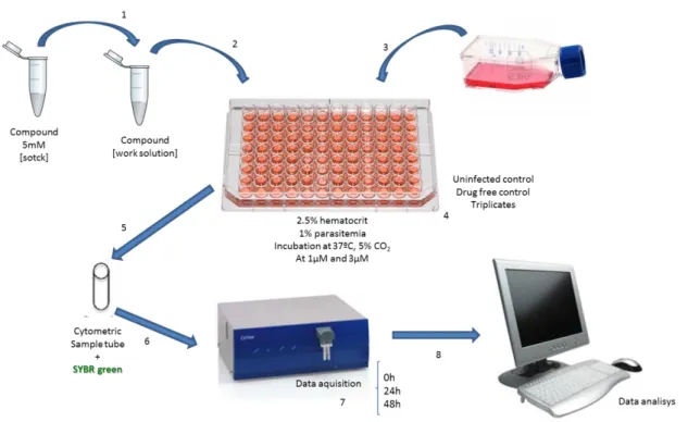
Figure 7 – Flow cytometric assay preparation. Work solutions were prepared from a 5 mM stock compound
(1) and distributed in a 96 well plate (2). Blood suspension in 1:1 proportion, was added to the wells (3). Uninfected and drug free controls were also included. The 96 well plate was set with a final 2.5% hematocrit and 1 % parasitemia was incubated at 37ºC in a 5 % CO2 atmosphere (4). Samples from each well stained with SYBR green I (5) before and analysed by flow cytometry (6). Sample analysis was performed at 0 h, 24 h and 48 h of incubation (7). Data was analysed using the FlowJo software (8).
3.3.3 Screening novel compounds from Faculdade de Farmácia
The inhibitory potential of 24 new compounds obtained from FFUL (Professor Rui Moreira’s group) was investigated. The compounds are quinazoline derivatives (Table 5). The action of these compounds against P. falciparum 3D7 and Dd2 strains was determined. The screening was performed using cut-off concentrations of 1 µM and 3 µM. Inhibitory activities of the compounds was assessed by flow cytometric and HRP2-ELISA assays, described in 3.3.2 and 3.3.6. Chloroquine at 100 nM, twice the determined IC50 concentration, was used as an internal control.
17
Table 5 – Chemical structure of the quinazoline derivative screened compounds as well as from chloroquine, artemisinin and pyrimethamine as comparison.Quinazoline
ABJ1 AD3 AD6
AD20 AD26 AD34
ASR 252 ASR 256 ASR 291
ASR 292 ASR 294 ASR 295
ASR 299 ASR 300 ASR 304
ASR 305 ASR 306 ASR 317
18
ASR 327 ASR 338 ASR 341
Example of antimalarial drugs structure:
Chloroquine Artemisinin Pyrimethamine
3.3.4 Flow cytometer
The flow cytometer, the CyFlow® (Partec®, Münster, Germany), used in the assay is a five parameter flow cytometer with a blue laser (488 nm), adapted to detect depolarizing events. The optical bench include detectors for: forward scatter (FSC), side scatter (SSC), green (FL1 - band pass 535/45 nm filter) and red fluorescence (FL3 - long pass 610 nm filter), as well as a detector for depolarized side scatter, used to detect infected red blood cells containing Hz (Figure 6).
Flow cytometry enables the counting and the analysis of multiple parameters of individual cells within heterogeneous populations. As a cell passes through the laser it will refract or scatter light at different angles. Light scattered in the forward direction (0º angle), is quantified in the FCS detector and usually a measure of particle size. Light that is scattered to the side (at 90º angle) indicates the granularity and structural complexity inside the cell. The depolarized side scatter detects light which has been scattered at 90º and depolarized. For this, a polarization filter, perpendicularly positioned to the incident polarized laser beam is placed in front of the detector blocking out scattered light which remains in its original polarization plane.
3.3.5 Flow cytometric assays - results analysis
Flow cytometric data was analysed by using FlowJo (version 9.6.2) software. The gating strategy was established by comparing the uninfected RBCs and P. falciparum infected RBCs populations at each time point (Figure 8). Drug effects were assessed by comparing the percentage of depolarizing events or percentage of SYBR green positive events between the drug free control and the drug treated samples. Depolarization assay results were assessed at 24 h, when most of the parasites are schizonts, which contain Hz. The SYBR green assay results were determined at 48 h, the time point when reinvasion of uninfected RBCs already occurred and the parasitemia has increased.
19
Figure 8 - Gating strategy. At 24 h of incubation, depolarization is measured and a gate was establishedaccording to the existing population of events that depolarize in the drug free control and that are absent in the uninfected control. At 48 h of incubation, the SYBR green positive (SG+) events were assessed by establishing a gate around the detected population in the drug free control that is absent in the uninfected control. After setting the gates they were applied to the data acquired for the tested compounds.
3.3.6 Histidine-rich protein-2 sensitivity assay
The histidine-rich protein 2 (HRP2) ELISA was performed to corroborate the flow cytometric results using the P. falciparum strain 3D7. For this assay a growth control (without compound) was used as well as an effective concentration of chloroquine at 100 nM as a control for inhibition.
3.3.2.1 Pre coating protocol:
Primary IgM antibody (MPFM-55A, Immunology Consultants Laboratories, Inc, Newberg, OR, USA) was diluted to 1 µg/mL concentration in PBS 1x, and 100 μL was transferred to each well of 96 flat-bottom well-plates. The plates were sealed and incubated at 4ºC overnight. After this, plates were pad-dried and 200 μL of blocking solution (2% bovine serum albumin (Sigma, CAS No. 9048-46-8) solution in PBS 1x) was added to each well and the plates were incubated for 2 h, at room temperature. Then, plates were washed 3x with washing solution (PBS/Tween 20 [0.05%]) and pad-dried. Finally, the plates were sealed airtight and frozen at -20ºC.
Uninfected Drug free
Depolarization detection at 24 h SYBR green detection at 48 h Dep o la ri z e d s id e s c a tt e r Side scatter Green fluorescence Red f lu o re s c e n c e ( 6 1 0 n m )
20
3.3.7 HRP2-ELISA Sample analysis
From a synchronized culture, a blood suspension with a final hematocrit of 1.5% and a parasitemia of 0.05% was prepared. The blood suspension was distributed in a 96 well plate, together with the compound at the concentration to be tested. The plate was incubated at 37ºC in a 5% CO2 atmosphere for 72 h. At the end of incubation plates were frozen at -20ºC until the HRP2 ELISA was performed.
To perform the ELISA , plates were freeze and thawed twice and 100 µL of the lysed sample was transferred to a 96 well-plate pre-coated with MPFM-55A antibody (Immunology Consultants Laboratories, Portland, USA) and incubated for one hour at room temperature. Plates were washed three times with washing solution (Tween 20 at 0.05%), then 100 µL of the secondary antibody at 0.1 µg/mL, MPFG-55P (Immunology Consultants Laboratories, Portland, USA) was added and plates were incubated for another hour. Plates were washed again and incubated with the 100 µL of chromogen, TMB One (Biotrend, Köln, Germany) for 5 to 10 min, at room temperature, in the dark. The reaction was stopped by adding 50 µL of sulphuric acid at 1 M (Merck, Darmstadt, Germany). Finally, the absorbance at a wavelength of 450 nm was immediately determined using the Infinite M200 plate reader (Tecan, Mannedorf, Switzerland).
3.3.8 50% inhibitory concentration determination of selected compounds
The 50% inhibitory concentration (IC50) was determined for chloroquine in both atmospheres, and for three of the FF compounds.
The IC50 values for chloroquine were calculated based on 2-fold serial dilutions (6, 12, 25, 50 and 100 nM). The inhibitory effect was assessed by the flow cytometric detection of Hz and SYBR green, as explain above in 3.3.2.
IC50 values were determined for the compound 321, which had been previously reported to have an inhibitory activity at 3 µM, and for two other compounds that were randomly selected (256 and 291). Concentrations of 1, 2, 4, 6, 8 µM were used for all three compounds. An additional concentration of 10 µM was used for compounds 256 and 291.
The SigmaPlot software from Systat Software Inc. (SSI) (San Jose, California) was used to trace the dose-response curves of the acquired data, in order to calculate the IC50 values.
3.4 Gametocyte cultures
Gametocytogenesis is only induced in in vitro cultures when the parasites present in the culture are “stressed”. For that, culture conditions were modified to induce stress in the asexual parasites, and two methods were experimented (A) and (B):
3.4.1 Culture Method A
Starting with a 5% hematocrit, culture medium was changed every day until a 2% - 3% parasitemia was reached. At this point the hematocrit was decreased to 2.5%, representing the “stressing factor” to induce the gametocyte culture. Every day until day ten, medium was changed
21
daily and cultures were monitored by flow cytometry, as well as by microscopic observation of thin blood smears.3.4.2 Culture Method B
In a recently synchronized culture, the hematocrit was reduced to 2.5% and the parasitemia to 0.7%, corresponding to day one. Additionally to this “stressing factor”, the medium was not changed until day four. On this day, culture medium was changed and replaced by a suspension of lysed RBCs that corresponded to twenty percent of the final culture volume. This suspension consisted of RPMI 1640 with lysed RBCs which were frozen and thawed at least twice, in 1:1 proportion. The medium was changed on days six and eight and thereafter on a daily basis. Flow cytometric acquisitions as well as blood smear were done every day until day ten.
3.4.3 Erythrocytes fixation using paraformaldehyde
Cells were centrifuged to remove the culture medium. A suspension of P. falciparum infected RBCs with a 50% hematocrit was prepared. A volume of 5.5 mL of paraformaldehyde (PFA) 2% (previously diluted 1:8 in PBS 1.2x) was added to 250 μL of the infected RBC suspension. The same procedure was performed using uninfected erythrocytes. Samples were incubated for 2 h at 37ºC in a water bath. Subsequently, they were centrifuged for 3-5 min at 1800 rpm. The supernatant was discarded and the pellet was re-suspended in PBS 1x.
3.4.4 Sorting Gametocyte cultures
The MoFlo (Beckman Coulter, Fort Collins, USA) cytometer is a high speed sorter with an open architecture. This enables many different configurations based on the modularity which meet the diverse sorting applications. For example, it is easy to include polarizing filters in any light-path and thus create depolarized SSC detectors.
After fixing the erythrocytes, the gametocytes were sorted in a MoFlo cytometer according to its SYBR green fluorescence and depolarization degree. First, the SYBR green positive population was selected and its depolarization was analysed. In the depolarization plot three gates based on the depolarization degree were established: (i) non-depolarizing events, (ii) medium depolarizing events and (iii) high depolarizing events (Figure 9). Cells within these gates were sorted into separate tubes. The purity of sorted cells was assessed by reanalysing each sorted population in the MoFlo.
Sorted cells were left to sediment overnight. Then, after removal of most of the supernatant, a small volume of cells were transferred onto a slide and analysed immediately by microscopy. Pictures of each population were taken using light microscopy and depolarizing microscopy.
22
Figure 9 – Sorting gating strategy.SYBR green positive population was plotted based on their depolarized side scatter and side scatter. Three gates were established according to the depolarization degree: i) non-depolarizing events, ii) medium depolarizing events and iii) high depolarizing events. Cells within these gates were sorted using a MoFlo (Beckman Coulter, Fort Collins, USA).
D e p o lar a z e d s ide s c a tt e r Side scatter iii) High ii) Medium i) Non-depolarizing