Braz. J. Phys. vol.39 número4
Texto
Imagem

Documentos relacionados
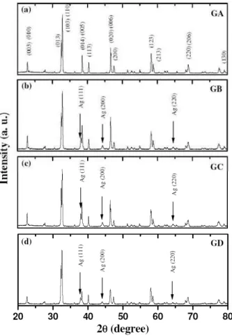
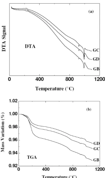
The characterization of this solid crystal structure using the Raman spectroscopy, X-ray Powder diffraction, Differential scanning calorimeter, thermogravimetric analysis and
Various physico-chemical methods like particle analysis, scanning electron microscopy, differential scanning calorimetry, differential thermal analysis, thermogravimetry analysis,
samples, prior to and after functionalization, were characterized by chemical analyses, thermogravimetric analysis (TGA), x ray diffraction (XRD), diffuse reflectance infrared
The characterization and chemical analysis of the synthesized biomimetic apatite powders were performed by scanning electron microscopy (SEM), powder X ray diffraction
The structural properties of particles were investigated by X-ray diffraction (XRD), atomic force microscopy (AFM), differential thermal analysis (DTA), and N 2
Starting from the scanning electronic microscopy characterizations, the chemical analysis for energy dispersive spectroscopy, and phase analysis by X-ray diffraction, the
The growth technique used was the Hot Wall Epitaxy (HWE) and the films were characterized by scanning electron micrographs, atomic force microscopy (AFM), x-ray diffraction and
The inclusion complexes obtained were characterized using X-ray powder diffraction (XRPD) and scanning electron microscopy (SEM), and the dissolution profile of the complex and
