Article
J. Braz. Chem. Soc., Vol. 25, No. 5, 861-866, 2014. Printed in Brazil - ©2014 Sociedade Brasileira de Química 0103 - 5053 $6.00+0.00
A
*e-mail: [email protected]; [email protected]
A New Triazene Ligand Immobilized on Triacetylcellulose Membrane for Selective
Determination of Mercury Ion
Hossein Tavallali,*,a Homa Shafiekhani,a Mohammad Kazem Rofouei,b and
Mahmood Payehghadra
aDepartment of Chemistry, Payame Noor University (PNU), 19395-4697 Tehran, Iran
bFaculty of Chemistry, Kharazmi University, P.O. Box 14911-15719 Tehran, Iran
Este trabalho apresenta um novo sensor químico óptico, simples e de baixo custo, para a determinação de íon mercúrio (II) em solução aquosa. A membrana sensível a mercúrio foi preparada pela incorporação de triazina ((E)-1-(2-etoxifenil)-3-(4-nitrofenil) triazol-1-ene como um ligante adequado para Hg(II) sobre uma membrana de triacetilcelulose. O estudo espectrofotométrico do complexo formado entre o ligante triazene (L) e íons Hg2+ em solução
de dimetilformamida (DMF) mostrou uma grande constante de estabilidade para o íon mercúrio complexado. A membrana responde ao íon mercúrio pela troca reversível da cor de laranja para verde em solução tamponada em pH 3,0. Uma relação linear foi observada entre a absorbância da membrana em 405 nm, na faixa de 7 a 90 µg mL-1, com um limite de detecção de 64 ng mL-1
em soluções aquosas, em pH 3,0. O sensor óptico for aplicado com sucesso na determinação de mercúrio em amostras de água enriquecidas com o analito.
A new simple and inexpensive optical chemical sensor for mercury(II) ion in aqueous solutions is presented. The mercury sensing membrane was prepared by incorporating of triazene ((E)-1-(2-ethoxyphenyl)-3-(4-nitrophenyl) triaze-1-ene) (L) as a suitable ligand for Hg(II) on triacetylcellulose membrane. Spectrophotometric study of complex formation between the triazene ligand L and Hg2+ in dimethyl formamide solution indicated a large stability constant for the mercury
ion complex. The membrane responds to mercury ion by changing color reversibly from orange to green in buffer solution at pH 3.0. A linear relationship was observed between the membrane absorbance at 405 nm in a range from 7 to 90 µg mL-1 with a limit of detection of 64 ng mL-1 in
aqueous solutions at pH 3.0. The optical sensor was successfully applied to the determination of mercury in spiked water samples.
Keywords: optical sensor, Hg2+ ion, triazene ((E)-1-(2-ethoxyphenyl)-3-(4-nitrophenyl)
triaze-1-ene) ligand
Introduction
Mercury is one of the most toxic elements in the world and represents a major toxicity to microorganisms and environment even in low concentrations.1 Accurate and
sensitive determination of mercury is now a challenging task. Inorganic mercury has been reported to produce harmful effects at concentrations as low as 5 µg L-1.2
Inorganic mercury may be converted to methyl mercury in marine environment that is even more toxic than inorganic mercury.3 Thus, the development of simple methods for
selective determination of mercury in trace amounts in different matrices is critical. A wide range of analytical
methods has been used for the determination of mercury in real samples. These include spectrophotometry,4 neutron
activation analysis,5 anodic stripping voltammetry,6 X-ray
fluorescence spectrometry,7 inductively coupled plasma
mass spectrometry,8 electro-thermal atomic absorption
spectrometry,9 fluorescence spectrometry,10 and cold vapor
atomic absorption spectrometry.11 However, most of these
techniques are rather expensive and time consuming and may not be available in all laboratories. A few works have been introduced on the development of optodes which are based on spectrophotometric method for Hg(II).12-16
over electrochemical sensors.17,18 Optical chemical
sensors (optodes) are considered to be better studied for monitoring of toxic ions/species.19-22 Optodes are easy to
prepare, selective for pre-concentration of target analyte in the sensing matrix, and there is no need to separate reference devices. Thus, in recent years, a number of optodes with different optical principles have been reported for the determination of mercury.23-29 The current paper
encompasses the fabrication of an optode for determination of low levels of mercury and it utilizes triazene ligand as the sensing reagent. Triazene compounds, characterized by having a diazoamino group (–N=N–N–), have been studied for over 130 years concerning their interesting structural, anticancer and reactivity properties. They have been used in medicinal and as organometallic ligands.30
Triazene compounds with two cis and trans conformers can behave like a microelectronic switch.31 Triazenes can bind
to metals in both monodentate and bidentate manners. The products are air and moisture stable and are easily prepared. However, despite extensive combinatorial chemistry scientific reports on the synthesis, characterization and crystalline structure of the transition metal triazenide complexes, reports on analytical application of these ligands are quite sparse. In the present study, a new triazene ligand (E )-1-(2-ethoxyphenyl)-3-(4-nitrophenyl)triaze-1-ene (L) is immobilized on a triacetylcellulose membrane to be used as an effective ligand for construction of a selective optical sensor for the spectrophotometric determination of Hg2+ in aqueous solutions.
Experimental
Material and instruments
All of the chemicals used were of analytical reagent grade (Merck, Darmstadt, Germany). De-ionized water (Abpakhsh, Tehran, Iran) was used throughout. A 1000 µg mL-1 Hg(II) stock solution was prepared by
dissolving 0.1708 g Hg(NO3)2.H2O in de-ionized water
and 0.5 mL of 1 mol L-1 HNO
3 and diluted to 100 mL in
a volumetric flask. Sample solutions were prepared by appropriate dilution. The triazene ligand was synthesized and its structure with Hg2+ ion has been confirmed
(Figure 1).32 The 1.166 × 10⁻³ mol L-1 triazene ligand
solution was prepared by dissolving 10.0 mg ligand L in 30.0 mL ethylenediamine. A Shimadzu 1601 (Kyoto, Japan) PC UV-Vis spectrophotometer with 1 cm cell quartz was used for recording all spectra and absorbance measurements. A Jenway 3510 pH-meter (Staffordishe, UK) with a combined glass electrode was used after calibration against standard Merck buffer for pH adjustment.
Determination of formation constant
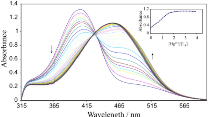
In order to determine stoichometry and stability of the resulting metal-ligand complexes, in a typical procedure, 2.0 mL of 5.0 × 10⁻⁵ mol L-1 ligand solution in dimethyl
formamide (DMF) were placed in the spectrophotometer cell and the absorbance of solution in range of 315-600 nm was measured. Then a known amount of the concentrated solution of Hg2+ in 1.3 × 10⁻³ mol L-1 DMF was added in
a stepwise manner using a 10 µL Hamilton syringe. The absorbance spectrum of the solution was recorded after each addition. The absorbance curves are shown in Figure 2. By addition of Hg2+, a decrease in absorbance is observed at
405 nm. By decreasing the absorbance at 405 nm, a new peak at about 465 nm is formed that corresponds to the formation of an Hg2+-L complex. The resulting plots of
the absorbance at 465 nm against metal ion/ligand mole ratios are shown in Figure 2. From the sharp inflection point observed for Hg2+ at a mole ratio of 2, it can be concluded
that a 2:1 complex of [L2-Hg] is formed in DMF solution.
The formation constant of the resulting complex between Hg2+ and L ligand was evaluated to be equal to 7.81, from
the absorbance versus [Hg2+]/[L] mole ratio data using
known equations and utilizing a non-linear least-squares curve fitting program, KINFIT.33
Figure 1. The molecular structure of [Hg(C14H13N4O3)2].
Preparation of the sensor membrane and measurement
For immobilization of the triazene ligand L, triacetylcellulose membrane was treated with a 1.166 × 10⁻³ mol L-1 ligand solution, in ethylenediamine
for 2-5 min at ambient temperature. The resulting orange color membranes were thoroughly washed with detergent solution and water. Prepared membranes were kept under water when not in use. Each time six membranes were made and they can still be used after 1 month when kept under water. A 1 cm × 3 cm piece of the fabricated membrane sensor was cut and placed inside the quartz cell of the spectrophotometer. All the measurements on the triacetylcellulose membranes were performed in aqueous medium. Tap water samples were collected from the laboratory water tap at Payame Noor Shiraz University, Iran. The tap water samples were spiked with different amounts of Hg2+ and pH adjusted
to 3.0 before analysis. For analysis, about 2.5 mL of the samples were transferred to a 1 cm quartz cell equipped with the membrane sensor. The absorbance was then measured at 405 nm and subtracted from an absorbance reading for a buffer solution at the same wavelength. The Hg2+ concentration was then derived using an ordinary
calibration curve method.
Results and discussion
The first extensive investigation of the coordination chemistry of a triazene derivative (1,3-diphenyltriazene) was carried out in 1887 by Meldola and Streatfield.34 The
interaction of triazene derivatives with mercury ion has been studied in the past few years by several authors.35-40
The affinity of the ligand L toward Hg2+ must be related
to the coordinate interaction between ligand L and Hg2+
ion. In a ligand molecule, the O and N atoms can play the role of an electron donor which can coordinate metal ions as electron acceptors. On the other hand, on the basis of Pearson’s hard-soft [Lewis] acid-base principle, Hg2+ is a
soft acid and triazene ligand is a soft Lewis base. Organic ligands containing unsaturated nitrogen atoms can be regarded as soft Lewis bases. As such they may reveal high tendency to form stable coordination complexes with numerous transition metal ions, particularly those that can be regarded as soft Lewis acids [e.g., Cu(I), Ag(I) and Hg(II)].41 Therefore, it was also known that
certain ligands formed the most stable complexes with Hg2+. Soft Lewis acid binds to soft Lewis base to give
covalent complexes. These interactions are dominated by the energies of the participating frontier molecular orbitals (FMO).
Spectral characteristics
Figure 3 shows the absorption spectra of immobilized triazene ligand, which was obtained after being equilibrated in pH 3.0 buffer solution containing different concentrations of mercury (0.0-5.0 µg mL-1 Hg2+, by addition of
0.5 µg mL-1 Hg2+ at each interval). The spectral change
(increase in absorption band at 433 nm and decrease in absorption band at 405 nm) is the result of increase of mercury ion concentration in the membrane and complex formation. The maximum absorbance of the immobilized ligand L is located at 405 nm. The wavelength of 405 nm was selected for further studies because of higher selectivity and sensitivity at this wavelength.
Effect of pH of test solution on the sensor response
The influence of test solution pH on the response of the proposed Hg2+ ion-selective optical sensor is illustrated in
Figure 4. The response characteristic of the membrane sensor was highly dependent on pH. The absorbance measurements were made for 10 µg mL-1 mercury ion in the pH range of
1-8 at 405 nm. As it is obvious from Figure 4, the absorbance
Figure 3. Absorption spectra of the optode film in the presence of 0.0-5.0 µg mL-1 Hg2+ at pH 3.0, by addition of 0.5 µg mL-1 Hg2+ at each interval.
increased rapidly by changing the pH from 2.0 to 3.0, while it decreased at pH values higher than 4.0. This phenomenon might be due to the fact that at lower pH values (pH < 3.0), complexation is weak. On the other hand, the reduced optical response of the proposed sensor at pH > 3.0 could be due to a possible hydroxide formation of mercury ions. Thus, in subsequent experiments, pH 3.0 was used for further studies.
Sensor response time
Figure 5 shows the profile of the response of Hg2+
optical sensor at 405 nm with time. The response time of the present optode was tested by recording the absorbance change at 405 nm from a pure pH 3.0 buffer to a buffered mercury solution of 10 µg mL-1 Hg2+. It can be seen that
the time taken to achieve to the 95% steady state response is within 15 minfor 10 µg mL-1 Hg2+ at pH = 3.0.
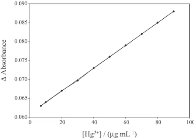
Dynamic range
Figure 6 shows the absorbance signals of the optode film to various concentrations of Hg2+ ions in the range of
7.0-90.0 µg mL-1 at pH 3.0 and 90.0 µg mL-1 was found as
the concentration of Hg2+ ion that saturates the film. The
regression equation was:
∆A = 0.0003 C(Hg2+) + 0.0609 (1)
with a correlation coefficient of 0.9998, where ∆A is the
increase in absorbance of the film at 405 nm for a fixed time of 15 min, and C is the concentration of Hg2+ in µg mL-1.
The detection limit which is based on 3σ of the blank was
calculated to be 64.0 ng mL-1.
Regeneration of the optode membrane
Some reagents, including HCl, NaOH, H2SO4, HNO3
and ethylenediamine, were studied as regenerating reagents. It was found that the best result was obtained by applying ethylenediamine which gave short membrane regeneration times (10-25 s). After this regeneration and for the next mercury concentration measurement, the optode must be placed in buffer pH 3.0 for 10-15 min.
Selectivity
The major property of the optode membrane, its selectivity, reflects its relative response to the analyte ion and to the other ions present in solution. Thus, the influence of a number of common metal ions on the absorbance of the proposed Hg2+ optical sensor was carried out. To determine
of selectivity of the optode membrane, the membrane was tested for the determination of 10.0 µg mL-1 of Hg2+ ions in
the presence of some metal ions, including Cd2+, Cu2+, Fe2+,
Pb2+, Zn2+, Ni2+, Co2+, and Fe3+. The species were considered
as interference if they caused an analytical variation of 5% or more when compared to the analytical signal obtained in the absence of the interfering species. At the applied pH value, no interference was observed from even 50-fold excess of the interfering ions.
Accuracy and analytical applications
The proposed optical sensor was found to work well under laboratory conditions. In order to test the practical application of the present sensor, the distilled water and tap water samples spiked with different amounts of mercury ions were measured by the proposed optode. The mercury content of water was analyzed by standard addition method and then determined by the proposed optode (Table 1). From the data given in Table 1, it is rapidly apparent that the present optode is useful for the determination of mercury in real samples.
Figure 5. Typical response curve of the optode film at 405 nm as a function of time when the film was exposed to 10 µg mL-1 Hg2+.
Conclusions
In this work, a new triazene ligand (E )-1-(2-ethoxyphenyl)-3-(4-nitrophenyl) triaze-1-ene was considered as a suitable complexing agent for construction of a triacetylcellulose optical membrane selective sensor for mercury(II) ion detection at low concentration levels. On the basis of the results presented in this work, the proposed Hg(II) ion-selective optode has many advantages including: easy preparation, low cost, fast response time, wide dynamic range, low detection limit, and good reproducibility. It was applied in order to determine the concentration of mercury(II) ions in water samples.
Acknowledgements
The authors wish to acknowledge the support of this work by Shiraz Payame Noor University research council.
References
1. Green, N. N.; Earnshaw, A.; Chemistry of Elements; Pergamon Press: New York, 1984.
2. Boening, D. W.; Chemosphere2000, 40, 1335.
3. Krishna, M. V. B.; Ranjit, M.; Karunasagar, D.;Arunachalam, J.; Talanta2005, 67, 70.
4. Park, S. M.; Choi, H. S.; Anal. Chim. Acta2002, 459, 75. 5. Yu, J. C.; Lo, J. M.; Wai, C. M.; Anal. Chim. Acta1983, 154,
307.
6. Ugo, P.; Moretto, L.; Bertoncello, P.; Wang, J.; Electroanalysis
1998, 10, 1017.
7. Bennun, L.; Gomez, J.; Spectrochim. Acta, Part B 1997, 52, 1195.
8. Powell, M. J.; Quan, E. S. K.; Boomer, D. W.; Wiederin, D. R.; Anal. Chem.1992, 64, 2253.
9. Burrini, C.; Cagnigni, A.; Talanta1997,44, 1219.
10. Safavi, A.; Eddon, L.; Foulkes, M.; Stockwell, P.; Corns, W.; Analyst1999, 124, 185.
11. Yamini, Y.; Alizadeh, N.; Shamsipur, M.; Anal. Chim. Acta
1997, 69, 355.
12. Czolk, R.; Reichert, J.; Ache, H. J.; Sens. Actuators, B1992, 7, 540.
13. Morales-Bahnik, A.; Czolk, R.; Reichert, J.; Ache, H. J.; Sens. Actuators, B1993, 13, 424.
14. Lerchi, M.; Reitter, E.; Simon, W.; Pretsch, E.; Chowdhury, D. A.; Kamata, S.; Anal. Chem.1994,66,1713.
15. Ensafi, A.; Fooladgar, M.; Sens. Actuators, B2006, 113, 88. 16. Nuriman, N.; Kuswandi, B.; Verboom, W.; Sens. Actuators, B
2011, 157, 438.
17. De-Silva, A. P.; Gunaratne, N.; Gunnlaugsson, T.; Huxley, A. J. M.; McCoy, C. P.; Rademacher, J. T.; Rice, T. E.; Chem. Rev.1997, 97, 1515.
18. Valeur, B.; Leray, I.; Coord. Chem. Rev. 2000, 205, 3. 19. Bakker, E.; Buhlmann, P.; Pretsch, E.; Chem. Rev. 1997, 97,
3083.
20. Pretsch, E.; Buhlmann, P.; Bakker, E.; Chem. Rev. 1998, 98, 1593.
21. Janata, J.; Josowicz, M.; Vanysek, P.; Devaney, D. M.; Anal. Chem. 1998, 70, 179R.
22. Wolfbeis, O. S.; Anal. Chem.2006, 78, 3859.
23. Sanchez-Pedreno, C.; Ortuno, J. A.; Albero, M. I.; Garcia, M. S.; Valerom, M. V.; Anal. Chim. Acta 2000, 414, 195.
24. Chan, W. H.; Yang, R. H.; Wang, K. M; Anal. Chim. Acta2001, 444, 261.
25. Kuswandi, B.; Narayanaswamy, R.; Sens. Actuators, B 2001, 74, 131.
26. Safavi, A.; Bagheri, M.; Sens. Actuators, B 2004, 99, 608. 27. Ensafi, A. A.; Fouladgar, M.; Sens. Actuators, B2006, 113, 88. 28. Cano-Raya, C.; Fernandez-Ramos, M. D.; Gomes-Sanchez, J.;
Capitan-Valley, L. F.; Sens. Actuators, B2006, 117, 135. 29. Khezri, B.; Amini, M. K.; Firooz, A. R.; Anal. Bioanal. Chem.
2008, 390, 1943.
30. Kimball, D. B.; Herges, R.; Haley, M. M.; J. Am. Chem. Soc.
2002, 124, 1572.
31. Martin, P. J.; Petty, M. C.; Bryce, M. R.; Bloor, D.; An Introduction to Molecular Electronics, Oxford University Press:
New York,1995.
Table1. Result of mercury (II) ion determination in spiked samples
Sample Mercury(II) added / (µg mL-1) Mercury(II) founda / (µg mL-1) RSD / % Recovery / %
Distilled water
0.0 NDb – –
10 9.54 0.097 95.4
30 29.53 0.014 98.4
Tap water
0.0 NDb – –
30 29.2 0.086 97.3
50 47.4 0.011 94.8
32. Rofouei, M. K.; Fereyduni, H.; Gharamaleki, J. A.; Bruno, G.; Amiri Rudbari, H.; Acta Crystallogr., Sect. E: Struct. Rep. Online2010,E66, m1082.
33. Alizadeh, N.; Ershad, S.; Naeimi, H.; Sharghi, H.; Shamsipur, M.; Pol. J. Chem.1999, 73, 915.
34. Meldola, R.; Streatfield, F. W.; J. Chem. Soc. 1887,52, 434. 35. Casagrande, G. A.; Lang, E. S.; Horner, G. M. M.; Broch, F.;
Inorg. Chim. Acta 2007, 360, 1776.
36. Horner, M.; De Oliveira, G. M.; Visentin, L. D.; Cezar, R. S.; Inorg. Chim. Acta2006, 359, 4667.
37. Melardi, M. R.; Rofouei, M. K.; Massomi, J; Anal. Sci. 2007, 23, 67.
38. Hematyar, M.; Rofouei, M. K.; Anal. Sci.2008, 24, 117. 39. Melardi, M. R.; Salemi, Y.; Kazemi, S. R.; Rofouei, M. K.; Acta
Crystallogr., Sect. E: Struct. Rep. Online 2009, E65, m302.
40. Rofouei, M. K.; Beizaee, A.; Gharamaleki, J. A.; Acta Crystallogr., Sect. E: Struct. Rep. Online 2009,E65, m1259.
41. Pearson, R. G.; Inorg. Chim. Acta1995, 240, 93.
Submitted: November 23, 2013