Article
J. Braz. Chem. Soc., Vol. 25, No. 5, 907-912, 2014. Printed in Brazil - ©2014 Sociedade Brasileira de Química 0103 - 5053 $6.00+0.00
A
*e-mail: [email protected]
Flavonoid Glycosides from
Hosta longipes
, Their Inhibition on NO Production, and
Nerve Growth Factor Inductive Effects
Chung Sub Kim,a Oh Wook Kwon,b Sun Yeou Kimc,d and Kang Ro Lee*,a
aNatural Products Laboratory, School of Pharmacy, Sungkyunkwan University, 440-746 Suwon, Republic of Korea
bGraduate School of East-West Medical Science, Kyung Hee University Global Campus, 446-701 Yongin, Republic of Korea
cCollege of Pharmacy, Gachon University, 406-799 Incheon, Republic of Korea
dGachon Medical Research Institute, Gil Medical Center, 405-760 Incheon, Republic of Korea
Investigação fitoquímica das folhas da Hosta longipes identificou um novo flavonóide glicosídeo, o caempferol-3-O-β-D-glucopiranosil-(1→2)-[6’’’-O-acetil-β -D-glucopiranósido]-7-O-β-D-glucopiranósido, e mais cinco derivados flavonóides conhecidos. As estruturas de dois compostos foram reveladas por vários métodos de RMN (1H e 13C RMN, 1H-1H COSY, HMQC HMBC) e hidrólise química. Dados de RMN de um deles são publicados pela primeira vez. As atividades biológicas de seis compostos revelaram que cinco inibiram fortemente a produção de óxido nítrico (NO), com valores de IC50 de 11,56-15,97 µm em células BV-2 estimuladas por lipopolissacarídeo (LPS), sem toxicidade celular. Dois compostos mostraram indução moderada da secreção no fator de crescimento do nervo (NGF) em linhagem de células C6 de glioma (124,70 ± 7,71% e 117,02 ± 3,60%, respectivamente).
An extended phytochemical investigation of the leaves of Hosta longipes identified the new flavonoid glycoside, kaempferol-3-O-β-D-glucopyranosyl-(1→2)-[6’’’-O-acetyl-β -D-glucopyranoside]-7-O-β-D-glucopyranoside and five known flavonoid derivatives. The structures of two compounds were revealed by extensive NMR methods (1H and 13C NMR, 1H-1HCOSY, HMQC and HMBC) and chemical hydrolysis. NMR data of one of them are published for the first time. Bioactivities of six compoundsrevealed that fivestrongly inhibited the production of nitric oxide (NO) with IC50 values of 11.56-15.97 µm in lipopolysaccharide (LPS)-stimulated BV-2 cells without cell toxicity. Two compounds showed moderate induction of secretion of nerve growth factor (NGF) in C6 glioma cells (124.70 ± 7.71% and 117.02 ± 3.60%, respectively).
Keywords:Hosta longipes, flavonoid glycoside, nitric oxide, nerve growth factor
Introduction
More than 8,000 flavonoids have been isolated from plant sources. Flavonoids have a variety of pharmacological effects that include anti-cancer, anti-microbial, anti-oxidant, and anti-inflammatory activities.1,2 Neuroprotective effects
of flavonoids from Citrus species are reportedly associated with their anti-inflammatory action, the ability to traverse the blood-brain barrier, and multiple neuroprotective
mechanisms.3 NO and NGF have important roles in
neuropathological conditions. These roles include
regulation of inflammatory response and recovery of tissue damage in brain injury.4,5 Regulation of NO production or/
and NGF secretion in microglia and astrocytes is a good target for the treatment of neurodegenerative disorders.
As part of our efforts to screen bioactive constituents of Korean medicinal plants with anti-neuroinflammatory activities, we found that the MeOH extract of the leaves of
Hosta longipes (FR. et SAV.) MATSUMURA (Liliaceae) inhibited NO production in murine microglia BV-2 cells.
H. longipes is an edible vegetable in Korea and has long been used as a traditional Korean medicine for treating cough, sputum, laryngopharyngitis, and burns.6,7 Previous
isolation of cytotoxic steroidal saponins.8,9 Our earlier
phytochemical investigation of H. longipes resulted in the isolation of steroidal constituents capable of inhibiting
NO production.10 Our continuing research for active
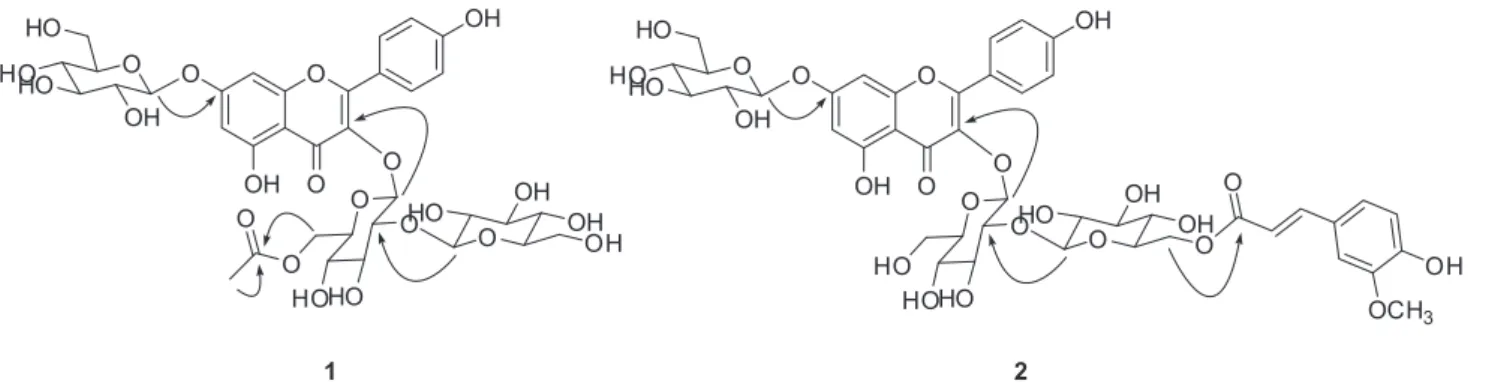
constituents in MeOH extracts led to the isolation of six flavonoid glycosides (1-6) (Figure 1), including one new
compound (1). All six compounds were tested for their
inhibitory effects on NO production in a LPS-activated murine microglial cells and their effects on NGF secretion from C6 glioma cells.
Experimental
General procedures
Optical rotations were measured on a Jasco P-1020 polarimeter in MeOH (Jasco, Easton, MD). IR spectra were recorded on a Bruker IFS-66/S FT-IR spectrometer (Bruker, Karlsruhe, Germany). UV spectra were recorded using a Schimadzu UV-1601 UV-Visible spectrophotometer (Shimadzu, Tokyo, Japan). HR-FAB and ESI mass spectra were obtained on a JEOL JMS700 mass spectrometer (JEOL, Peabody, MA). NMR spectra, including COSY, HMQC, and HMBC experiments were recorded on a Varian UNITY INOVA 500 and 900 NMR spectrometer with chemical shifts given in ppm (Varian, Palo Alto, CA). Preparative high performance liquid chromatography (HPLC) was conducted using a Gilson 306 pump (Gilson, Middleton, WI) with Shodex refractive index detector
(Shodex, New York, NY). Silica gel 60 and RP-C18 silica
gel (230-400 mesh, Merck, Darmstadt, Germany) were used for column chromatography. The packing material for molecular sieve column chromatography was Sephadex
LH-20 (Pharmacia, Uppsala, Sweden). Spots were detected by thin layer chromatography (TLC) under UV light or by heating after spraying with 10% H2SO4 in C2H5OH (v/v).
Plant materials
Leaves ofH. longipes were collected in Taebaek City, Korea, in June 2010. The plant was identified by one of the authors (K. R. Lee). A voucher specimen (SKKU-NPL 1103) of the plant has been deposited at the herbarium of the School of Pharmacy, Sungkyunkwan University, Suwon, Korea.
Extraction and isolation
Leaves of H. longipes (2.5 kg) were extracted with
80% MeOH at room temperature and filtered. The filtrate was evaporated under reduced pressure to give a MeOH extract (190 g), which was suspended in water (800 mL) and solvent-partitioned to give n-hexane (3 g), CHCl3 (14 g),
EtOAc (3 g), and BuOH (24 g) layers. The BuOH soluble layer (24 g) was separated over a RP-C18 silica gel column
(600 g) with a MeOH-H2O gradient (1:1, 3:1, 5:1, and 1:0,
v/v) to give six fractions (Fractions A to F). Fraction A (10 g) was chromatographed on a Diaion HP-20 column (Sigma, St. Louis, MO) eluting with a gradient solvent system
of 100% H2O and 100% MeOH, yielding subfractions
A1 and A2. Subfraction A2 (2.3 g) was separated over a silica gel column (40 g) with CHCl3-MeOH-H2O (2:1:0.2,
v/v) to afford nine subfractions (A21-A29). Subfraction
A26 (350 mg) was separated on a RP-C18 silica gel
column (10 g) with 45% MeOH to give five subfractions (A261-A265). Subfraction A262 (50 mg) was purified by RP-C18 preparative HPLC with MeOH-H2O (2:1, v/v) at a
flow rate of 2.0 mL/min (Econosil RP-18 10 µm column; 250 × 10 mm; 10 µm particle size; Shodex refractive index detector) to give 1 (3 mg, Rt = 13.0 min) and 6 (3 mg,
Rt = 15.2 min). Compounds 2 (50 mg, MeOH-H2O, 1:1.5,
v/v, Rt = 9.2 min) and 4 (40 mg, MeOH-H2O, 1:1.5, v/v,
Rt = 11.6 min) were isolated from subfractions A264 and
A265, respectively, through HPLC purification. Subfraction A28 (420 mg) was separated over a Sephadex LH-20
(150 g) with MeOH-H2O (1:1, v/v) to give 5 (200 mg).
Fraction B (2.5 g) was separated over a Sephadex LH-20
(150 g) with MeOH-H2O (4:1, v/v), chromatographed
over a silica gel column (30 g) with CHCl3-MeOH-H2O
(2:1:0.2, v/v), and further purified with a silica gel prep. HPLC with CHCl3-MeOH-H2O (2:1:0.2, v/v) at a flow rate
of 2.0 mL min-1 (Apollo Silica column; 250 mm × 10 mm
i.d., 5 µm, Alltech; Shodex refractive index detector) to yield 3 (6 mg, Rt = 13.6 min).
Compound 1
Yellow gum; [α]D
25 –6.5° (c 0.5, MeOH); IR (KBr)
νmax/cm-1 3729, 3395, 2931, 1656, 1531, 1518, 1240, 1058,
669; UV (MeOH) λmax/nm (log ε) 261 (4.1), 339 (4.5); 1H
and 13C NMR (see Table 1); HR-ESI-MS (positive mode)
m/z 837.2086 [M + Na]+ (calcd for C
35H42NaO22, 837.2065).
Compound 2
Yellow gum; [α]D25 –74.0° (c 0.5, MeOH); IR (KBr)
νmax/cm
-1 3729, 3395, 2931, 1656, 1531, 1518, 1240, 1058,
669; UV (MeOH) λmax/nm (log ε) 268 (4.2), 328 (4.0); 1H
and 13C NMR (see Table 1); HR-FAB-MS (positive mode)
m/z 949.2612 [M + H]+ (calcd for C
43H49O24, 949.2614).
Alkaline hydrolysis of 1 and 2
A solution of compound 1 (2.0 mg) in 0.1 N KOH (3 mL) was stirred at room temperature for 24 h. The reaction
mixture was neutralized with Dowex HCR W2 (H+ form)
and the resin was removed by filtration. A portion of the reaction product was partitioned between CHCl3/H2O (1 mL
each) and kaempferol-3-O-β-D-glucopyranosyl-(1→2)-β -D-glucopyranoside-7-O-β-D-glucopyranoside (5, 1.1 mg) was obtained from the H2O layer. Through a similar procedure,
5 (1.0 mg) and (E)-ferulic acid (0.5 mg) were obtained from H2O and CHCl3 layer, respectively, from 2 (2.0 mg).
Compound 5
Yellow gum; 1H NMR (500 MHz, CD
3OD) d 8.07
(d, 2H, J 9.0 Hz), 6.90 (d, 2H, J 9.0 Hz), 6.77 (d, 1H,
J 2.0 Hz), 6.48 (br s, 1H), 5.48 (d, 1H, J 7.5 Hz), 5.06 (d, 1H, J 7.0 Hz), 4.76 (d, 1H, J 7.5 Hz); FAB-MS m/z 773.2 [M + H]+.
Acid hydrolysis of 5 and sugar determination
Compound 5 (1.0 mg) was refluxed with 1 mL of
1 N HCl for 4 h at 100 °C. The hydrolysate was extracted
with EtOAc and the extract was evaporated in vacuo to
yield the aglycone kaempferol (0.5 mg) as a yellow gum.
The H2O layer was neutralized by passage through an
Amberlite IRA-67 column (Sigma, St. Louis, MO) and was repeatedly evaporated to give D-glucose identified by co-TLC (CHCl3:MeOH:H2O = 2:1:0.2, Rf value: 0.2)
with an authentic sample and optical rotation {[α]D25 +61.0
(c = 0.10, H2O)}.
Kaempferol
Yellow gum; 1H NMR (500 MHz, CD
3OD) d 8.08 (d,
2H, J 8.5 Hz), 6.90 (d, 2H, J 8.5 Hz), 6.39 (br s, 1H), 6.18 (br s, 1H).
Measurement of NO production and cell viability in LPS-activated BV-2 cells
BV-2 cells were maintained in Dulbecco’s modified Eagle’s medium (DMEM) supplemented with 5% fetal bovine serum (FBS) and 1% penicillin-streptomycin. To measure NO production, BV-2 cells were dispensed in wells of a 96-well plate (3 × 10⁴ cells/well). After 24 h,
the cells were pretreated with compounds for 30 min and stimulated with 100 ng/mL LPS for 24 h. Nitrite, a soluble oxidation product of NO, was measured in the culture media using the Griess reaction. The supernatant was harvested and mixed with an equal volume of Griess reagent (1%
sulphanilamide, 0.1% N-1-naphthylethylenediamine
dihydrochloride in 5% phosphoric acid). After 10 min, the absorbance at 540 nm was measured using an Emax microplate reader (molecular devices). Sodium nitrite was used as a standard to calculate the nitrite concentration. Cell viability was measured using a 3-[4,5-dimethylthiazol-2-yl]-2,5-diphenyl-tetrazolium bromide (MTT) assay.
NG-Monomethyl-L-arginine (L-NMMA), a well-known
NO synthase inhibitor, was tested as a positive control.
NGF and cell viability assays
C6 glioma cells were used to measure NGF release into
the medium.11 C6 cells were purchased from the Korean
Cell Line Bank and maintained in DMEM supplemented with 10% FBS and 1% penicillin-streptomycin in a
humidified incubator with 5% CO2. To measure NGF
content in medium and cell viability, C6 cells were seeded into 24-well plates (1 × 10⁵ cells/well). After 24 h, the cells
were treated with DMEM containing 2% FBS and 1% penicillin-streptomycin with 20 µM of each sample for one day. Media supernatant was used for the NGF assay using an ELISA development kit (R&D Systems). Cell viability was assessed by the MTT assay.
Results and Discussion
Leaves of H. longipes (2.5 kg) were extracted with
80% MeOH and the extract was partitioned with n-hexane,
CHCl3, EtOAc, and BuOH. The BuOH layer (24 g) was
successively chromatographed over silica gel, Sephadex LH-20, and preparative HPLC to give one new flavonoid glycoside (1), together with five known flavonol glycosides
(2-6). The known compounds, kaempferol 3-O-β
-D-glucopyranosyl-(1→2)-β-D-glucopyranoside (3),12
kaempferol 3-O-β-D-glucopyranosyl-(1→2)-[α
-rhamnopyranosyl-(1→6)]-β-D-glucopyranoside (4),13
2)-glucopyranoside]-7-O-β-D-glucopyranoside (5),14 and
kaempferol 3-O-(2G-glucosylrutinoside)-7-O-glucoside
(6)15 were identified in comparison with previously
published data. Although compound 2
(kaempferol-3-O-[6-(E)-feruloyl]-β-D-glucopyranosyl-(1→2)-β
-D-glucopyranoside-7-O-β-D-glucopyranoside) was
previously reported from Brassica rapa L. Ssp. chinensis
L. (Hanelt.) by LC-MS/MS,16 NMR spectral data have
not been reported. By extensive NMR studies (1H and
13C NMR, 1H-1HCOSY, HMQC, and HMBC), full NMR
data were assigned for the first time.
Compound 1 was obtained as a yellow gum, whose
molecular formula was determined to be C35H42O22 from
its positive-ion mode HR-ESI-MS data at m/z 837.2086
[M + Na]+ (calcd for C
35H42NaO22, 837.2065). The 1H NMR
spectrum of 1 (Table 1) showed two pairs of NMR signals at dH 8.05 (d, 2H, J 9.0 Hz, H-2’ and H-6’) and 6.90 (d, 2H, J 9.0 Hz, H-3’ and H-5’), and dH 6.79 (d, 1H, J 1.8 Hz, H-8)
and 6.50 (d, 1H, J 1.8 Hz, H-6), which were characteristic of the B ring and A ring of kaempferol derivatives, respectively. The 1H and 13C NMR data of 1 were very
similar to those of 5,14 except for presence of an acetyl
group resonance [dH1.75 (s, 3H); dC 172.4 and 20.5] and
up-field shift of H-6’’ [dH 4.16 (dd, 1H, dd, J 11.7, 1.8 Hz)
and 4.02 (dd, 1H, J 11.7, 5.4 Hz)] and C-6’’ (dC 64.0)
signals. The position of the acetyl group was deduced to be at C-6’’ by analysis of the HMBC data showing correlation from H-6’’ to C=O (Figure 2). Glucose connectivities of
1 were confirmed by HMBC correlations of H-1’’/C-3,
H-1’’’/C-2’’ and H-1’’’’/C-7 (Figure 2). The J values of anomeric protons [dH 5.40 (d, 1H, J 8.1 Hz, H-1’’), 5.06
(d, 1H, J 7.2 Hz, H-1’’’’), 4.77 (d, 1H, J 8.1 Hz, H-1’’’)] of glucose indicated the β-configuration. Alkaline hydrolysis of 1 afforded kaempferol-3-O-β-D-glucopyranosyl-(1→
2)-β-D-glucopyranoside-7-O-β-D-glucopyranoside, which
was identified to be 5 by co-TLC (CHCl3:MeOH:H2O =
1:1:0.2, Rf value: 0.4) with the similar natural compound
5, 1H NMR and MS.14 Acid hydrolysis of 5 yielded the
aglycone and D-glucose. The aglycone was confirmed as
kaempferol by comparison of its 1H NMR data with an
authentic sample, whereas D-glucose was identified by co-TLC (CHCl3:MeOH:H2O = 2:1:0.2, Rf value: 0.2) with
an authentic sample and by optical rotation {[α]D 25 +61.0
(c = 0.10, H2O)}. The structure of 1 was thus established
as kaempferol-3-O-β-D-glucopyranosyl-(1→2)-[6’’’-O -acetyl-β-D-glucopyranoside]-7-O-β-D-glucopyranoside.
Anti-neuroinflammatory activities of the isolated
compounds (1-6) were evaluated by examining NO
production in LPS-activated microglia BV-2 cells (Table 2). Among the tested compounds, 1-5 significantly inhibited LPS-stimulated NO production with IC50 values of 11.56,
14.86, 13.63, 15.97, and 15.30 µM, respectively, which
displayed more activity than L-NMMA, a well-known
NOS inhibitor. Compound 6 showed moderate activity
(29.26 µM) and all isolates (1-6) had no influence on cell viability (84.29-99.22%) at concentrations up to 20 µM. These results showed that kaempferol derivatives with 3-O-β-D-glucopyranosyl-(1→2)-β-D-glucopyranose unit significantly inhibited LPS-stimulated NO production, while additional substitution of β-D-glucopyranose at C-7 and α-L-rhamnopyranose at C-6’’ reduced the activity.
Compounds 1-6 were tested using an ELISA
development kit for their influence on secretion of NGF
from C6 glioma cells into the medium.17 As shown in
Table 3, compounds 4 and 5 were moderate stimulants
of NGF release (124.70 ± 7.71% and 117.02 ± 3.60%, respectively) without cell toxicity (97.49% and 89.21% survival, respectively) at a concentration of 20 µM.
Conclusion
From the leaves of H. longipes a new flavonoid glycoside (1) and five known flavonoid derivatives (2-6) were isolated by chromatographic methods. The structure of compounds
1 and 2 was revealed by extensive NMR methods (1H and
13C NMR, 1H-1HCOSY, HMQC and HMBC) and chemical
hydrolysis. The NMR data of compound 2 are published
for the first time. Compounds 4 and 5 exhibited significant
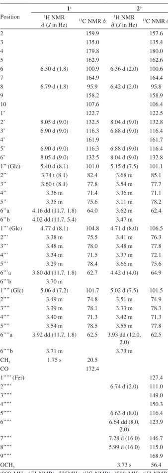
Table 1.1H and 13C NMR data for compounds 1 and 2 in CD
3OD (δ in ppm)
Position
1a 2b
1H NMR
d (J in Hz)
13C NMR d 1H NMR
d (J in Hz)
13C NMR d
2 159.9 157.6
3 135.0 135.4
4 179.8 180.0
5 162.9 162.6
6 6.50 d (1.8) 100.9 6.36 d (2.0) 100.6
7 164.9 164.4
8 6.79 d (1.8) 95.9 6.42 d (2.0) 95.8
9 158.2 158.9
10 107.6 106.4
1’ 122.7 122.5
2’ 8.05 d (9.0) 132.5 8.04 d (9.0) 132.8 3’ 6.90 d (9.0) 116.3 6.88 d (9.0) 116.4
4’ 161.9 161.7
5’ 6.90 d (9.0) 116.3 6.88 d (9.0) 116.4 6’ 8.05 d (9.0) 132.5 8.04 d (9.0) 132.8 1’’ (Glc) 5.40 d (8.1) 101.0 5.15 d (7.5) 101.1
2’’ 3.74 t (8.1) 82.4 3.68 m 85.1
3’’ 3.60 t (8.1) 77.8 3.54 m 77.7
4’’ 3.36 m 71.4 3.36 m 71.1
5’’ 3.35 m 75.6 3.11 m 78.2
6’’a 4.16 dd (11.7, 1.8) 64.0 3.62 m 62.4
6’’b 4.02 dd (11.7, 5.4) 3.47 m
1’’’ (Glc) 4.77 d (8.1) 104.8 4.71 d (8.0) 106.5
2’’’ 3.38 m 75.5 3.41 m 76.3
3’’’ 3.48 m 78.0 3.48 m 77.8
4’’’ 3.34 m 71.5 3.37 m 72.1
5’’’ 3.29 m 78.4 3.66 m 75.6
6’’’a 3.80 dd (11.7, 1.8) 62.7 4.42 d (4.0) 64.9
6’’’b 3.70 m
1’’’’ (Glc) 5.06 d (7.2) 101.7 5.02 d (7.5) 101.5
2’’’’ 3.49 m 74.8 3.51 m 74.9
3’’’’ 3.39 m 78.1 3.33 m 78.3
4’’’’ 3.40 m 71.3 3.42 m 71.3
5’’’’ 3.54 m 78.5 3.55 m 77.8
6’’’’a 3.92 dd (11.7, 1.8) 62.5 3.93 dd (12.0, 2.0)
62.5
6’’’’b 3.71 m 3.73 m
CH3 1.75 s 20.5
CO 172.4
1’’’’’ (Fer) 127.4
2’’’’’ 6.74 d (2.0) 111.0
3’’’’’ 149.0
4’’’’’ 150.3
5’’’’’ 6.63 d (8.0) 116.4
6’’’’’ 6.64 dd (8.0,
2.0)
123.9
7’’’’’ 7.28 d (16.0) 146.7
8’’’’’ 5.99 d (16.0) 115.0
9’’’’’ 168.9
OCH3 3.73 s 56.4
a900 MHz (1H NMR), 225MHz (13C NMR); b500 MHz (1H NMR), 125 MHz (13C NMR).
Table 2. Effects of compounds 1-6 and L-NMMA on LPS-induced NO production in BV-2 microglia cells
Compound IC50a / µM Cell viabilityb / %
1 11.56 98.12 ± 0.51
2 14.86 84.96 ± 0.65
3 13.63 99.22 ± 3.81
4 15.97 91.42 ± 5.36
5 15.30 85.12 ± 3.15
6 29.26 84.29 ± 6.07
L-NMMAc 16.23 101.54 ± 3.59 aIC
50 value of each compound was defined as the concentration (µM) that caused 50% inhibition of NO production in LPS-activated BV-2 cells; bcell viability after treatment with 20 µM of each compound was determined by MTT assay and is expressed in percentage (%). The results are averages of three independent experiments, and the data are expressed as mean ± SD; cL-NMMA as positive control.
Table 3. Effects of compounds 1-6 on NGF secretion in C6 cellsa Compound NGF secretion / % Cell viabilityb / %
1 97.76 ± 6.76 93.07 ± 1.77
2 103.38 ± 0.27 97.18 ± 1.37
3 101.64 ± 1.18 98.06 ± 1.04
4 124.70 ± 7.71 97.49 ± 0.30
5 117.02 ± 3.60 89.21 ± 0.64
6 93.67 ± 1.02 95.26 ± 3.94
6-Shoc 129.54 ± 11.23 98.12 ± 3.27 aC6 cells were treated with 20 µM of compounds 1-6. After 24 h, the content of NGF secretion in C6-conditioned media was measured by ELISA. The level of secreted NGF cells is expressed as percentage of the untreated control. The data shown represent the means ± SD of three independent experiments performed in triplicate; bcell viability after treatment with 20µM of each compound was determined by MTT assay and is expressed in percentage (%). The results are averages of three independent experiments, and the data are expressed as mean ± SD; c 6-Shogaol as positive control.
anti-neuroinflammatory activity by suppressing the release of NO in LPS-stimulated microglial cells and by inducing NGF secretion in C6 glioma cells. These results suggest that these compounds might be promising candidates for treatment of Alzheimer’s disease, Parkinson’s disease, and other neurodegenerative diseases.
Supplementary Information
Supplementary data are available free of charge at
http://jbcs.sbq.org.br as a PDF file.
Acknowledgments
This research was supported by the Basic Science Research Program through the National Research Foundation of Korea (NRF) funded by the Ministry of Education,
are thankful to the Korea Basic Science Institute (KBSI) for the measurements of NMR and MS spectra.
References
1. Benavente-García, O.; Castillo, J.; J. Agric. Food Chem.2008, 56, 6185.
2. Harborne, J. B.; Williams, C. A.; Phytochemistry 2000, 55, 481. 3. Hwang, S. L.; Shih, P. H.; Yen, G. C.; J. Agric. Food Chem.
2012, 60, 877.
4. Dawson, T. M.; Dawson, V. L.; Snyder, S. H.; Ann. Neurol.
1992, 32, 297.
5. Wyman, T. C.; Rohrer, D. C.; Kirigiti, P.; Nichols, H. V.; Pilcher, K. Y.; Nilaver, G.; Machida, C. A.; Gene Ther. 1999,6, 1648. 6. Park, J. H.; An Illustrated Guide to Korean Medicinal Plants;
Shinil Books Co.: Seoul, 2012.
7. Ahn, D. K.; A Coloured Ilustrated Guide to Korean Herbs; Kyohaksa: Seoul, 2003.
8. Mimaki, Y.; Kanmoto, T.; Kuroda, M.; Sashida, Y.; Nishino, A.; Satomi, Y.; Nishino, H.; Chem. Pharm. Bull. 1995, 43, 1190.
9. Mimaki, Y.; Kanmoto, T.; Kuroda, M.; Sashida, Y.; Satomi, Y.; Nishino, A.; Nishino, H.; Phytochemistry 1996, 42, 1065. 10. Kim, C. S.; Kim, S. Y.; Moon, E.; Lee, M. K.; Lee, K. R.; Bioorg.
Med. Chem. Lett. 2013, 23, 1771.
11. Mosmann, T.; J. Immunol. Methods 1983, 65, 55.
12. Schliemann, W.; Schneider, B.; Wray, V.; Schmidt, J.; Nimtz, M.; Porzel, A.; Böhm, H.; Phytochemistry 2006, 67, 191. 13. Kite, G. C.; Veitch, N. C.; Boalch, M. E.; Lewis, G. P.; Leon,
C. J.; Simmonds, M. S. J.; Phytochemistry 2009, 70, 785. 14. Nielsen, J. K.; Olsen, C. E.; Petersen, M. K.; Phytochemistry
1993, 34, 539.
15. Budzianowski, J.; Phytochemsitry1990, 29, 3643.
16. Rochfort, S. J.; Imsic, M.; Jones, R.; Trenerry, V. C.; Tomkins, B.; J. Agric. Food Chem. 2006, 54,4855.
17. Schwartz, J. P.; Costa, E.; Naunyn-Schmiedeberg’s Arch. Pharmacol. 1977, 300, 123.
Submitted: December 12, 2013