Angle Class II, subdivision, with agenesis of mandibular
second molars and extrusion of maxillary second molars*
How to cite this article: Tavares RR. Angle Class II, subdivision, with agenesis of mandibular second molars and extrusion of maxillary second mo-lars. Dental Press J Orthod. 2015 Mar-Apr;20(2):110-8. DOI: http://dx.doi. org/10.1590/2176-9451.20.2.110-118.bbo
» Patients displayed in this article previously approved the use of their facial and intraoral photographs.
Contact address: Rubens Rodrigues Tavares
Rua 6, no 370, sala 907, Edifício Empire Center, Setor Oeste,
Goiânia (GO) Brazil, CEP:74115-070 - E-mail: [email protected] » The author reports no commercial, proprietary or financial interest in the
prod-ucts or companies described in this article.
Submitted: February 9, 2015 - Revised and accepted: February 27, 2015.
1Specialist in Orthodontics and Facial Orthopedics, Universidade Paulista (UNIP).
Certified by the Brazilian Board of Orthodontics and Dentofacial Orthopedics (BBO).
* Case report, approved by the Brazilian Board of Orthodontics and Dentofacial
Orthopedics (BBO). INTRODUCTION
A female patient presented for initial examination at the age of 14 years and three months and was found to be in good general health. No significant informa-tion was found in her past medical and dental records. She did not have, nor did she report having, any del-eterious oral habits. As chief complaint she reported that some mandibular teeth were missing, which re-sulted in the presence of spaces, rotations and diffi-culty chewing in the posterior region. She had little growth potential, as she reported that her menarche had occurred when she was about 12 years old. While in many subjects the hereditary component is in-volved in determining partial anodontia, this aspect was not investigated in this case.
DIAGNOSIS
She had a rather well-balanced mesofacial pat-tern without any serious neuromuscular function-al changes, as well as a slightly convex profile and slightly protrusive maxillary and mandibular lips (UL-S line = 3mm LL-S line = 2 mm). This feature seemed fully compatible with the patient’s age group (Fig 1 and Table 1).
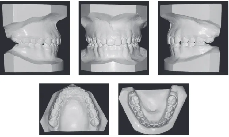
Dental analysis (Figs 1 and 2) disclosed Angle Class II malocclusion, subdivision right, aggravated by the ab-sence of second and third mandibular molars, distal mi-gration of mandibular posterior teeth, and extrusion of second maxillary molars. In addition to the aforemen-tioned teeth, tooth #18 was also missing. She presented asymmetry of maxillary canines in the anteroposterior
Rubens Rodrigues Tavares1
This clinical case reports the treatment of an Angle Class II malocclusion in a young woman with a balanced face affected by agenesis of second and third mandibular molars and subsequent extrusion of second maxillary molars. The atypical and peculiar occlusal anomaly led to individualized treatment proposed in order to normalize dental malpositions, with subsequent rehabilitation of edentulous areas by means of a multidisciplinary approach. This case was presented to the Brazilian Board of Orthodontics and Dentofacial Orthopedics (BBO) in partial fulfillment of the requirements for obtaining the title of certified by the BBO.
Keywords: Angle Class II malocclusion. Partial anodontia. Corrective Orthodontics. Dental implant.
DOI: http://dx.doi.org/10.1590/2176-9451.20.2.110-118.bbo
O presente caso clínico relata o tratamento de uma má oclusão de Classe II de Angle, em uma jovem com face harmo-niosa, porém agravada por agenesias de segundos e terceiros molares inferiores e consequente extrusão dos segundos molares superiores. A anomalia oclusal atípica e peculiar levou a uma proposta de tratamento individualizada, visando normalizar os maus posicionamentos dentários e uma posterior reabilitação das áreas edêntulas, por meio de uma abor-dagem multidisciplinar. O presente caso foi apresentado à Diretoria do Board Brasileiro de Ortodontia e Ortopedia Facial (BBO), como parte dos requisitos para a obtenção do título de Diplomado pelo BBO.
Figure 1 - Initial facial and intraoral photographs.
only tooth #28 was going through early stages of forma-tion, about Nolla stage 4, with all other teeth missing.
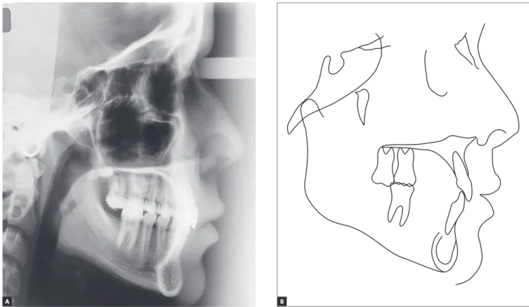
Proile cephalometric radiograph and cephalometric tracing (Fig 4) revealed good maxillomandibular rela-tionship in the vertical (SN-GoGn = 31o; FMA = 26o), and anteroposterior direction, with Class I skeletal pat-tern (SNA = 78o; SNB = 76o; ANB = 2o). Maxillary and mandibular incisors were slightly upright (1.NA = 17o; 1.NB = 20o), thereby increasing the interincisal angle (1/1 = 145o). These and other cephalometric values are shown in Table 1.
direction and no coincidence between maxillary and mandibular midlines and the midsagittal plane. The max-illary midline was shited to the let while the mandibu-lar one was shited to the right.1,2 She had an increased overbite with sharp incisal disocclusion and well-adjusted anterior centric stop. In the mandibular dental arch, there was generalized diastema, pronounced in the region be-tween canines and irst premolars.
Figure 2 - Initial casts.
Figure 4 - Initial profile cephalometric radiograph (A) and cephalometric tracing (B).
TREATMENT PLAN
Due to dental asymmetry, treatment planning aimed to produce a distal movement of maxillary molars on the right side and, at the same time, maintain vertical control of maxillary second molars already extruded due to absence of antagonists. This could allow retraction of the maxillary right quadrant so as to correct the anteroposterior asym-metry of canines and gain space to correct the deviation in the maxillary midline. As anchorage, one alternative would be to use mini-implants, which would allow a more efec-tive control of distalization of maxillary teeth. However, the patient’s legal guardians rejected this alternative, per-haps because it was not popular at that time. A removable appliance was therefore used encapsulating teeth #17 and 27 to prevent extrusion, along with an expansion screw for distalization (Fig 5). A hook was also placed on the right side to deploy Class II mechanics as soon as the mandibular arch had been leveled. Thereater, a ixed orthodontic ap-pliance would be placed with a stop on the already distal-ized posterior teeth, and mechanics applied to retract tooth #13, thereby achieving symmetry with its antagonist and space for midline correction.
In the mandibular arch, the ixed orthodontic appli-ance would allow not only the leveling of the occlusal plane, a necessary step to correct severe overbite, but also the mesialization of posterior teeth, especially on the right side, to correct the most distal position of the right canine relative to the let. As a result, diastemata would be eliminated, providing ideal canine and irst molar occlusion and adjusting the spaces for rehabili-tation with dental implants osseointegrated in the re-gion of second molars, in addition to correcting the mandibular midline.
TREATMENT PROGRESS
Treatment began eight months after completion of the initial examination, when the patient was almost 15 years old. This waiting time was meant to post-pone, albeit slightly, the completion of treatment, bringing it a little closer to the end of patient’s overall growth, when other rehabilitation resources, includ-ing dental implants, would be available.
For the maxillary arch, a removable orthodontic appliance was fabricated and installed.3 It consisted
of a Hawley retainer in the anterior region, Adams clasps on the first molars and bilateral screws to dis-talize teeth #17 and 27 (Fig 5). These teeth were kept encapsulated in the acrylic to prevent further extru-sion during distalization. The appliance also featured a hook on the right side for Class II elastics. The pa-tient was instructed to wear the appliance full time, removing it only to eat, engage in extreme sports and learn foreign languages. The recommended activa-tion was ¼ of a turn, in each screw, every five days. To ensure better vertical control, the maxillary sec-ond molars were replenished with self-curing acrylic resin every six weeks.
Orthodontic bands were placed on the mandibular first molars, and Roth prescription brackets with 0.018 x 0.030-in slots were bonded to all other teeth. Alignment and leveling were then achieved using up to 0.016-in round stainless steel archwires. Class II elastics were thereafter introduced to be worn on the right side, anchored on the removable appliance.
After creating spaces between first and second molars, maxillary fixed orthodontic appliance (Roth prescription, 0.018 x 0.030-in slot) was bonded after alignment and leveling, using the same sequence of round stainless steel archwires. All teeth received a mesial stop after distalization to progressively move
first molars, premolars and canines distally; more so on the right side, to ensure symmetry between homologous teeth. A 0.016 x 0.022-in TMA arch-wire with T loops was used to intrude and level the second molars.
Then, 0.016 x 0,016-in and 0.016 x 0,022-in Elgil-oy archwires were used for both maxillary and man-dibular arches, while intrusive steps4 were incorporat-ed to second molars (Fig 6). At this treatment stage, Class II elastics were used bilaterally to finish the relationship between molars and canines in an ideal occlusion. After obtaining the interocclusal space needed for rehabilitation with dental implants osseo-integrated in the region of mandibular second molars, the appliance was kept passive. It is noteworthy that implant surgery was delayed by about six months in order to make it coincide, as much as possible, with the end of patient’s growth. After the osseointegra-tion period, the prosthetic phase was performed con-currently with the removal of the fixed orthodontic appliance.
A removable plate with a Hawley retainer was used for retention in the maxillary arch in the ante-rior region, and a fixed intercanine retainer made of round 0.028-in stainless steel wire was used in the mandibular arch.
Figure 5 - Occlusal and right lateral views of the removable orthodontic appliance.
RESULTS
In assessing the patient’s final records (Figs 7-10) it is clear that all intended objectives were achieved. Given that the patient did not grow during this pe-riod and no significant changes were implemented in the anterior region, only subtle facial changes were noted. In correcting the dental problems, such as in-trusion of maxillary second molars and correction of the curve of Spee, the occlusal plane was leveled. Ideal occlusion was achieved between canines and molars at the expense of distal migration of maxillary teeth, and especially the mesial migration of mandibular
teeth, particularly on the right side. Correction of maxillary and mandibular canine asymmetry in the anteroposterior direction, midline deviations, extru-sion of mandibular second molars, reduced overbite, closing of mandibular spaces, and rotations were all solved in stages by means of specific mechanics.
By correcting deep overbite, the mandible was probably moved to a more anterior position, thus contributing to a mild improvement in facial harmony and providing, cephalometrically, a slight decrease in the value of the ANB angle (Steiner) from 2 to 1.5° (Table 1).
Figure 8 - Final casts.
Figure 10 - Final profile cephalometric radiograph (A), and final cephalometric tracing (B).
Figure 11 - Total (A) and partial (B) superimpositions of initial (black) and final (red) cephalometric tracings.
A
A
B
1. Sing G. Textbook of Orthodontics. 2nd ed. New Delli: Jaypee Brothers Medical; 2007.
2. Bruhn C, Hofrath H, Korkhaus G. Ortodoncia. Barcelona: Labor; 1944. 3. Roriz NB, Tavares RR. Distalização de molares superiores com aparelho
ortodôntico removível: um estudo piloto [monografia]. Goiânia (GO): Associação Brasileira de Odontologia; 2013.
4. Langlade M. Terapêutica Ortodôntica. 3a ed. São Paulo: Ed. Santos; 1995. 5. Ainamo J, Talari A. Eruptive movements of teeth in human adults. In:
Poole DFG, Stack MV editors. The eruption and occlusion of teeth. Boston: Butterworth; 1976.
6. Bjork A, Skieller V. Normal and abnormal growth of the mandible: a synthesis of longitudinal cephalometric implant studies over a period of 25 years. Eur J Orthod. 1983;5(1):1-46.
7. Brugnolo E, Cordioll G, Majzoub Z. Clinical and radiographic findings following placement of single-tooth implants in young patients: Case reports. Int J Periodontics Restorative Dent. 1996;16(5):421-33. 8. Bernard JP, Schatz JP, Christou P, Belser U, Kiliaridis S. Long-term vertical
changes of the anterior maxillary teeth adjacent to single implants in young and mature adults: a retrospective study. J Clin Periodontol. 2004;31(11):1024-8.
9. Thilander B, Odman J, Gröndahl K, Friberg B. Osseointegrated implants in adolescents: an alternative in replacing missing teeth? Eur J Orthod. 1994;16(2):84-95.
10. Thilander B, Odman J, Lekholm U. Orthodontic aspects of the use of oral implants in adolescents: a 10-year follow-up study. Eur J Orthod. 2001;23(6):715-31.
11. Mota PS, Freitas JF. Erupção continuada [monografia]. Goiânia (GO): Associação Brasileira de Odontlogia; 2007.
REFERENCES
FINAL CONSIDERATIONS
As previously mentioned, treatment was delib-erately delayed by approximately eight months. However, it was later found that this delay should have lasted longer, given that in the final phase, in agreement with the implant dentist, it proved more advisable to wait another six months before per-forming surgery, which increased treatment time unnecessarily. On the other hand, delaying the pro-cess might probably mean increased extrusion of maxillary second molars.5-11
Despite treatment time increase, patients and legal guardians were very pleased with the end result, espe-cially with regard to pleasant smile and balanced face. The goals initially set were met especially thanks to proper planning and use of biomechanical and rehabil-itation resources based on individualized and thorough diagnosis as required by all atypical cases. Treatment of these cases should not follow predetermined classi-cal protocols, but rather prompt professionals to hone their diagnostic skills in planning and carrying out a treatment tailored to suit individual peculiarities. Table 1 - Initial (A) and final (B) cephalometric values.
Measurements Normal A B Dif. A/B
Skeletal pattern
SNA (Steiner) 82° 78° 78° 0
SNB (Steiner) 80° 76° 76.5° 0.5
ANB (Steiner) 2° 2° 1.5° 0.5
Angle of convexity (Downs) 0° 2° 1° 1
Y axis (Downs) 59° 61° 62° 1
Facial angle (Downs) 87° 87° 87° 0
SN-GoGn (Steiner) 32° 31° 30° 1
FMA (Tweed) 25° 26° 24° 2
Dental pattern
IMPA (Tweed) 90° 85° 89° 4
1.NA (degrees) (Steiner) 22° 17° 18° 1 1-NA (mm) (Steiner) 4 mm 3.5 mm 2 mm 1.5 1.NB (degrees) (Steiner) 25° 20° 20° 0 1-NB (mm) (Steiner) 4 mm 3.5 mm 3 mm 0.5
1
1- Interincisal angle (Downs) 130° 145° 147° 2
1-APo (Ricketts) 1 mm 0.5 mm 0 mm 0.5
Profile Maxillary lip — S-line (Steiner) 0 mm 3 mm 2.5 mm 0.5