Universidade do Minho Escola de Ciências da Saúde
Sofia Pereira das Neves
STUDYING THE ROLE OF LIPOCALIN-2 IN THE
PATHOPHYSIOLOGY OF MULTIPLE SCLEROSIS
– LOOKING BEYOND THE BRAIN
O PAPEL DA LIPOCALIN-2 NA PATOFISIOLOGIA
DA ESCLEROSE MÚLTIPLA
– UM OLHAR PARA ALÉM DO CÉREBRO
Universidade do Minho Escola de Ciências da Saúde
Sofia Pereira das Neves
STUDYING THE ROLE OF LIPOCALIN-2 IN THE
PATHOPHYSIOLOGY OF MULTIPLE SCLEROSIS
– LOOKING BEYOND THE BRAIN
O PAPEL DA LIPOCALIN-2 NA PATOFISIOLOGIA
DA ESCLEROSE MÚLTIPLA
– UM OLHAR PARA ALÉM DO CÉREBRO
Maio 2015
Dissertação de Mestrado
Mestrado em Ciências da Saúde
Trabalho realizado sob a orientação de
Professora Doutora Fernanda Marques
e do
iii
Nome: Sofia Pereira das Neves
Endereço eletrónico: [email protected] Telefone: 913372844
Bilhete de Identidade/Cartão do Cidadão: 13364864
Título da dissertação: Studying the role of lipocalin-2 in the pathophysiology of multiple sclerosis – Looking beyond the brain
Orientadores:
Professora doutora Fernanda Marques Professor doutor João José Cerqueira
Ano de conclusão: 2015
Mestrado em Ciências da Saúde
É AUTORIZADA A REPRODUÇÃO INTEGRAL DESTA DISSERTAÇÃO APENAS PARA EFEITOS DE INVESTIGAÇÃO, MEDIANTE DECLARAÇÃO ESCRITA DO INTERESSADO, QUE A TAL SE COMPROMETE.
Universidade do Minho, _____/_____/_________
v
vi
AGRADECIMENTOS
A presente dissertação de mestrado resulta do trabalho que desenvolvi no ICVS, mas nunca poderia ter sido feita sem a ajuda de todos aqueles a quem passo a agradecer. Para quem me conhece sabe que eu não sou de grandes discursos, daí que os meus agradecimentos possam parecer pobres, mas saibam que valorizo muito todos os mencionados.
À Fernanda, uma grande orientadora, que esteve sempre presente para me ensinar e para me ajudar sempre que alguma coisa não corria tão bem, tanto a nível profissional como pessoal. Também me deu os merecidos raspanetes, que me ajudaram a melhorar o meu método de trabalho, e a tentar relativizar mais as coisas. Obrigado!
Ao João, pelas sugestões e ajuda, e por ter apostado em mim e me ter dado a minha primeira oportunidade de trabalho, o que me permitiu continuar no ICVS e percorrer todo este longo caminho. Aos meus amiguinhos de grupo, a Catarina e o Sandro, que tiveram a paciência para partilhar comigo os seus conhecimentos, que estiveram presentes para me ajudar, e tiveram uma palavra de carinho, sempre que precisei.
À Cláudia Miranda, pelo imenso apoio que me deu numa área da qual eu sabia muito pouco.
Ao Nuno Sousa, à Margarida Correia-Neves e à Joana Palha, pelas discussões científicas e sugestões dadas.
À Susana Roque, ao Bruno e à Cláudia Nobrega, pela ajuda com a parte imunológica do trabalho. A todos os membros do restante grupo, Ashley, João Sousa, João Costa, Leonor, Diana e Ana pelas sugestões em todas as reuniões.
À Sónia Gomes por sermos as umas choramingas que se apoiam uma à outra. Serás sempre a minha companheira de cativeiro.
À Gabriela e ao restante astrogang pela ajuda com a parte dos astrócitos. À Cristina e à Susana Monteiro, por serem boas companheiras de meu 2º grupo. Às minhas companheiras de almoço, a Fátima Lopes, a Liliana e a Sónia Borges.
A todos aqueles que foram em busca de novos desafios longe de Braga, mas que deixaram a sua marca: Ana Oliveira, Sara, Daniela, Filipa, Filipe
vii
Às restantes companheiras do I201, sempre as mais animadas: Dulce, Fátima Ramalhosa, Rita, Cláudia Antunes, Vanessa, Neide, Marta, Francisca. E Eduardo, o homem no meio das mulheres. À minha amiga para a vida, Diana, que também esteve sempre disponível para discutir comigo e ouvir-me.
A todos os meus amigos, da terrinha ou antigos colegas de curso. Aprendi algo com todos vocês. Aos meus companheiros de mestrado, que partilharam alguns dos meus problemas e angústias, principalmente à minha colega da frente, a Isabel, com quem convivi vários vezes fora do horário de trabalho.
A todos os NERD’s, pelo ambiente de trabalho que criaram e me faz querer continuar cá.
Por fim, às 4 pessoas mais importantes da minha vida: a minha irmã Carla e o meu namorado Ricardo, que sempre acreditaram em mim e nas minhas capacidades, mesmo quando eu própria não acreditava; e aos meus grandes PAIS, porque sem eles eu não estaria aqui, e por todos os sacrifícios que fizeram desde sempre.
Esta tese de mestrado decorreu no âmbito do projeto financiado pela Fundação para a Ciência e Tecnologia (FCT) e COMPETE através do projeto EXPL/NEU-OSD/2196/2013.
ix
Studying the Role of Lipocalin-2 in the Pathophysiology of Multiple Sclerosis – Looking beyond the brain
ABSTRACT
Multiple sclerosis (MS) is an immune-mediated demyelinating disease of the central nervous system
(CNS), characterized by the presence of demyelination plaques, inflammation and gliosis that
consequently lead to axonal damage. The sequence of events that leads to demyelination remains unclear and the pathophysiological mechanisms are diverse. Also, although this is a disease of the CNS, there is no doubt that, in terms of peripheral organs, the thymus, as the organ of T cell differentiation and maturation, plays an important role in the pathophysiology of the disease.
Recently, the levels of lipocalin 2 (LCN2), an acute phase protein that is part of the defense system against bacteria, by binding to iron-loaded siderophores, were found to be increased in cerebrospinal fluid (CSF) and serum of MS patients, when compared to control subjects. Similarly, using the MS animal model of experimental autoimmune encephalomyelitis (EAE), LCN2 was detected in brain parenchyma astrocytes, in regions typically affected in MS patients. This expression by astrocytes, together with an increased LCN2 level in the CSF, occurs during the active phases of the disease, which could point towards a role for LCN2 secreted by astrocytes in the mediation of inflammatory responses in the EAE model. Altogether, these findings support LCN2 as a valuable molecule for the diagnostic/monitoring of MS and suggest its potential involvement as a disease modulator. Of relevance, the exact role of LCN2 in the pathophysiology of the disease remains largely unknown and contradictory data exists on its potential protective or deleterious effect. Therefore, we sought to investigate the role of LCN2 in the onset and progression of the disease. Herein, we tackled the disease, by evaluating the role of LCN2, not only in the perspective of the CNS, but also on the perspective of peripheral organs such as the thymus.
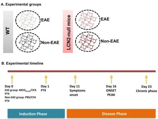
First we intended to perform a characterization of the thymus regarding thymocyte populations and histological morphology, in wild-type (WT) animals induced with EAE, in the onset and chronic phases of disease. Next, to further understand the role of LCN2 in MS pathology, we induced EAE both in LCN2-null mice and in WT littermates. Non-induced EAE animals were used as controls.
The thymus of EAE animals was atrophied, as assessed by its weight, normalized for total body weight, and by the number of total cells. Also, we found a decrease in total cell number of all thymocyte populations, during the onset and chronic phases of EAE. In relative terms, the percentage of double positive cells was decreased, and the percentages of the cluster of differentiation (CD)4 and CD8 single positive cells were increased, during the onset phase. At the chronic phase, the proportions between the different populations were restored.
LCN2-null mice induced with EAE did not present major alterations in terms of the clinical score, when compared with WT littermates also induced with EAE. Likewise, their thymic alterations were similar to the ones observed in WT EAE animals. Of relevance, as for the inflammatory profile in the cerebellum, LCN2-null mice presented less inflammation, as assessed by decreased expression levels of pro-inflammatory cytokines interferon (Ifn)-gamma, interleukin (Il)12a and Il17a. Also of interest, the cerebellum of LCN2-null mice presented a decrease in the percentage of lesioned areas. Finally, EAE animals, from both genotypes, presented an increase in the area positive for glial fibrillary acidic protein (GFAP), in the white matter of the cerebellum, in both the onset and chronic phases of disease. On the
contrary, the expression levels of Gfap in the cerebellum were only increased at the onset phase of
disease.
KEYWORDS: MULTIPLE SCLEROSIS, LIPOCALIN-2, NEUROINFLAMMATION, ASTROCYTES, THYMUS, EXPERIMENTAL
xi
O papel da lipocalin-2 na patofisiologia da Esclerose Múltipla – Um olhar para além do cérebro
RESUMO
A Esclerose Múltipla (EM) é uma doença autoimune desmielinizante do sistema nervosa central (SNC), caracterizada pela presença de placas de desmielinização, inflamação e gliose, que tem como consequência dano axonal. A sequência de eventos que induzem desmielinização permanecem desconhecidos, e os mecanismos patofisiológicos são diversos. Embora esta seja uma doença do SNC, não há dúvidas que, em termos de órgãos periféricos, o timo, sendo o órgão de maturação e diferenciação das células T, desempenha um papel importante na patofisiologia da doença.
Recentemente, os níveis de lipocalin-2 (LCN2), uma proteína de fase aguda que participa no sistema de defesa contra infeções bacterianas, através da ligação a sideróforos, foram encontrados como estando elevados no líquido cefalorraquidiano (LCR) e no soro de doentes com EM, comparativamente aos controlos. Da mesma maneira, usando o modelo animal de EM de encefalomielite autoimune experimental (EAE), a LCN2 foi detetada em astrócitos do parênquima, em regiões tipicamente afetadas em doentes com EM. Esta expressão pelos astrócitos, associada a um aumento de LCN2 no LCR, ocorreu durante as fases ativas da doença, o que aponta para um papel da LCN2 secretada pelos astrócitos na mediação da response inflamatória no modelo de EAE. No seu conjunto, estas evidências suportam o papel da LCN2 como uma molécula importante no diagnóstico e/ou monitorização da EM, e sugere o seu possível envolvimento como moduladora da doença. É relevante dizer que o papel exato da LCN2 na patofisiologia da doença permanece desconhecido, e existem dados contraditórios no que diz respeito ao seu potencial efeito protetor ou deletério. Por isso, nós procurámos perceber o papel da LCN2 no onset e na progressão da doença. Assim, nós investigámos o papel da LCN2 na doença, não só na perspetiva do SNC, mas também dos órgãos periféricos, nomeadamente do timo.
Primeiro pretendemos caracterizar o timo em relação às populações de timócitos e morfologia
histológica, em animais wild-type (WT) induzidos com EAE, no onset e na fase crónica da doença. De
seguida, para melhor entender o papel da LCN2 na patologia da EM, induzimos EAE em animais LCN2-null e em WT da mesma ninhada. Para além disso, usámos animais não induzidos como controlos. Nós observámos que o timo dos animais induzidos com EAE estava atrofiado, com base no seu peso, após normalização para o peso total do animal, e no número total de células. Para além disso, encontrámos uma diminuição no número total de células de todas as principais populações de timócitos, durante o onset e fase crónica da doença. No que diz respeito à percentagem de cada uma
das populações de timócitos, durante o onset da doença, a percentagem de células duplas positivas
encontrava-se diminuída, enquanto as percentagens das populações CD4+CD8- e CD4-CD8+ se encontrava aumentada. Na fase crónica da doença, as proporções entre as diferentes populações foram reestabelecidas.
Os animais LCN2-null induzidos com EAE não apresentaram grandes alterações em termos de score clínico, quando comparados com os animais WT da mesma ninhada também induzidos com EAE. Para além disso, as alterações observadas no timo foram semelhantes às encontradas nos animais WT EAE. De relevância, no que diz respeito ao perfil inflamatório no cerebelo, os animais LCN2-null apresentaram menos inflamação, o que é suportado por níveis diminuídos dos níveis de expressão das citoquinas pró-inflamatórias interferão-gama, e interleucinas 12 e 17. É importante também referir que os cerebelos de animais LCN2-null apresentaram uma diminuição na percentagem de áreas com lesões. Os animais EAE, de ambos os genótipos, apresentaram um aumento na área positiva para
GFAP, na substância branca do cerebelo, no onset e na fase crónica da doença. Pelo contrário, os
níveis de expressão de Gfap no cerebelo só foram encontrados elevados no onset da doença.
PALAVRAS-CHAVE: ESCLEROSE MÚLTIPLA, LIPOCALINA-2,NEUROINFLAMAÇÃO, ASTRÓCITOS, TIMO, ENCEFALOMIELITE AUTOIMUNE EXPERIMENTAL
xii
I
NDEXAgradecimentos ... vi
Abstract... ix
Resumo... xi
Abbreviations and acronyms list ... xiv
Figures list... xvi
Tables list ... xvii
1. Introduction ... 1
1.1 Multiple Sclerosis ... 2
1.2 Etiology of MS disease ... 3
1.3 MS symptoms ... 6
1.4 MS animal models – Experimental autoimmune encephalomyelitis ... 6
1.5 The role of glial cells in EAE ... 10
1.5.1 Oligodendrocytes ... 10 1.5.2 Microglia ... 11 1.5.3 Astrocytes... 12 1.6 Lipocalin-2 ... 13 1.6.1 Lipocalin-2 and CNS ... 14 1.6.2 Lipocalin-2 and MS ... 15
1.7 Role of the thymus in MS and EAE development ... 16
1.8 Research objectives ... 19 2. Experimental procedures ... 21 2.1 Mice ... 22 2.1.1 Genotyping ... 22 2.1.2 EAE induction ... 24 2.1.3 Sample collection ... 25 2.2 Flow cytometry ... 26
2.3 Gene expression analysis ... 27
2.4 Histological analysis ... 29
2.4.1 Thymic morphology ... 29
2.4.2 Immunofluorescence for GFAP ... 29
xiii
2.5 Serum corticosterone quantification ... 30
2.6 Statistical analysis ... 31
3. Results ... 33
3.1 Chronic disease course in MOG35-55 induced EAE ... 34
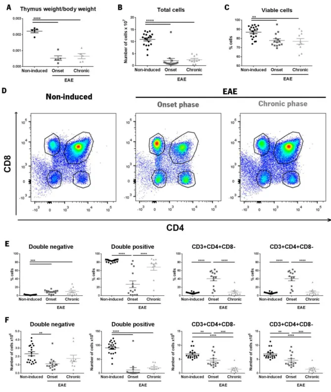
3.2 The proportion of the four main populations of the thymus was altered in WT animals during the onset phase EAE but was restored in the chronic phase of disease ... 34
3.2.1 Corticosteroid measurement ... 37
3.3 Thymic histological morphology was altered in WT EAE animals ... 37
3.4 Disease course in LCN2-null mice was similar to WT animals ... 38
3.5 LCN2-null mice presented similar alterations to the WT animals regarding thymic populations, during EAE development ... 39
3.5.1 Corticosteroid measurements ... 42
3.6 LCN2 deficiency results in a decreased inflammatory profile in the cerebellum of EAE animals 43 3.6.1 Inflammatory cytokines expression levels ... 43
3.6.2 Inflammatory infiltrates in the cerebellum white matter ... 45
3.7 Astrogliosis is increased in the cerebellum during EAE ... 47
4. Discussion ... 49
4.1 Thymus characterization in the context of a chronic EAE model ... 50
4.1.1 Influence of the hypothalamic-pituitary-adrenal (HPA) axis in thymic alterations in the EAE animal model induced in WT ... 52
4.1.2 Histological alterations in the thymus ... 53
4.2 Role of LCN2 in the pathophysiology of EAE ... 53
4.2.1 Disease development in LCN2-null mice ... 53
4.2.2 Thymus alterations and the HPA axis in LCN2-null mice ... 55
4.2.3 Inflammation in the cerebellum ... 56
4.2.4 Astrogliosis in the cerebellum... 58
5. Concluding remarks ... 59
6. References ... 63
xiv
ABBREVIATIONS AND ACRONYMS LIST
2,5-DHBA - 2,5-dihydroxybenzoic acid 7-ADD – 7-Aminoactinomycin D μL – Microliter
A
APC – Antigen presenting cell
B
BBB – Blood-brain barrier Bcl-2 – B cell lymphoma 2
BIM – Bcl-2 interacting mediator of cell death
C
CD – Cluster of differentiation
cDNA – Complementary deoxyribonucleic acid
CFA – Freund’s complete adjuvant
CIS – Clinically isolated syndrome CNPase – 2’,cyclic-nucleotide 3’-phosphodiesterase
CNS – Central nervous system
CP – Choroid plexus
CSF – Cerebrospinal fluid D
DAPI – 4’, 6-diamino-2-phenylindole
DMEM – Dulbecco’s modified eagle’s medium
DN – Double negative DNA – Deoxyribonucleic acid DP – Double positive
E
EAE – Experimental autoimmune encephalomyelitis
EDTA - Ethylenediaminetetraacetic ELISA – Enzyme-linked immune assay F
FACS – Fluorescence-activated cell sorting
FBS – Fetal bovine serum
Foxp3 – Forkhead box p3 G
G - Gauge
GFAP – Glial fibrillary acid protein
H
HE – Hematoxylin and eosin staining I
IFN – Interferon
Ig – Immunoglobulin
IL – Interleukin
iNOS – Inducible nitric oxide synthase L
LCN2 – Lipocalin-2
LPS – Lipopolysaccharide M
MBP – Myelin basic protein
MHC – Major histocompatibility complex
mL – Millilitre
MMP – Metalloproteinase
MOG – Myelin oligodendrocyte protein
MRI – Magnetic resonance imaging
MS – Multiple sclerosis
N
Neo – Neomycin
NG2 – neural/glial antigen 2
NO – Nitric oxide
NOD/Lt – Nonobese diabetic O
OD – Optical density
OPCs – Oligodendrocyte precursor cells
P
PB – Phosphate buffer
PBS – Phosphate buffered Saline PCR – Polymerase chain reaction
PDGFRα – Platelet-derived growth factor
receptor α
PFA - Paraformaldehyde PLP – Proteolipid protein PP – Primary progressive PTX – Pertussis toxin
xv
Q
qRT-PCR – Real time quantitative polymerase
chain reaction R
RNA – Ribonucleic acid
ROS – Reactive oxygen species
RR – Relapse-remitting RT – Room temperature RT-PCR – Reverse-transcription polymerase chain reaction S SP – Secondary progressive T TCR – T cell receptor
TGF – Transforming growth factor
Th – T helper
TIMP – Tissue inhibitors of metalloproteinases TMEV – Theiler’s murine encephalomyelitis virus
TNFα – Tumor necrosis factor α
Treg – regulatory T cells V
VCAM – Vascular cell adhesion protein VLA – Very late protein
W
xvi
FIGURES LIST
Figure 1 - Proposed pathogenic pathway involved in MS.
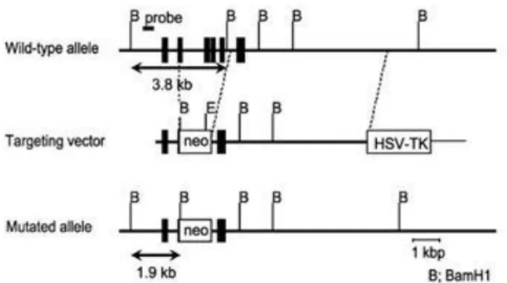
Figure 2 - Generation of LCN2-null mice.
Figure 3 - Schematic representation of the genotype strategy for LCN2-null mice.
Figure 4 - Schematic representation of the experimental groups and the EAE induction protocol. Figure 5 - Gating strategy used in the flow cytometry analysis.
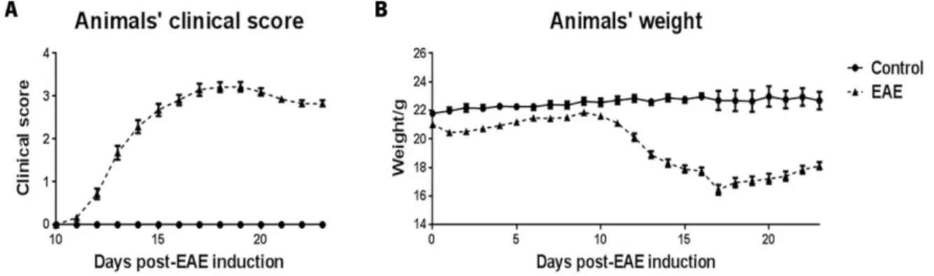
Figure 6 - Disease course of MOG35-55-induced EAE in WT animals.
Figure 7 – Thymus flow cytometry results.
Figure 8 – Corticosterone serum concentration at the sacrifice day.
Figure 9 – Histological morphology of the thymus in WT non-induced and EAE animals at the onset phase of disease.
Figure 10 – Disease course of MOG35-55--induced EAE in LCN2-null mice and WT littermates.
Figure 11 – Flow cytometry results in LCN2-null and WT mice.
Figure 12 – Serum corticosterone concentration in LCN2-null and WT mice at the sacrifice day.
Figure 13 - Expression levels of inflammatory cytokines in the cerebellum. Figure 14 – Luxol fast blue staining for myelin in cerebellum slices.
xvii
TABLES LIST
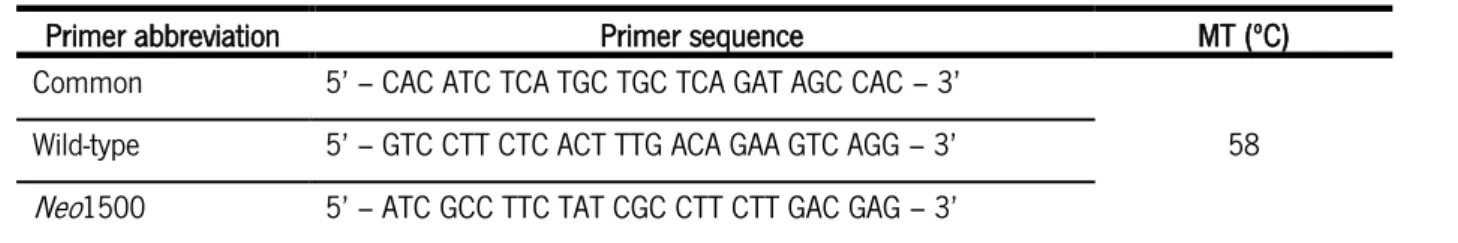
Table 1 – Primers’ sequence for genotyping LCN2-null animals and littermates.
Table 2 – Volume of reagents and PCR reaction conditions used for the genotyping PCR reaction. Table 3 - Mix and reaction conditions used for the RT-PCR.
Table 4 - Reaction conditions used for the qRT-PCR.
Table 5 – EAE progression in LCN2-null mice and WT littermate mice.
Table 6 - Two-Way ANOVA results for the comparison of thymus populations percentages between LCN2-null and WT mice.
Table 7 – Two-Way ANOVA results for the comparison of thymus cell numbers between LCN2-null and
WT mice.
1
2
1.
I
NTRODUCTION
1.1
Multiple Sclerosis
Multiple sclerosis (MS) is a chronic immune mediated demyelinating disease of the central nervous system (CNS) (Lassmann & van Horssen, 2011; Noseworthy et al., 2000). In developed countries it is the second cause of neurological disability in young adults, with high burden for the patient, the family and the resources of the health system (Borreani et al., 2014). MS is a complex disease in which there is a strong immune response against the myelin sheath of CNS axons, but its underlying mechanisms are only partially understood. Most patients initially present with a clinically isolated syndrome (CIS) in early adulthood. These CIS patients present an acute episode, which typically affects one brain region, being the clinical symptoms variable depending on the involvement of motor, sensory, visual or autonomic systems. (Compston & Coles, 2008). Some CIS patients will evolve to definite MS disease, while others won’t. Nowadays, the diagnosis of definite MS is based on recognized clinical criteria, with the support of magnetic resonance imaging (MRI) data and cerebrospinal fluid (CSF) analysis (Noseworthy et al., 2000), and can only be done when there is dissemination of neurologic dysfunction in space and time (Compston & Coles, 2008; Noseworthy et al., 2000; Polman et al., 2011), and after differential diagnosis have been excluded (Polman et al., 2011). Concerning the MRI findings, the presence of multifocal demyelinating lesions at different timepoints involving preferentially the periventricular white matter, the brain stem, the cerebellum and the spinal cord are indicative of MS (Noseworthy et al., 2000). Besides this, the presence of oligoclonal bands or increased concentration of immunoglobulin (Ig)G in the patients’ CSF are widely used to support MS diagnosis, but are not MS-specific (Fossey et al., 2007; Noseworthy et al., 2000).
Patients with definite MS can develop different profiles of the disease. Taking in consideration the different profiles of the disease, MS can be classified as relapse-remitting (RR)-MS, primary progressive (PP)-MS or secondary progressive (SP)-MS. RR-MS represents about 80-85% of MS cases (Sospedra & Martin, 2005) and is characterized by transient symptoms (relapse) that in most of the times improve within weeks (remission). However, the ability to fully recover from relapse episodes diminishes with time, and irreversible damage accumulates in the CNS, giving rise to SP-MS. The remaining 15-20% of patients has PP-MS, and does not show this relapse-remitting pattern; rather, their symptoms become gradually worst along the course of the disease. The RR-MS presents a female to male ratio of 2:1, while in the PP-MS the incidence is similar between both genders (Noseworthy et al., 2000).
3
Noticeably, some CIS patients never progress to definite MS. But in those patients that do evolve, an early treatment might reduce disease severity. Taking this into account, it is crucial to find new therapeutics to reduce not only the strong pro-inflammatory reaction, but also the relapse rate and, consequently, delay irreversible damage (Comi et al., 2000). Another challenge regarding MS has been to identify the CIS patients that present a high risk of having future episodes, which would confirm the diagnosis of definite MS (Brettschneider et al., 2010). This identification could be done based on disease biomarkers, measured in the patients’ blood or CSF. However, the etiology and the pathogenesis of MS are poorly understood, which makes it harder to find appropriate biomarkers for disease initiation and progression. (Lassmann & van Horssen, 2011).
1.2
Etiology of MS disease
1.2.1 Environmental and genetic factors
Regarding the etiology of MS, both environmental factors, such as infectious agents and lifestyle, and genetic factors have been proposed to induce or contribute to disease appearance (Compston & Coles, 2008; Sospedra & Martin, 2005). Among the environmental factors, the influence of viral infections is being largely addressed but until now nothing was proved. Regarding viral infections, it was shown that human herpesvirus-6 or Epstein-Barr viruses, may trigger a cascade of inflammatory events that lead to MS. In addition, because a higher proportion of women suffer from MS it was suggested that changes in hormone levels may influence disease initiation (Sospedra & Martin, 2005). Moreover, variations in sunlight exposure were also strongly associated with increased MS risk (Ebers, 2008), mostly due to changes in melatonin production. Specifically, it was shown that an excess of melatonin induced by decreased sunlight exposure enhances the Th1 response, boosting the inflammatory response (Sospedra & Martin, 2005). Finally, in the field of the genetic factors, genome-wide association studies have identified the allele for human leukocyte antigen (HLA)-DRB1*15:01 as the haplotype that represents the highest risk factor for MS (Nylander & Hafler, 2012; Sawcer et al., 2014). This haplotype explains between 14-50% of the genetic risk for MS (Hafler et al., 2005; Sawcer et al., 2014). Other susceptibility in non-HLA loci were also identified as being associated with MS, being most of them involved in the immunological response, especially in T cell immunity, like the interleukin (IL)2 receptor α gene (Il2Rα) and the IL7 receptor α (Il7Rα) gene (Berge et al., 2013; Hafler et al., 2007).
4
1.2.2 Onset mechanism
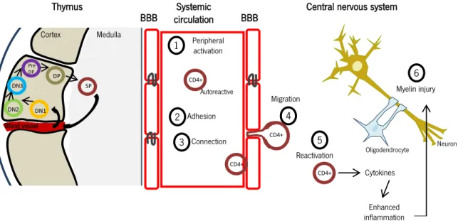
MS pathogenesis is fostered by a dysregulated autoimmune response. Initially, naïve myelin-specific T cells are primed in the lymph nodes by antigen presenting cells (APCs) that present myelin cross-reactive epitopes (Nakahara et al., 2010). During this process, an unknown mechanism triggers the activation of autoreactive T cells, which start to show cross-reactivity to self-antigens, such as proteins found in the myelin sheath, namely myelin basic protein (MBP), myelin oligodendrocyte protein (MOG) and proteolipid protein (PLP) (Steinman, 2001) (phase 1, Figure 1). After the initial priming of T cells, they migrate to the CNS and cross the endothelium, breaching the blood-brain barrier (BBB) (phases 2-4, Figure 1). The crossing is facilitated by the expression of the integrin very late antigen (VLA)-4 by activated T cells, which will interact with the vascular cell adhesion protein (VCAM) expressed by
inflamed endothelial cells (Reboldi et al., 2009; Steinman, 2001). Likewise, activated T cells express
members of the immunoglobulin superfamily, such as cluster of differentiation (CD)4 and CD8, which will interact with endothelial cells via major histocompatibility complex (MHC) class II and class I
molecules, respectively. Moreover, the transendothelial migration of T cells is facilitated by
metalloproteinases (MMP)2 and 9, which play a role in the degradation of the extracellular matrix, and are detectable in the CSF of MS patients (Steinman, 2001). Besides entering through the BBB, activated autoimmune T cells are also able to enter the CNS through the choroid plexus (CP), which
forms the blood-CSF barrier. (Reboldi et al., 2009) Once the barriers of the brain have been breached,
inflammatory cells spread into the white matter of the CNS, and are re-activated by myelin epitopes (Nakahara et al., 2010) (phase 5, Figure 1). The re-activated T cells in particular will produce cytokines and chemokines, which will promote the recruitment of other immune cells, like B cells (Miller, 2012), monocytes, and mast cells (Sospedra & Martin, 2005), from the periphery, and activate resident cells, namely microglia and astrocytes, inducing the production of nitric oxide (NO) and osteopontin (Sospedra & Martin, 2005; Steinman, 2001). Increased levels of NO are a major contributor for the death of oligodendrocytes and osteopontin induces the production of T helper (Th)1 cytokines, namely interferon (IFN)- and interleukin (IL)-12, and down-regulates Th2 cytokine production, contributing to an exacerbation of MS. The end result of this combined response by innate and adaptive immune cells, and respective inflammatory mediators, is the formation of an inflammatory lesion (phase 6, Figure 1), characterized by demyelinated axons, apoptotic oligodendrocytes, macrophages loaded with phagocytized myelin lipids, and activation and proliferation of astrocytes (Sospedra & Martin, 2005). It is believed that, upon lesion formation, brain cells are recruited to the injury site to promote regeneration of the tissue. In particular, oligodendrocyte precursor cells (OPCs) are recruited to the
5
lesion sites and differentiate into myelinating oligodendrocytes that are able to remyelinate denuded internode areas. In these areas the nerve conduction velocity is slower than before, because the myelin sheath produced is less thick than the original one, which also leads to the redistribution of sodium channels as a compensation measure (Sospedra & Martin, 2005).
Figure 1 - Proposed pathogenic pathway involved in MS.
Another proposed theory for the pathogenesis of MS is the oligodendrogliopathy hypothesis. According to this hypothesis, the inflammation is not the cause of demyelination, but instead is the result of oligodendroglial apoptosis, myelin degeneration and impaired myelin regeneration. The lesions result from apoptosis of oligodendrocytes, which induce myelin degeneration and the recruitment of macrophages to eliminate the myelin debris. Consequently, immune reactions involving T and B cells occur along with a slower and poorly effective oligodendroglial regeneration and remyelination (Nakahara et al., 2010). However, considering that most of the susceptible loci identified for MS are located in genes involved in the immunological response (Berge et al., 2013), this favours the first theory, i.e. an initial immune dysregulation followed by secondary degenerative processes (Slavin et al., 2010). Nevertheless, and considering that the clinical, genetic, MRI and pathological data is heterogeneous in MS, it is highly possible that more than one pathogenetic mechanism contributes to disease establishment (Noseworthy et al., 2000), which rises the necessity for the development of therapies that not only modulate the immune response, but can also act on other disease targets, like oligodendrocytes and axons (Lucchinetti et al., 2005).
DN1 DN2 DN3 Pre DP DP SP Cortex Medulla Blood vessel Thymus Systemic circulation 1 Peripheral activation CD4+ Autoreactive BBB 2 Adhesion 3 Connection CD4+ 4 Migration CD4+ CD4+ 5 Reactivation 6 Myelin injury Cytokines
Central nervous system
Enhanced inflammation
Oligodendrocyte Neuron
6
1.3
MS symptoms
MS patients usually present the first symptoms of disease between the ages of 20 and 40. Disease activity results in reversible but also in irreversible sequelae. Irreversible sequelae ultimately lead to progressive impairment and disability in the majority of patients. Common symptoms among MS patients include sensory problems, such as numbness or tingling, weakness, difficulties walking, double vision and poor coordination. However, no two patients have exactly the same symptoms. With disease progression patients start to develop increased disability, showing problems in bladder and bowel control, cognitive performance and sexual performance. They may also present fatigue, even after a good night’s sleep, and walking problems. Ultimately, MS patients need wheelchair and can become bedridden (Joy & Johnston Jr., 2001).
The first symptoms presented by MS patients are widely believed to result from the exacerbated inflammatory process, which is associated with reduction of the nerve conduction velocity, and, on the other hand, the regression of symptoms correlates with the resolution of the inflammatory lesion and partial remyelination. As already mentioned, the axonal injury resultant from repeated episodes can become irreversible. In this case there is the formation of a glial scar and also there is an exhaustion of the OPCs pool, leading to progressive loss of neurological function, and to the formation of demyelinating plaques (Noseworthy et al., 2000). Altogether, the presence of focal demyelination, inflammation, glial scar formation and varying axonal damage are considered the hallmarks of MS disease (Lucchinetti et al., 2005).
Over the last years, the use of animal models has provided important insights into MS pathophysiology of MS and possible therapeutic targets. In the next section we will present the main characteristics of the animal models used to study MS, with special attention given to the experimental autoimmune
encephalomyelitis (EAE) model, which is the most common animal model for studying MS.
1.4
MS animal models – Experimental autoimmune encephalomyelitis
Human studies have some limitations, such as limited access to human tissue, especially from living patients, the fact that the experimental design of clinical trials cannot be easily modified, and the fact that studies of disease mechanisms are difficult to perform in humans (Denic et al., 2011). For that reason, the importance of animal studies relies on the fact that they provide flexible, potent and rapid platforms for MS research (Ransohoff, 2012). As already mentioned, the etiology and pathogenesis of MS are still unclear, which makes it difficult to find one animal model that accurately represents all
7
aspects of this disease (Denic et al., 2011). Instead, there are currently three main models that are used to study different pathological characteristics of MS: (1) viral models of inflammatory demyelination; (2) demyelination induced by toxic agents; (3) inducible models of EAE (Batoulis et al., 2011; Procaccini et al., 2015). Besides these three models, there are T-cell receptors (TCR) transgenic mice models that spontaneously develop EAE (Croxford et al., 2011; Wekerle & Kurschus, 2006). The Theiler’s murine encephalomyelitis virus (TMEV) is the best characterized model of virus-induced demyelination. It is used to study how viral infections are able to induce CNS autoimmunity (Batoulis et al., 2011). In this model, the animals are inoculated with a virus, which will induce acute inflammation in the CNS, and ultimately lead to demyelination and axonal damage in the CNS (Batoulis et al., 2011; Denic et al., 2011). Susceptible mice always develop a chronic-progressive form of disease (Batoulis et al., 2011). One disadvantage of this model is the induction of a special inflammatory environment, by the virus, which is unlikely to reflect what happens in typical MS lesions (Wekerle & Linington, 2006). Toxin-induced models of demyelination include the cuprizone and the lysolecithin models. Cuprizone is a copper chelator, which causes demyelination, with minor inflammation or axonal damage, predominantly in the cerebellar cortex and peduncle, when fed to the animals. This is a good model to study the main characteristics of focal demyelination and also remyelination (Batoulis et al., 2011; Denic et al., 2011; Fossey et al., 2007), since spontaneous remyelination is observed after withdrawal of cuprizone (Mix et al., 2010). Lysolecithin is used to induce focal demyelination, after injection directly in the white matter, which allows the control of the lesion size, nature and anatomical location (Woodruff & Franklin, 1999).
The EAE animal model has been used to study MS since the initial observation of paralysis, accompanied by perivascular infiltration and demyelination, in the pons and cerebellum of monkeys that were given repeated intramuscular injections of normal rabbit brain extracts (Rivers et al., 1933). Since then, a variety of mammals have been induced with EAE, from mice and rats, to goats and sheep (Denic et al., 2011). This is a model of antigen-specific autoimmunity against myelin, mediated by CD4+ T cells, which results in inflammatory demyelination of brain and spinal cord of induced animals (Dal Canto et al., 1995). Due to the clinical, immunopathological and histopathological similarities with MS, the EAE model is the most commonly used to study this human disease (Swanborg, 1995). This model has numerous applications, especially in the study of mechanisms involved in autoimmune-mediated inflammation, demyelination, axonal damage, immune cell migration across the BBB and regulatory mechanisms that can supress autoimmunity in the CNS (Stromnes & Goverman, 2006).
8
There is not one single model of EAE, but instead, the different combinations of animal species and strains, type of antigen used for immunization and the type of adjuvant employed give rise to different models, that differ from each other in the type of disease course (chronic vs. monophasic vs.
relapse-remitting) (Swanborg, 1995), lesion location and involvement of B cell produced antibodies vs. T cell
responses, sustaining the notion that antigens and immunogenetic background contribute to disease phenotype (Sospedra & Martin, 2005). As an example, Slavin and co-workers (1998) have shown that
immunization of different strains of mice with the same peptide leads to different outcomes – after EAE
induction with MOG35-55, nonobese diabetic (NOD/Lt) mice presented a relapse-remitting course of
disease, while C57BL/6 mice showed a chronic non-remitting disease, and BALB/c mice were not susceptible to disease induction (Slavin et al., 1998).
EAE can be induced actively, when the animals are immunized with CNS antigens, or passively, if autoreactive CD4+ T cells obtained from immunized animals are transferred into naïve animals (Batoulis et al., 2011; Dal Canto et al., 1995). Some of the CNS antigens used to induce EAE are purified MBP or PLP; whole spinal cord homogenates; or specific peptides, which correspond to the
encephalitogenic portion of a protein for a given strain (Dal Canto et al., 1995), such as the MOG35-55
peptide. Unlike MBP and PLP, which together represent about 80-90% of CNS myelin proteins, MOG only accounts for 0.01-0.05% of these proteins, but its location at the outermost lamellae of the oligodendrocyte membrane makes it easily accessible to antibodies, hence its importance as a cellular
and humoral target in MS and EAE (Slavin et al., 1998; Sospedra & Martin, 2005). Since the MOG35-55
-induced mice model, specifically in C57BL/6 mice, will be used in the context of the present thesis we will further characterize it in the next paragraph.
As previously shown, C57BL/6 mice induced with MOG35-55 present a chronic course of disease (Slavin
et al., 1998; Sospedra & Martin, 2005), characterized by ascending paralysis resultant from the preferential attack to the spinal cord (Batoulis et al., 2011). The disease is characterized in the beginning by a limp tail, which progresses to hind and forelimbs paralysis (Stromnes & Goverman, 2006). Also multifocal and confluent areas of mononuclear inflammatory infiltrates and perivascular inflammatory cuffing in the cerebellum and hindbrain white matter are observed. The chronic disease
course makes this a good model to study SP-MS (Constantinescu et al., 2011). The MOG35-55-induced
C57BL/6 model is particularly important in MS studies due to the increased availability of gene-modified strains on this background (Kuerten et al., 2007; Ransohoff, 2012). One disadvantage of the EAE model arises from the use of Freund’s complete adjuvant (CFA) and pertussis toxin (PTX) for active disease induction. CFA contains bacterial components that are able to activate the innate immune
9
system, via pattern recognition receptors (Constantinescu et al., 2011), and, consequently, misrepresent the animals’ general immune reactivity and confound the findings related with regulatory mechanisms (Wekerle & Kurschus, 2006). In the case of PTX, it will contribute to BBB permeabilization and facilitate autoantigen recognition in the CNS, by activating tissue-resident APCs (Kuerten & Lehmann, 2011). When it comes to all EAE models, other limitations can be pointed out, namely, the early detection of clinical signs in EAE, as compared with MS, which allows an early therapeutic intervention in the case of the animal model that cannot be translated into the human disease. Moreover, several therapies tested in EAE have proven ineffective or even harmful in human patients. These differences might be explained by the lower disruption of the BBB in MS patients, as compared with EAE-induced animals, which could prevent the therapeutic molecules from reaching their target in the CNS (Mix et al., 2010; Procaccini et al., 2015). Additionally, EAE is induced on highly inbred strains that are kept in a pathogen-free environment, with controlled temperature, light and humidity conditions, which is not representative of the various environmental and genetic factors that contribute to MS susceptibility (Handel et al., 2011). Besides, if we take into account that in the EAE model a CD4+ T cell response is induced, it becomes difficult to study the role of CD8+ T cells, which have been shown to be key players in MS pathogenesis (Procaccini et al., 2015; Ransohoff, 2012). Another difference that has been pointed out is the preferential involvement of the spinal cord in the EAE model, unlike the human disease, in which the cerebral and cerebellar cortex are more affected (Brown & Sawchenko, 2007; Procaccini et al., 2015; Ransohoff, 2012). Nevertheless, other brain structures have been proven to also be involved in the EAE model, namely the cerebellum and optic tract (Brown & Sawchenko, 2007).
Briefly, both in MS and in MS animal models there are four key pathological features: inflammation, demyelination, axonal loss or damage and gliosis (Constantinescu et al., 2011; Lucchinetti et al., 2005). So it is not surprising that glial cells also play a key role in MS pathophysiology. It was already mentioned that after T cell re-activation, in the CNS, there is the production of cytokines and chemokines which will activate microglial cells and astrocytes, leading to oligodendrocyte damage (Miller, 2012; Sospedra & Martin, 2005; Steinman, 2001). In the next section we will discuss the main characteristics of CNS glial cells and their participation in MS/EAE pathophysiology.
10
1.5
The role of glial cells in EAE
There are 3 types of glial cells in the mature CNS: astrocytes, oligodendrocytes and microglial cells (Purves et al., 2001), and all of them have been reported as being involved in MS pathogenesis.
1.5.1 Oligodendrocytes
Oligodendrocytes are responsible for producing myelin that wraps around some, but not all, neuronal axons. The oligodendrocyte-myelin-axon unit represent a unique structure, with specialized functions, within the CNS. Myelin contributes not only to increase the cross-sectional diameter of axons, increasing the velocity of action potential conduction, through saltatory conduction (Frohman et al., 2006; Purves et al., 2001), but also for the axonal stability, and normal neuronal functioning, by inducing sodium channel clustering, via oligodendrocyte-derived soluble factors, even in the absence of direct axon-glial contact (Chandran et al., 2008; Kuerten & Lehmann, 2011). During the course of MS and EAE, oligodendrocyte death occurs, mediated by endogenous factors, such as the activation of cell death by caspase-11 and caspase-3, via specific pathways (Hisahara et al., 2001), or by the release of
cytotoxic cytokines by activated microglia and astrocytes, namely IFN- (Vartanian et al., 1995).
New oligodendrocytes that participate in remyelination could arise either from surviving oligodendrocytes or OPCs at the lesion sites, or from OPCs that migrate to the lesion (Reynolds et al., 2002). Taking into account that mature oligodendrocytes are post-mitotic cells, it is very unlikely that they could originate new functional oligodendrocytes. OPCs have been identified in chronic lesions, although in a relatively quiescent state (Chari & Blakemore, 2002), and have also been shown to proliferate after acute demyelination (Reynolds et al., 2002), hence they are the most probable candidates involved in remyelination. This idea is supported by studies in which, after labelling OPCs in normal white matter and inducing demyelination, it was possible to locate these cells as remyelinating oligodendrocytes. Also, the transplantation of OPCs, isolated from the adult CNS, was found to be able to remyelinate areas of demyelination (Franklin, 2002). Consequently to an acute demyelination episode, induced by chemical or immunological mechanisms, neural/glial antigen 2 (NG2)+ OPCs become reactive, proliferate, and migrate to the lesion sites (Reynolds et al., 2002). At the lesion sites, OPCs have to be able to differentiate into functional myelinating oligodendrocytes (Chari & Blakemore, 2002).
Different theories try to explain why remyelination fails in MS. One of the most popular theories claims that failure in remyelination arises from exhaustion of the pool of OPCs, induced by repeated episodes of inflammation and demyelination (Reynolds et al., 2002). It could also be that the local OPCs present
11
are not able to further differentiate into myelinating oligodendrocytes, due to the lack of differentiation signals or to the presence of inhibitory signals (Back et al., 2005; Franklin, 2002), such as hyaluronan (Williams et al., 2007).
In addition, studies using organotypic cultures of cerebellum, challenged with lipopolysaccharide (LPS), have shown microglia activation, characterized by morphological changes from ramified to amoeboid shaped cells, the production of pro-inflammatory cytokines, like IL-1 , IL-6 and tumor necrosis factor (TNF)-α, and the induction of oxidative stress, via production of reactive oxygen species (ROS) and inducible nitric oxide synthase (iNOS) (di Penta et al., 2013). This microglia activation was associated with oligodendrocyte death, and myelin and axonal damage (di Penta et al., 2013).
1.5.2 Microglia
Microglial cells are considered to be the macrophages of the brain, due to their myeloid origin, ability to circulate and migrate within different regions of the brain, and to phagocyte, process and present antigens. In this sense, microglial cells are the CNS-resident immune cells, functioning as the first line of defense against immune and inflammatory stimuli (Lee et al., 2007). Under physiological conditions, resting microglia play a crucial role in the immune surveillance of the brain, interacting with the other brain cells and actively monitoring and remodelling impaired synapses. Subsequently to CNS damage, the number of microglial cells increases dramatically at the lesioned site, due to proliferation of resident microglia and migration of monocytes, from circulation (Purves et al., 2001). These cells share many properties with tissue macrophages, namely they are scavenger cells that remove cellular debris from injury sites (Purves et al., 2001). This phagocytic removal of apoptotic cellular material, without induction of inflammation, is one of the main beneficial roles of microglia. However, microglial cells are unable to efficiently remove myelin debris when Wallerian degeneration is associated with acute injury, preventing axonal regeneration. Principally after activation, these cells are able to secrete important pro- or anti-inflammatory mediators that act as paracrine mediators of neural cell migration and survival, but may also promote the autocrine polarization of microglia into different states of activation, the M1 and M2 microglia. In the first case microglial activation is more harmful, and in the second is more protective. Furthermore, microglial cells are involved in CNS repair, by producing cytokines and chemokines, but can also induce oligodendrocyte death through the production of cytotoxic molecules. Another beneficial function that has been attributed to microglia is the ability to recruit stem cells and induce neurogenesis (Napoli & Neumann, 2010). In response to an inflammatory stimuli, microglia upregulate CD45, CD86, CD80, CD40 and MHC-class II, enhancing their ability to function as APC
12
(Minagar et al., 2002; Murphy et al., 2010). Moreover, persistent activation of microglia contributes to increase the permeability of the BBB, and promotes the infiltration of peripheral macrophages (Miller, 2012). It was already referred that MMP2 and MMP9 are important in MS pathogenesis, more specifically, for facilitating T cell transendothelial migration (Steinman, 2001). In the CNS, these two MMPs are secreted by microglia and astrocytes as active forms (Nakanishi, 2003). Thus, microglia can exert both beneficial and detrimental functions during disease development, depending on the stage and context of a given lesion (Correale, 2014).
1.5.3 Astrocytes
Astrocytic involvement in MS has long been recognized. A few years before Charcot described a
disease, which he called “Sclérose en plaques”, in 1868, Rindfleisch had already described the
presence of multinucleated cells with processes in a CNS pathology named “grey degeneration”. These cells were later identified as astrocytes (Williams et al., 2007). In physiological conditions, astrocytes play an essential role in maintaining survival of neurons and other glial cells (Miller, 2012), by providing structural support to neurons (Nair et al., 2008), maintaining the extracellular ionic environment and pH and secreting metabolic substrates for neurons (Sofroniew, 2005). They are also able to actively alter the synaptic transmission by clearing and releasing extracellular glutamate from/to the synaptic cleft (Nair et al., 2008; Sofroniew, 2005). In addition, perivascular astrocytes are tightly associated with the basal lamina of blood vessels, contributing for the maintenance of the BBB (Brambilla et al., 2014). Following a CNS insult, like injury, ischemia and neurodegeneration, astrocytes become reactive and suffer morphological changes, such as cytoplasm enlargement, increased size and ramification of processes (Lee et al., 2009), upregulation of glial fibrillary acidic protein (GFAP) and vimentin, and re-expression of nestin (Williams et al., 2007). Astrocytes, along with microglial cells, play a dual role in protection and injury in MS (Minagar et al., 2002).
In situations of CNS insult, astrocytes upregulate the expression of glutamate transporters and glutamine synthetase, preventing the extracellular accumulation of glutamate, and consequently its excitotoxic effects on neurons and oligodendrocytes (Nair et al., 2008; Sofroniew, 2005). Also, astrocytes produce chemokines, that are able to recruit OPCs to demyelination sites (Williams et al., 2007), and anti-inflammatory cytokines, such as IL-10, IL-4, IL-5, IL-27 and transforming growth factor
(TGF)- (Nair et al., 2008). The formation of a reactive scar by astrocytes, on one hand, helps to
surround and separate the damaged tissue from normal white matter, but on the other hand, presents as a major obstacle for axon regeneration (Sofroniew, 2005). The gliotic scar could also result from a
13
failed attempt of remyelination (Franklin, 2002). In addition, astrocytes further contribute for the permeabilization of the BBB by secreting IL-6, TNF-α and IL-1β, which will act specifically on endothelial cells and tight junctions (Nair et al., 2008). As mentioned previously, astrocytes can express MMPs (Nakanishi, 2003), but they are also able to express tissue inhibitors of metallopeptidases (TIMP)-1, that regulate negatively the activity of MMPs (Nair et al., 2008; Williams et al., 2007).
Reactive GFAP+ astrocytes were found in the spinal cords of Lewis rats induced with EAE, even before the appearance of disease clinical symptoms (Smith et al., 1983). Also, in a mice model of EAE with a chronic relapsing disease course [model characterized by both relapsing-remitting episodes, with inflammatory-mediated demyelination, and progressive disease, with axonal and neuronal loss (Ramaglia et al., 2015)], astrocytic activation was found at lesion sites, and Gfap expression levels were found to fluctuate according to the symptomatic stage of disease (Kothavale et al., 1995). Of relevance, when a drug that targets glial activation was used, it seems that there was a reduction in both clinical and pathological severities of EAE, namely there was a partial rescue of oligodendrocytes from cell death (Guo et al., 2007). Previously, GFAP-null mice induced with EAE presented a more severe clinical course, supporting the importance of GFAP in structural stabilization of the white matter (Liedtke et al., 1998). Altogether, these findings suggest that an excessive glial response is deleterious for EAE recovery, but a defective glial response is also unable to delimitate lesion sites, and protect the healthy surrounding tissue from secondary degeneration.
Previous in vitro and in vivo studies from our laboratory have shown the production by astrocytes of an acute-phase protein, lipocalin-2 (LCN2), in response to different inflammatory stimuli, namely in the EAE context. We will further assess this subject and describe the possible involvement of LCN2 in MS and EAE pathogenesis.
1.6
Lipocalin-2
Lipocalin-2 (LCN2), also known as neutrophil gelatinase-associated lipocalin (NGAL) or 24p3, was first identified by Kjeldsen and colleagues (1993) as a protein present in neutrophil granules, partly covalently associated with human neutrophil gelatinase (Kjeldsen et al., 1993, 1994). LCN2 is a member of the lipocalin family, whose members are characterized by a compact tertiary structure, with a central hydrophobic core that allows the binding to small lipophilic substances (e.g. retinol), along with an ability to bind specific cell-surface receptors and to covalently associate with other proteins, leading to the formation of molecular complexes (Flower, 1996; Kjeldsen et al., 1994).
14
Initial reports described LCN2 as an acute phase protein in mice, involved in acute responses to various inflammatory or stressful stimuli (Q. Liu & Nilsen-Hamilton, 1995; Marques et al., 2008).
The particular production of LCN2 upon bacterial infection is suggested to occur as part of the innate immune response (Flo et al., 2004). Invading pathogens, when confronted with the low levels of iron present in the host, and in order to proliferate, secrete siderophores, small binding molecules that have higher-affinity to iron than the endogenous host iron-trafficking proteins. To cope with this, and at the site of infection, neutrophils release LCN2, which will then bind specifically to the bacterial ferric siderophores, preventing iron from being transported into the bacteria (Flo et al., 2004; Goetz et al., 2002). The importance of this mechanism is demonstrated by the fact that LCN2-null mice have increased susceptibility to bacterial infection (Flo et al., 2004).
More recently, LCN2 was also found to interact with endogenous iron (Yang et al., 2002) using an endogenous mammalian siderophore [2,5-dihydroxybenzoic acid (2,5-DHBA)] (Bao et al., 2010; Devireddy et al., 2010) and to be able to deliver this iron to cells in culture, independently of transferrin delivery system (Yang et al., 2002). In fact, a model of iron trafficking by LCN2 was proposed, where LCN2 lacking iron (apo-LCN2) binds to 24p3 receptor (24p3r), is internalized and binds to an intracellular mammalian siderophore, chelates iron and is exocytosed from the cell, decreasing intracellular iron concentration. On the contrary, holo-LCN2 binds to 24p3r, is internalized, traffics to endosomes and releases iron from the complex, thereby increasing intracellular iron concentration. Importantly, this LCN2-mediated modulation of cell iron content has been shown to impact on cell proliferation and apoptosis , since apo-LCN2 internalization results in apoptosis and cell death through the pro-apoptotic B cell lymphoma 2 (Bcl-2)-interacting mediator of cell death (BIM) protein (Devireddy et al., 2005).
1.6.1 Lipocalin-2 and CNS
In what concerns LCN2 and CNS, current literature is not consensual in defining the real function of LCN2, neither by which cells it is produced. In response to a peripheral immune challenge, such as the
intraperitoneal injection of LPS, Lcn2 expression is quickly upregulated in the CP, which is a thin
membrane present in the brain ventricles that produces the majority of the CSF. As a result of this upregulation, LCN2 protein levels in the CSF are also increased. Immunohistochemical stainings in the CP have shown positive labelling for LCN2 in some, but not all, epithelial cells, suggesting that only certain cells express detectable amounts of LCN2, at least at a given moment. In this study, also the endothelial cells seemed to be able to produce LCN2 (Marques et al., 2008). In addition, using the
15
same LPS-injection model, another group confirmed the LCN2 production by CP-associated cells and by endothelial cells, but they also showed that microglial cells were able to produce it, which is controversial in the literature (Ip et al., 2011). The production by astrocytes is more accepted, in several inflammatory conditions, and even in physiological conditions (Chia et al., 2011). Cultured astrocytes
stimulated with LPS, TNF-α and amyloid- 1-42 are able to upregulate LCN2 expression and secretion (Lee
et al., 2009; Mesquita et al., 2014). Regarding the role of LCN2 in astrocytes, it seems that LCN2 is able to polarize astrocytes into a more inflammatory phenotype (Jang et al., 2013). In accordance, in vitro studies, using astrocyte cultures, have reported increased sensitivity of these cells for NO-induced
apoptotic death, as well as H2O2- and paraquat-induced necrotic cell death, mediated by LCN2.
In addition, the absence of LCN2 has been reported to affect emotional and cognitive behaviour. Specifically, when compared to wild-type (WT) animals, 10-weeks old male LCN2-null mice presented anxiety and depressive-like behaviours, which were associated with increased levels of corticosterone production, along with mild spatial reference memory impairments. This behavioural phenotype was correlated with overall alterations in hippocampal neuronal morphology, suggesting a role for LCN2 in neuronal plasticity and behaviour (Ferreira et al., 2013).
1.6.2 Lipocalin-2 and MS
As for CNS diseases, LCN2 has been implicated in the pathophysiology of Alzheimer’s disease (Choi et al., 2011; Naude et al., 2012), pain (Jha et al., 2014) and MS (Berard et al., 2012; Marques et al., 2012; Nam et al., 2014). Moreover, in a relapse-remitting EAE mice model, the analysis of transcriptome of the CP showed that Lcn2 was one of the most significantly up-regulated genes during the onset and relapse phases of disease, with a reduced expression during the remission phase. These results were correlated with high LCN2 CSF levels during the active phases of the disease, and low levels in the remission phase. Interestingly, aside from CP infiltrating neutrophils, astrocytes were found to be the only cells expressing LCN2. The production of LCN2 by the astrocytes was restricted to the brain areas that are typically affected in MS patients (Marques et al., 2012). Of relevance, LCN2 levels were also found to be elevated in CSF (Marques et al., 2012) and serum samples (Berard et al., 2012) from MS patients, when compared with healthy controls.
Specifically for MOG35-55 induced EAE in LCN2-null mice, the published results are contradictory. Berard
and colleagues (2012) have shown that LCN2-null mice induced with EAE present a more severe disease and pro-inflammatory response, when compared to WT induced animals (Berard et al., 2012), while Nam and co-workers (2014) reported that induced LCN2-null mice presented a significant
16
decrease in disease severity accompanied by reduced inflammatory infiltration, glial activation, inflammatory cytokine/chemokine expression, demyelination in the spinal cord and autoreactive T cell proliferation (Nam et al., 2014).
Nevertheless, taking into account all the known functions of LCN2, there is no doubt that it plays a role in CNS inflammation, specifically in autoimmune diseases like MS. For that reason, in our study we
intend to further understand and clarify if LCN2 plays a detrimental or protective role in MOG35-55-induced
EAE. Contrary to the mentioned published data, we will give special attention to the cerebellum, which is a brain region known to be affected in MS, and to the thymus, not often looked at, but no less
important if we consider that it is the organ were T cells mature. In the next section we will make a
description of the thymus, and present the data that is known regarding its involvement in MS and EAE development.
1.7
Role of the thymus in MS and EAE development
The thymus is a primary lymphoid organ, where bone marrow derived progenitor cells undergo differentiation and maturation, to originate a functional T cell repertoire (Pearse, 2006). This organ is surrounded by a thin layer of connective tissue, which gives rise to septae, in some species, that partially subdivide the thymus into lobules that interconnect with each other. In mice there is no distinct sublobulation. The cortex is constituted by packed, small, immature lymphocytes, few epithelial cells and phagocytic macrophages (Pearse, 2006). These macrophages phagocyte the lymphocytes that present high avidity for self-antigens, and that are negatively selected to die (Takahama, 2006). The medulla presents less cellular density, staining paler that the cortex, and contains more mature T cells, which are larger and have more cytoplasm than cortical T cells (Pearse, 2006). Between the cortex and the medulla, it is possible to recognize a cortico-medullary region, rich in blood vessels, thought which the prothymocytes enter the thymus (Schuurman et al., 1997).
During fetal development, hematopoietic stem cells migrate from the yolk sac and/or fetal liver to the thymus to begin thymic differentiation. In the adult thymus the differentiation process is also recapitulated by bone-marrow-derived stem cells that continually migrate to the thymus. However, in this last case there is a dynamic equilibrium between the stem cells that enter the thymus, and the mature cells that leave it. Early differentiation processes occur in the cortex of the thymus, and later ones in the medulla (Zuniga-Pflucker & Lenardo, 1996).
17
One of the first events taking place during thymocyte differentiation is the induction of CD25 expression in CD117+/CD44+/CD4-/CD8- thymocytes (pro-T cell stage). Thymocytes lose the expression of both CD44 and CD117, and increase the expression of CD25 (early pre-T cell stage). During this phase the first checkpoint of thymocyte differentiation occurs: the selection, during which there is the Tcrb gene
rearrangement, and the cells begin to assemble TCR- and pre-TCRα chains to form the pre-TCR
complex (Zuniga-Pflucker & Lenardo, 1996). If TCR gene rearrangement has been successful, thymocytes differentiate to double positive (DP) cells, which lose expression of CD25 and gain expression of either CD4 or CD8; if not, thymocytes die by apoptosis (late pre-T cell phase) (Michie & Zuniga-Pflucker, 2002; Takahama, 2006). Still in the cortical layer of the thymus, there is TCR engagement with MHC class I or class II molecules, resulting in positive selection (Goldrath & Bevan, 1999). The positive selection process ensures that thymocytes have a minimal reactivity for self-peptides, allowing only the further maturation of cells restricted to self-MHC. On the other hand, thymocytes that present high-avidity interactions to self-peptides are negatively selected and are further eliminated by apoptosis (Savage & Davis, 2001). This process of negative selection is very important for preventing autoimmunity, since it allows the elimination of the self-reactive T cells. There are also DP thymocytes that fail to receive TCR signals and are also destined to die at this stage (Takahama, 2006). The low-affinity recognition of self-MHC during the positive selection is also responsible for determining commitment to either the CD4 or CD8 lineage. Recognition of MHC class I results in CD8+ T cells, while the recognition of MHC class II results in CD4+ T cells (Goldrath & Bevan, 1999). Thymocytes that have been positively selected, and have committed to a CD4 or CD8 lineage, go to the medullary layer and interact with thymic epithelial cells to complete thymocyte maturation (Takahama, 2006). At the medulla, self-reactive thymocytes that have escaped negative selection in the cortex are further deleted. This additional deletion is particularly important in establishing central tolerance to tissue-specific antigens. The survival single positive cells are then exported from the thymus into circulation (Takahama, 2006).
Even after two negative selection checkpoints, at the cortex and the medulla of the thymus, some auto-reactive T cells are able to escape to the periphery. For example, encephalitogenic peptides, like the MBP NAc1-11 peptide, were found to bind weakly to MHC, forming unstable peptide/MHC complexes. In this case the negative selection of the T cells specific for this peptide is not efficiently mediated, allowing some of them to escape to the periphery (Song et al., 2006). Nevertheless, in the periphery the activity of auto-reactive T cells is controlled by regulatory T (Treg) cells, avoiding autoimmune
18
antigens – they are in an anergic state. Instead, when activated, Treg cells inhibit the proliferation of
effector T cells in their surroundings (Rose, 1994). Hence, the thymus plays an important role in the control of organ-specific autoimmunity by limiting the development of autoreactive T cells, for which the tolerance to self-antigens is essential, and also by generating Treg cells. Of notice, an inverse correlation between MBP expression in the thymus and EAE susceptibility was found (H. Liu et al., 2001).
The role of thymus in EAE has previously been addressed by different authors with thymectomized animals. The EAE induction in animals thymectomized soon after birth or at a young age showed a significant delay in disease onset. In contrary, animals thymectomized 10 days after EAE induction did not present differences in the course or characteristics of disease when compared with sham thymectomized animals (Flechter et al., 1984). In another study, after thymectomy or sham surgery performed at 6 weeks of age, both groups of animals developed a similar disease course. However, after day 16 post-disease induction, the animals subjected to sham surgery presented an improvement in their clinical score, unlike thymectomized animals. This recovery was probably due to accelerated differentiation and proliferation of thymic CD4+ forkhead box p3 (Foxp3)+ Treg cells. In thymectomized animals, the recovery from EAE was severely impaired due to the absence of new Treg cell emigrants, which suggested that the pre-existing peripheral Treg pool alone was not sufficient for complete regulation of pathogenic T cells (Chen et al., 2009). So it seems that the thymus plays a pivotal role in EAE development, and that the age at the time of thymectomy influences the resistance or susceptibility for EAE establishment.
Although, it is widely recognized the vital role of the thymus in MS/EAE pathogenesis, it is still lacking a characterization of this organ in the WT C57BL/6 mice induced with EAE, namely regarding thymocyte populations and thymus histological morphology during disease onset and development, which we will also address in this thesis. In addition, the impact of LCN2 on this process will be evaluated.
19
1.8
Research objectives
Although, the initial and principal aim of the present thesis is to assess whether and how LCN2 modulates the severity of MS, because there is still limited amount of information regarding the alterations that occur in the thymus during disease development, even in WT mice, our first objective is to:
1. Characterize the main thymic populations and the histological morphology of the thymus, in WT
C57BL/6 mice induced with EAE, at the onset and chronic phases disease.
To address the need to better understand the role of LCN2 in the pathophysiology of MS we will take
advantage of the EAE model and of the mouse targeted deletion of Lcn2, and considering that most
EAE studies are focused on the spinal cord, and few have studied the cerebellum, an area known to be affected in MS disease, we proposed to:
2. Study, in the cerebellum, the expression of inflammatory cytokines, evaluate the presence of
inflammatory infiltrates and explore astrocytic activation, at the onset and chronic phases of disease, after inducing EAE in LCN2-null mice and WT littermates. Also, we will address the thymus to unravel the impact of LCN2 in the T cell differentiation and maturation.
21