Article
Printed in Brazil - ©2017 Sociedade Brasileira de Química 0103 - 5053 $6.00+0.00
*e-mail: [email protected]
Study of Reactions of Two Mannich Bases Derived of 4’-Hydroxychalcones with
Glutathione by RP-TLC, RP-HPLC and RP-HPLC-ESI-MS Analysis
Aline Bernardes,a,b Caridad N. Pérez,a Mátyás Mayer,c Cameron C. da Silva,a
Felipe T. Martinsa and Pál Perjési*,b
aInstituto de Química, Universidade Federal de Goiás, 74690-900 Goiânia-GO, Brazil
bInstitute of Pharmaceutical Chemistry and cDepartment of Forensic Medicine, University of Pécs,
H-7624 Pécs, Hungary
4’-Hydroxychalcones have been reported to possess several beneficial biological effects. Several lines of evidence accumulated to demonstrate increased biological activities of the Mannich base derivatives of the parent 4’-hydroxychalcones. Bioactivities of chalcones and related
α,β-unsaturated ketones are frequently associated with their reactivity with cellular thiols, such as GSH. For comparison of GSH reactivity, two bis Mannich bases of two 4’-hydroxychalcones were synthesized and reacted with GSH under non-cellular conditions. Reversed-phase thin layer chromatography (RP-TLC) and reversed-phase high performance liquid chromatography (RP-HPLC) analysis showed formation of two polar products which structures were confirmed by RP-HPLC-ESI-MS (RP-HPLC-electrospray ionization mass spectrometry) as 1:1 chalcone-GSH adducts in each case. At pH values below 8.0, the two bis Mannich bases showed higher GSH reactivity than two 4’-hydroxychalcones. Influence of the nature of the amino groups, the ring-B substituents and pH of the medium on reactivity was also investigated. The findings could serve as useful structure-activity information for subsequent molecular modification of thiol-reactive 4’-hydroxychalcones.
Keywords: hydroxychalcones, Mannich bases, glutathione, RP-TLC, RP-HPLC-ESI-MS
Introduction
Hydroxychalcones are intermediary compounds of the
biosynthetic pathway of the natural flavonoids.1 The wide
range of biological activities of both naturally occurring and
synthetic analogues, among other cytotoxic,2,3 antitumor,
anti-inflammatory4,5 and chemopreventive (antioxidant)5,6
properties are well documented in the literature.7-9
Chalcones are α,β-unsaturated ketones and representatives
of this class of compounds have demonstrated a preferential reactivity towards thiols in contrast to amino and hydroxyl
groups.7 Thus interactions with nucleic acids may be absent
which could eliminate the important genotoxic side effect
which have been associated with certain anticancer drugs.10
GSH is a water-soluble tripeptide and the prevalent non-protein thiol present in mammalian systems at
concentrations as high as 10 mmol L-1.11 Besides that, the
reduced form of GSH plays central roles in the antioxidant defense mechanism and it may be depleted by four primary
mechanisms: (i) enzymatic conjugation, (ii) increased
oxidation to glutathione disulfide (GSSG), (iii) inhibition
of its synthesis and (iv) non-enzymatic reactions with
electrophilic species.12 Electrophilic compounds such
as α,β-unsaturated ketones can be converted to GSH
conjugates by nucleophilic attack of sulfhydryl (−SH)
group, which pKa in aqueous solution is about 9.0 and the low-energy d orbitals of the cysteinyl sulfur atom favors
the spontaneous Michael reaction.13 Reduced GSH levels
are related to cytotoxic effects resulted from a wide range of signaling pathways as activation of apoptosis through mitochondrial pathways or by rendering sensitized cells
particularly vulnerable to oxidative damage.14
Aminoalkylation reactions can change lipophilicity of a drug leading to an improved delivery of a drug to the
active sites in the human body.15 Solubility of the Mannich
bases in water could be further increased as a result of
their protonation to form the respective ammonium salts.16
They can also act as prodrugs forming reactive derivatives
via deaminomethylation or deamination processes.17 It is
tool to improve the biological activity of many drugs as
tetracyclines and doxorubicin.18,19
Recent literature has shown phenolic Mannich bases derived from chalcones analogues to have remarkable cytotoxic potencies towards cancer cell lines.20-24 However,
the exact mechanism by which chalcone compounds and derivatives exert their cytotoxic effects in cancer cells remains unclear. In order to investigate how GSH-reactivity is affected by Mannich-type modification
of 4’-hydroxychalcones, bis Mannich derivatives (3 and 4)
of 4’-hydroxychalcones 1 and 2 have been synthesized.
Previous investigations showed 1 as a potent inhibitor of
MDA-MB-231 cells (with IC50 of 3.8 µM).25 Furthermore,
chalcone 2 have been reported as a relatively potent
inhibitor (with IC50 of 17.9 µM) of PC-3 cell.17 In the same
study the authors could not determine effective inhibitory
action of compound 1. Since the 13C nuclear magnetic
resonance (NMR) shifts, reflecting electron density, of the
β-carbon atoms of the two compounds are rather similar
(∆d = 1.5 ppm), the compounds are expected to show similar
reactivity towards GSH. Whether or not GSH-reactivity of the compounds can play an important role in determining the related cytotoxic activities, comparative cell-free GSH reactivity of the two compounds was planned to perform.
Furthermore, to the best of our knowledge, there are no studies reporting on non-enzymatic GSH-adducts formation of Mannich bases of 4’-hydroxychalcones. In order to investigate the nature of such Michael addition reactions, the present study was carried out with the aim of detecting and identifying GSH conjugates of the bis Mannich bases
3 and 4 in cell free incubations carried out at different pH
and also comparing them to GSH-reactivity of the parent
hydroxychalcones 1 and 2.
In order to separate and monitor formation of the expected GSH-adducts, a reversed-phase thin layer chromatographic (RP-TLC) method with double (fluorescence and ninhydrin) visualization and a reversed-phase high performance liquid chromatographic (RP-HPLC) method with ultraviolet-visible (UV-Vis) detection has been developed and applied. The structure of the expected chalcone-GSH conjugates was confirmed by HPLC-ESI-MS (HPLC-electrospray ionization mass
spectrometric)analysis.
Experimental
Material and methods
All commercially available reagents were purchased from Sigma-Aldrich (St. Louis, MO, USA). Infrared (IR) spectra were performed on a Bruker IFS-55 FT
spectrometer (Bruker Optik GmbH, Leipzig, Germany).
1H and 13C NMR spectra were recorded on a Bruker Avance
III 500 (500.15/125.77 MHz for 1H/13C) spectrometer
(Bruker Optik GmbH, Ettlingen, Germany). The melting points were measured on a Barnstead Electrothermal 9100 melting point apparatus (San Francisco, USA).
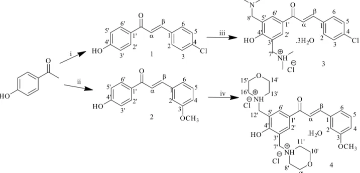
Synthesis of compounds
4’-Hydroxychalcones 1 and 2 as well as their bis
Mannich derivatives 3 and 4 (Figure 1) were synthesized in
Department of Chemistry, Universidade Federal de Goiás, Goiânia-GO, Brazil.
Hydroxychalcones 1 and 2 were synthesized by
Claisen-Schmidt condensation as previously described
by Kumar et al.26 with slight modifications. Equimolar
portions of 4-hydroxyacetophenone (10 mmol) and 4-chlorobenzaldehyde (10 mmol) or 3-methoxybenzaldehyde (10 mmol) were added to 15 mL of 40% NaOH methanolic solution. The reaction mixtures were stirred at room temperature for 24 h, when TLC indicated the end of the reactions. Then, cold water (5 mL) was added to the media and acidified with 10% hydrocloric acid until pH 3. The resulting yellow solids were crystallized from methanol to
obtain the expected hydroxychalcones 1 and 2 in 87 and
63% yields, respectively. The structures of synthesized chalcones were confirmed based on melting point measurement and spectroscopic methods by comparison with literature data.26-28
(2E )-3-(4-Chlorophenyl)-1-(4-hydroxyphenyl)prop-2-en-1-one (1)
Yield: 87%; mp 186-188 °C; IR (KBr) ν / cm-1 3436,
1647, 1564; 1H NMR (500 MHz, DMSO-d
6) d 8.09 (d, 2H,
J 8.80 Hz, H2’, H6’), 7.96 (d, 1H, J 15.59 Hz, Hβ), 7.92
(d, 2H, J 8.50 Hz, H6, H2), 7.69 (d, 1H, J 15.59 Hz, Hα),
7.53 (d, 2H, J 8.50 Hz, H3, H5), 6.92 (d, 2H, J 8.80 Hz,
H3’, H5’); 13C NMR (126 MHz, DMSO-d
6) d 187.5 (C=O),
162.8 (C4’), 141.7 (Cβ), 135.2 (C4), 134.4 (C1), 131.7
(C2’, C6’) 130.9 (C2, C6), 129.5 (C1’), 129.4 (C3, C5),
123.4 (Cα), 115.9 (C3’, C5’).
(2E )-1-(4-Hydroxyphenyl)-3-(3-methoxyphenyl)prop-2-en-1-one (2)
Yield: 63%; mp 160-162 °C; IR (KBr) ν / cm-1 3299,
1653, 1610, 1279; 1H NMR (500 MHz, DMSO-d
6) d 10.43
(s, 1H, OH), 8.08 (d, 2H, J 8.69 Hz, H2’, H6’), 7.91 (d, 1H,
J 15.58 Hz, Hβ), 7.66 (d, 1H, J 15.58 Hz, Hα), 7.44-7.01
(m, 4H, H2, H4, H5, H6), 7.40 (ddd, 1H, J 8.17, 7.30,
1.75 Hz, H4’), 7.35 (d, 2H, J 8.17 Hz, H3, H5), 7.01 (m, 2H,
OCH3); 13C NMR (126 MHz, DMSO-d6) d 187.6 (C=O),
162.7 (C4’), 160.1 (C3), 143.2 (Cβ), 136.8 (C1), 131.7
(C2’, C6’), 130.4 (C5), 129.6 (C1’), 122.9 (Cα), 122.0 (C6),
116.9 (C2), 115.9 (C3’, C5’), 113.7 (C4), 55.8 (OCH3).
The bis Mannich chalcone 3 was synthesized
from the corresponding hydroxychalcone 1 using the
Mannich reagent N,N-dimethylmethyleneiminium
chloride, also known as Eschenmoser’s salt, previously
prepared by a literature procedure.2 The Mannich reagent
N,N-dimethylmethyleneiminium chloride (2.0 mmol) was
dissolved in a solution of hydroxychalcone 1 (1.0 mmol)
in acetonitrile (10 mL) and the mixture was heated under reflux for 78 h. The solution was concentrated followed by the addition of a hydrogen chloride solution in diethyl
ether to form the corresponding hydrochloride salt 3 in 78%
yield. The yellow solid was crystallized from a mixture of water and ethyl acetate.
In order to obtain compound 2 as a Mannich base
containing morpholino group as the amine reactants, synthesis of the compound 4 followed the classical conditions
of the Mannich reaction for phenolic compounds.29 First,
morpholine (2.4 mmol) in ethanol (15 mL) was heated under reflux with formaldehyde (3.0 mmol) for 2 h and
then, hydroxychalcone 2 (1.2 mmol) and few drops of 37%
hydrochloric acid (0.15 mL) were added to the reaction medium and stirred for 96 h. The solution was concentrated followed by addition of a hydrogen chloride solution in dry diethyl ether to form the corresponding hydrochloride salt
4 in 52% yield. The yellow solid was crystallized from a
mixture of water and methanol.
(2E )-1-[3,5-Bis[(dimethylamino)methyl]-4-hydroxyphenyl]-3-(4-chlorophenyl)prop-2-en-1-one hydrochloride (3)
Yield: 78%; mp 110-113 °C; IR (KBr) ν / cm-1 3418,
2703, 1657, 1568; 1H NMR (500 MHz, DMSO-d
6) d 8.12
(s, 2H, H2’, H6’), 7.99 (d, 1H, J 15.41 Hz, Hβ), 7.92 (d,
2H, H2, H6), 7.65 (d, 1H, J 15.41 Hz, Hα), 7.52 (d, 2H,
J 8.43 Hz, H3, H5), 4.14 (s, 4H, H7’, H8’), 2.64 (s, 12H,
H9’, H10’, H11’, H12’); 13C NMR (126 MHz, DMSO-d
6)
d 195.3 (C=O), 172.0 (C4’), 143.4 (Cβ), 136.0 (C4), 135.5
(C2, C6), 135.5 (C1’), 130.0 (C3, C5), 130.0 (C1), 129.0
(C2’, C6’), 121.5 (C3’, C5’), 119.1 (Cα), 58.0 (C9’, C10’,
C11’, C12’), 42.4 (C7’, C8’).
(2E )-1-[4-Hydroxy-3,5-bis[(morpholin-4-ylmethyl)phenyl]-3-(3-methoxyphenyl)prop-2-en-1-one dihydrochloride (4)
Yield: 52%; mp 195-199 °C; IR (KBr) ν / cm-1 3140,
2449, 1663, 1577, 1271; 1H NMR (500 MHz, DMSO-d
6)
d 8.49 (s, 2H, H2’, H6’), 8.16 (d, 1H, J 15.41 Hz, Hβ), 7.73
(d, 1H, J 15.41 Hz, Hα), 7.57-7.03 (m, 4H, H2, H4, H5,
H6), 4.46 (s, 4H, H7’, H12’), 3.87 (s, 3H, OCH3), 3.79 (t,
4H, H9’, H10’, H14’, H15’), 3.08 (t, 4H, H8’, H11’, H13’,
H16’); 13C NMR (126 MHz, DMSO-d
6) d 198.1 (C=O),
162.8 (C4’), 159.3 (C3), 145.5 (Cβ), 136.4 (C2’, C6’) 136.0
(C1), 130.4 (C5), 128.8 (C1’), 122.1 (Cα), 117.9 (C3’, C5’),
117.1 (C2), 113.6 (C4), 63.7 (C8’, C11’, C13’, C16’), 55.7
(OCH3), 51.6 (C9’, C10’, C14’, C15’), 43.3 (C7’, C12’).
Single crystal X-ray diffraction
Single crystals of compounds 3 and 4 were selected for
the X-ray diffraction experiment. The data were collected on a Bruker-AXS Kappa Duo diffractometer with an APEX
II CCD detector using graphite monochromated Mo Kα
radiation. After data acquisition, the crystallographic softwares were used as follows: Bruker programs SAINT and
SADABS were used for cell refinement and data reduction.30
Multi-scan absorption correction was performed with the ration between the minimum and maximum apparent
transmission of 0.36 and 0.91 for compounds 3 and 4,
respectively. The structures were solved and refined using
the programs SHELXS-97 and SHELXL-97, respectively.31
Non-hydrogen and hydrogen atoms were refined by
full-matrix least-squares method on F2, adopting free
anisotropic and constrained isotropic atomic displacement parameters, respectively. Water and NH hydrogens were firstly located on the difference Fourier map and then fixed with that coordinates set. All other CH hydrogens were added to the parent carbon following a riding model. The
Uiso values for hydrogen atoms bonded to either carbon
(except methyl groups) or nitrogen were set to 1.2Uiso. In
the case of hydrogens bonded to either oxygen or methyl carbon atoms their isotropic thermal parameters were set to 1.5Uiso. The structural data of compounds 3 and 4 are
available at CCDC numbers 1479638, 1479615, respectively. At last, MERCURY and ORTEP-3 were used for structure analysis and graphical representations.32,33 Crystal data and
refinement parameters of the Mannich bases 3 and 4 are
given in Table S1 (Supplementary Information).
Reaction of compounds 1-4 with GSH
To evaluate reactivity of the Mannich bases 3 and 4
with GSH at four different pH, a freshly prepared solution
of potassium phosphate buffers (i) 0.01 mol L-1, pH 7.4,
and (ii) 0.2 mol L-1, pH 6.1, 7.4 and 8.0, containing GSH
(0.03 mmol, 3 equiv.) were mixed with compounds 3 and
4 in a final volume of 1 mL. The final concentrations of
GSH and the Mannich bases were 30 and 10 mmol L-1,
respectively. The pH of the GSH solution in the 0.01 mol L-1
(pH 7.4) buffer was pH 3.2. The pH of the more concentrated
(0.2 mol L-1 ) buffers (pH 6.1, 7.4 and 8.0) did not change
on dissolving GSH.
To evaluate reactivity of chalcones 1 and 2 with GSH,
reactions were performed according to Perjési et al.34 with
slight modifications. The pH of a solution of 0.03 mmol (3 equiv.) GSH in 250 µL of distilled water was set at pH (i) 3.2, (ii) 6.1, (iii) 7.4 and (iv) 8.0 by addition of appropriate
volume of 1 mol L-1 NaOH solution. The aqueous solutions
were mixed with a freshly prepared methanol solution of 0.01 mmol (1 equiv.) of compounds 1 or 2 in a final volume of
1 mL. The final concentrations of GSH and tested compounds
were 30 and 10 mmol L-1, respectively.
All reactions were incubated at 37 °C in a water bath and the progress of reactions were followed by HPLC at the 0, 30, 60, 120, 180 minutes and 7-day time points.
RP-TLC and HPLC-UV-Vis investigations of the incubates
RP-TLC was performed on Merck TLC glass plates
precoated with RP-18 F254 using methanol-0.05%
trifluoroacetic acid:water-0.05% trifluoroacetic acid
(i) 60:40 v/v% and (ii) 50:50 v/v% mixture as eluent for
investigations of the above incubations of (i) compounds 1
and 2, and (ii) for compounds 3 and 4, respectively. For
visualization, UV illumination at 254 nm and ninhydrin staining were used.
HPLC analysis were performed with an Agilent 1100 (Agilent Technologies, Waldbronn, Germany) integrated HPLC system equipped with a quaternary pump, a degasser, an autosampler, and a variable wavelength detector (UV-Vis/VWD). Data were recorded and evaluated by using Agilent Chem Station (Rev. B.03.01) software.
For monitoring each incubation, the components were separated on a Zorbax Eclipse XDB-C8 (150 × 4.6 mm, particle size 5 µm) column (Agilent Technologies, Waldbronn, Germany). Chromatography was performed at (i) 20 °C (1 and 2) or (ii) 25 °C (3 and 4). The flow rate
was (i) 1.2 or (ii) 1.0 cm3 min-1 for chalcones (1 and 2) and
Mannich derivatives (3 and 4), respectively. Detection was
UV (λ = 254 nm). The injection volume was 20 µL. A binary
gradient consisting of mobile phases A and B (A: water-0.1% trifluoroacetic acid; B: methanol-0.1% trifluoroacetic acid) was applied for the chromatographic separation. For separation of compounds of the hydroxychalcone-GSH reactions, the following gradient profile was used: 62% A for 17 min followed by a decrease to 47% in 11 min and a further decrease to 20% for 8 min. The column was equilibrated to the initial conditions with a 5.5-min linear gradient to 38% A and an isocratic period of 5 min. The compounds from the Mannich-chalcone-GSH reactions were separated with the following gradient profile: 65% A for 5 min followed by a decrease to 35% in 6 min. The column was equilibrated to the initial conditions with a 1-min linear gradient to 65% A and an isocratic period of 7 min.
HPLC-ESI-MS investigation of the incubates
injector (Rheodyne 7725i) with a 5 µL loop and a diode array detector (DAD) system (MD-2010) (ABL&E-Jasco Magyarország Kft., Budapest, Hungary). Data were evaluated with ChromNAV software (version 1.21) using
an analytical Thermo ScientificTM AccucoreTM RP-MS
(150 × 2.1 mm, particle size 2.6 µm) column. The binary gradient applied consisted of mobile phases A and B (A: 15% methanol-85% water-0.05% formic acid; B: 90% methanol-10% water-0.05% formic acid). Chromatography
was performed at 40 °C and flow rate was 0.250 mL min-1.
The detection was performed at 260 nm.
A Waters TQ-S triple-quadrupole mass spectrometer with electrospray ionization in the positive ion mode (Manchester, United Kingdom), specifically with a capillary voltage of 3.00 kV at 150 °C was used for mass detection. The following electrospray parameters were
kept constant during the analysis: cone gas (N2) flow rate
of 150 L h-1 and desolvation gas of 500 L h-1; desolvation
temperature of 500 °C. The scan was monitoring m/z values
ranging from 100-1500. Data acquisition was carried out with MassLynx V4.1 software.
Determination of GSH and GSSG levels
Measurement of GSH and GSSG content of the samples was performed by a slight modification of the fluorimetric assay of Rozmer et al.35 which was based on the
quantification of glutathione levels by RP-HPLC method
after derivatization with ortho phtalic aldehyde (OPA).
In this assay, the investigated samples were prepared
by diluting 10 µL of the incubated samples at 0.2 mol L-1
phosphate buffer pH 7.4 or 3.2 to a final volume of 1 mL. For GSH determination, to 100 µL of the sample,
1.0 mL of 0.1% EDTA in 0.1 mol L-1 sodium hydrogen
phosphate, pH 8.0, was added. To 20 µL portion of this
mixture, 300 µL of 0.1% EDTA in 0.1 mol L-1 sodium
hydrogen phosphate and 20 µL of 0.1% OPA in methanol, was added. The reaction mixture was incubated at 25 °C in dark for 15 minutes.
For GSSG determination, the thiol group of GSH had to
be modified by N-ethylmaleimide (NEM) reaction to form
non-OPA-reactive product. Then, 100 µL of the sample was
incubated at 25 °C with 100 µL of 40 mmol L-1 of NEM
for 25 min in dark. To this mixture, 375 µL of 0.1 mol L-1
of NaOH was added. To 20 µL portion of this mixture,
300 µL of 0.1 mol L-1 of NaOH and 20 µL of 0.1% OPA in
methanol, was added. The reaction mixture was incubated at 25 °C in dark for 15 minutes. Standard solutions of GSH
(0.4-0.025 mmol L-1) and GSSG (0.3-0.00015 mmol L-1)
freshly prepared dissolved in 0.1 mol L-1 phosphate buffer
pH 7.4 or 3.2 were used for determining the calibration curves.
Lack of interference of NEM-treated GSH and GSSG measurement was checked using the highest concentration
(0.4 mmol L-1) GSH standard.
HPLC analysis of GSH and GSSG after their derivatization with OPA to form a fluorescent derivative was accomplished with an Agilent 1100 (Agilent Technologies, Waldbronn, Germany) integrated HPLC system equipped with a quaternary pump, a degasser, an autosampler and a fluorescent detector. The derivatives were quantified using isocratic elution on a Zorbax Eclipse XDB-C8 (150 × 4.6 mm, particle size 5 µm) column (Agilent Technologies, Waldbronn, Germany) at 37 °C. The mobile
phase consisted of 15% (v/v) methanol in 25 mmol L-1
sodium hydrogen phosphate pH 6.0. The flow rate was
1.0 cm3 min-1. The injection volume was 10 µL. The
response of fluorescence was observed with an excitation and emission wavelengths of 350 and 420 nm, respectively. Data were recorded by the use of Agilent Chem Station (Rev.A.10.02) software. The GSH and GSSG levels were quantified from the standard calibration curve using the corresponding peak areas. Each value is the average of two independent experiments. The data are presented as mean values.
Results and Discussion
Mannich bases 3 and 4 were prepared from the
appropriate 4’-hydroxylchalcones (1 and 2) using
Eschenmoser’s salt (for 3) or formaline and morpholine
(for 4). The obtained compounds were isolated as
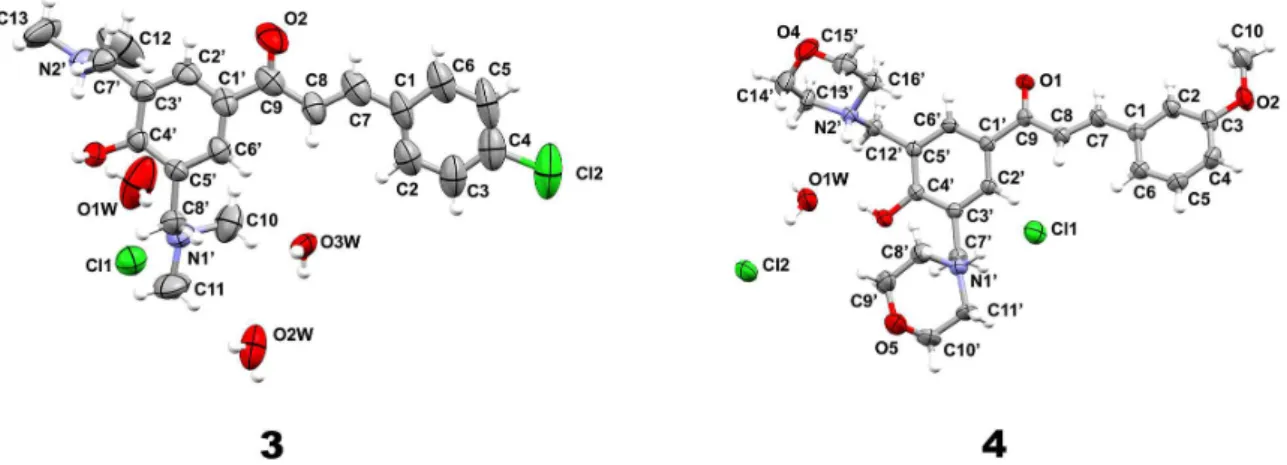
hydrochloride hydrate salts, as shown in Figure 2.
Compound 3 was solved in the orthorhombic space group
C2221 with one base unit, three water molecules and
one chloride anion per asymmetry unit. The structure of
compound 4 crystallized in the triclinic space group P-1
containing one molecule of Mannich base prepared from the corresponding hydroxychalcone, one water molecule and two chloride anions in the asymmetric unit. In the structure
of 3, one of the dimethylamino moiety is protonated, while
the other one is neutral. On the other hand, in 4, both amino
groups as morpholino groups are protonated.
The hydrogen bonding pattern in the three
dimension-network of 3 and 4 plays an important role in the assembly
of the crystal structures reported in this study. The analysis
of the hydrogen bonding pattern in 3 revealed that there is
an intramolecular interaction by means of N2’−H2’1···O1
contact between the hydrogen H2’1 of the protonated dimethylamino group and the oxygen O1 of the hydroxyl
moiety of the phenol ring forming a S11(6) motif. Also, the
hydroxyl group of the phenol ring donates one hydrogen
O1−H1OH···N1’. In this case, the water O1W and the chloride anion also assist in the connection of these two base units. The water O1W is bridging the dimethylamino group of one base to the chloride anion Cl1 by means of
O1W−H11W···N1’, while this same anion is connecting
the two base units by being an acceptor of hydrogen bond
through the contact N2’−H2’1···Cl1. Thus, this complex
hydrogen bonding network gives rise to a ribbon made up of two base units connected to each other where the water and chloride hydrogen bonding functionalities help in the stabilization of the crystal structure (Figure 3a). In addition,
another characteristic of this crystal structure is that, there is formation of channels filled with water molecules and chloride anions between the ribbons (Figure 3b). At last, the water molecules O2W and O3W, which are not involved in the ribbon assembly, are engaged in hydrogen bonding interactions with each other and with O1W in the
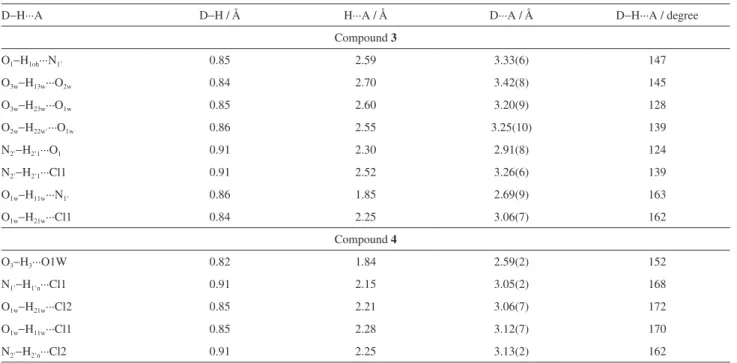
channels. The metrics of hydrogen bonds found in 3 is
shown in Table 1.
In the case of compound 4, due to the fact that both amino
functions are protonated, there is no classical intramolecular hydrogen bond. Only intermolecular interactions of this type
Figure 2. Asymmetric units of compounds 3 and 4. Only non-hydrogen atoms are labeled and their ellipsoids are at 50% probability level. The hydrogen atoms are depicted as spheres of arbitrary radius.
occur in the crystal structure. To assemble this compound, the hydroxyl group of the phenol ring is donating one hydrogen bond to the water molecule O1W through the interaction
O3−H3···O1W. Besides being engaged in hydrogen bonding
as acceptor, the water molecule is also a hydrogen bond donor
to both chloride anions by means of O1W−H11W···Cl1
and O1W−H21W···Cl2 contacts. In addition, the amino
nitrogen N1’ of one morpholine ring is hydrogen bonded
to the chloride anion through the N1’−H1’N···Cl1 contact
(Figure 3c). Likewise, the nitrogen N2’ of the second morpholine ring donates a hydrogen bond to the second
chloride anion Cl2 by means of N2’−H2’N···Cl2. In this way,
this set of hydrogen bonding interaction leads to the formation
of chains which connect the units of Mannich base 4 using
the water molecules and chloride anions as bridge between them (Figure 3c). On the whole, this crystal structure exhibit layers (Figure 3d) made up of chain motifs involving both morpholine rings of this Mannich base. The metrics of
hydrogen bonds found in compound 4 is shown in Table 1.
It is also important to mention that the crystal structure of the Mannich bases 3 and 4 is reported for the first time here,
while the crystal structure of the precursor of compound 3,
chalcone 1, has been already known.36 The planarity of the
chalcone skeleton (Figure 4) of compounds 3 and 4 is very
similar to the one reported by Jasinski et al.36 (the root mean
square deviation (RMSD) for their non-hydrogen atoms are 0.331 and 0.259, respectively).
As previously shown, aminomethylation of phenols occurs preferably at the ortho position.21H NMR, 13C NMR
and IR spectra of compounds 1, 2, 3 and 4 are in accordance
with their chemical structures. In the 1H NMR spectra,
a coupling constant higher than 15.4 Hz for the vinyl
H-atoms confirmed the (E)-configuration of compounds.
The methylene protons appeared as singlets at d 4.14 and
4.46 ppm, respectively. In the 13C NMR spectra, besides
the appearance of four more carbons of the morpholino and dimethylamino groups, the carbon peaks of olefinic
group were also observed at d 143.4 and 119.1 ppm (3) and
d 145.5 and 122.1 ppm (4). In the IR spectra, the presence
of the C=C functional group was confirmed by the υC=C
peaks appearing about 1600 cm-1. Conjugation of the
C=C to the carbonyl group decreases the C=O stretching
frequencies below 1710 cm-1. Therefore, the strong υC=O
peaks at 1657 (3) and 1663 cm-1 (4) in the IR spectra support
the α,β-unsaturated ketone structure of the compounds.
Table 1. Hydrogen-bond geometry in compounds 3 and 4
D−H···A D−H / Å H···A / Å D···A / Å D−H···A / degree
Compound 3
O1−H1oh···N1’ 0.85 2.59 3.33(6) 147
O3w−H13w···O2w 0.84 2.70 3.42(8) 145
O3w−H23w···O1w 0.85 2.60 3.20(9) 128
O2w−H22w’···O1w 0.86 2.55 3.25(10) 139
N2’−H2’1···O1 0.91 2.30 2.91(8) 124
N2’−H2’1···Cl1 0.91 2.52 3.26(6) 139
O1w−H11w···N1’ 0.86 1.85 2.69(9) 163
O1w−H21w···Cl1 0.84 2.25 3.06(7) 162
Compound 4
O3−H3···O1W 0.82 1.84 2.59(2) 152
N1’−H1’n···Cl1 0.91 2.15 3.05(2) 168
O1w−H21w···Cl2 0.85 2.21 3.06(7) 172
O1w−H11w···Cl1 0.85 2.28 3.12(7) 170
N2’−H2’n···Cl2 0.91 2.25 3.13(2) 162
The O−H stretching of the phenolic groups was observed at 3418 (3) and 3140 cm-1 (4).
In the reactions, the sulfur of GSH is added to
the β-carbon, followed by proton transfer reaction to
the α-carbon generating a chiral center.37 RP-TLC
of the incubates supported formation of the expected diastereomeric conjugates in each case. Visualization of the plates by UV (254 nm) illumination indicated presence
of the chalcones (1 and 2) and the Mannich derivatives
(3 and 4) with lower retention factors (Rf): 1: 0.05, 2: 0.08,
3: 0.21 and 4: 0.24. Besides, there could be seen a new
spot in each case: 1: Rf 0.31, 2: Rf 0.45, 3: Rf 0.53 and 4: Rf 0.56, indicating the more polar character of the latter.
The new spots, along with the GSH spots, were proved to
be ninhydrin-positive spots as well. This latter observation was in accordance with formation of the expected GSH conjugates.
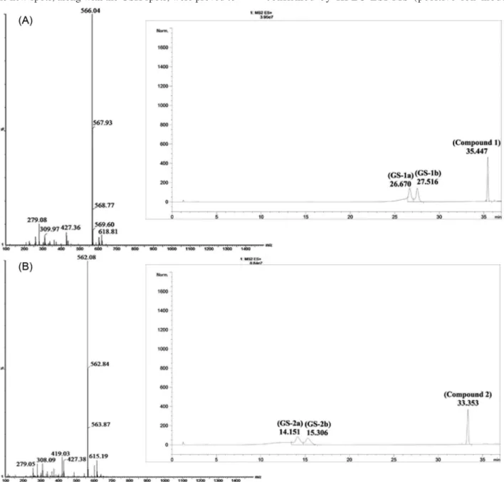
Formation of the expected two diastereomeric adducts on the prochiral faces of the enone moiety was confirmed by RP-HPLC. The RP-HPLC chromatograms of the incubates showed three major peaks attributed to two isomeric adducts and the starting chalcones with the following
retention times: GS-1a: 26.67, GS-1b: 27.52, 1: 35.45;
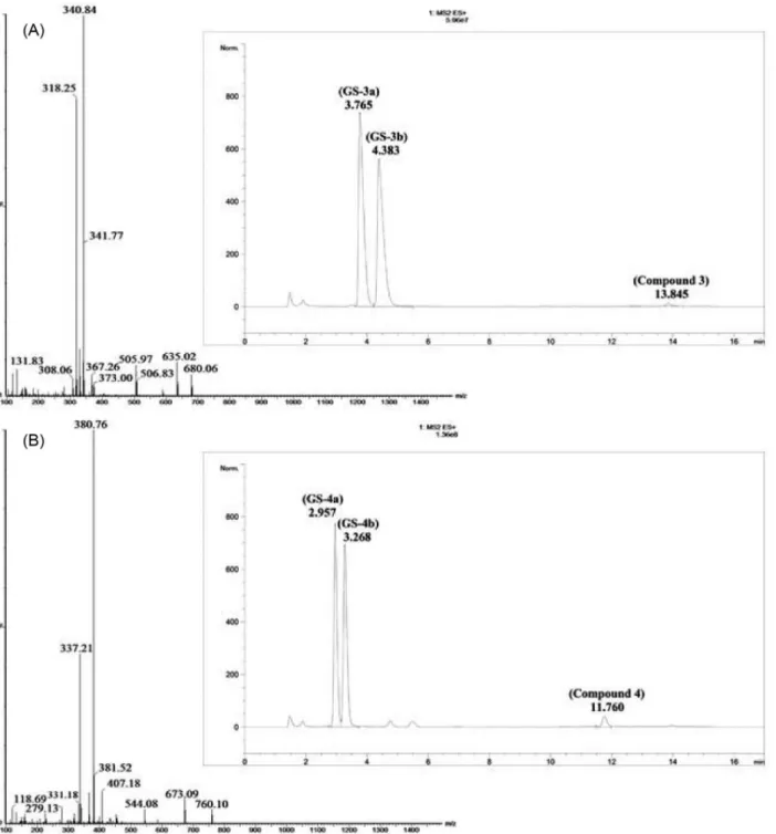
GS-2a: 14.15, GS-2b: 15.31, 2: 33.35; GS-3a: 3.94, GS-3b: 4.59, 3:13.55; GS-4a: 3.03, GS-4b: 3.35, 4: 11.63
(Figures 5 and 6).
The structures of the expected GSH adducts were confirmed by HPLC-ESI-MS (positive ion mode)
analysis of the 180 min incubates of compounds 1, 2, 3
and 4 (Figures 5 and 6). Besides the MH+ peaks of GSH
(m/z 308.02) and the starting compounds: 1 (m/z 259.05),
2 (m/z 255.15), 3 (m/z 373.02), 4 (m/z 453.12), ESI-MS
analysis of each incubation confirmed presence of the
respective GSH conjugates. These latter showed MH+ peaks
of m/z attributed to the respective molecular ion peaks of
GS-1 (m/z 566.04), GS-2 (m/z 562.08), GS-3 (m/z 680.06)
and GS-4 (m/z 760.10) adducts. In addition, in the case of
GS-3 and GS-4 double charged [M + 2H]2+ ions displayed
at m/z 340.63 and 380.74, respectively (Figure 6).
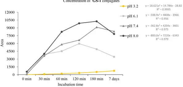
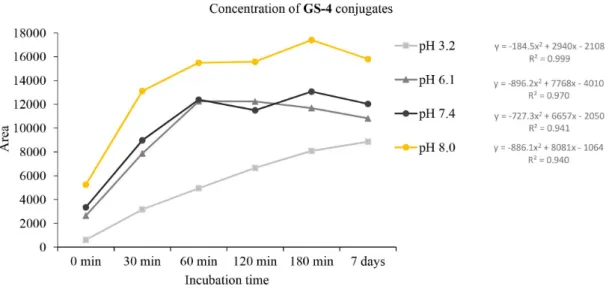
The rate of reactions of the compounds with GSH was found to be affected by the different pH values. As it is shown on Figure 6, a rather similar hyperbolic-type profile of decrease of the area of the HPLC peaks of each chalcone could be observed at pH 6.1, 7.4 and 8.0. On the contrary, a striking difference in the reactivity of chalcones 1 and 2,
and the Mannich derivatives 3 and 4 could be observed:
the Mannich derivatives showed much higher reactivity than the respective parent hydroxychalcones. The rate of the reactions was found to be not affected by the ring B substituents (4-chloro and 3-methoxy) at any pH (Figure 7). On the other hand, a similar increase of the peak areas corresponding to the formed GSH-adducts could be recorded (Figures 8-11).
The obtained data are in accordance with the 13C NMR
data of the compounds. It is known that the 13C NMR
shift values are sensitive probes of electron density.38 The
13C NMR chemical shifts of the β-carbon atoms of the
parent 1 and 2 are very similar: 141.7 and 143.2 ppm,
respectively. The corresponding chemical shift values for 3
and 4 are 143.4 and 145.5 ppm, respectively. The difference
in the 13C shift values between 2 and 1, and 4 and 3 is 1.5 and
2.1 ppm, respectively. Introduction of the Mannich-base moiety into the molecules increased the electrophilic
character of the β-carbon atoms of hydroxychalcones
practically by the same degree. The difference in the 13C
shift values between 3 and 1, and 4 and 2 is 1.7 and 2.3 ppm,
respectively. Such spectroscopic data are in accordance
with the similar reactivity of the two parent chalcones (1
and 2) and the two Mannich derivatives 3 and 4.
As previously mentioned, bioactivities of Mannich bases, e.g. cytotoxicity, is frequently attributed to the
high reactivity with thiols.2,18,22 Thus, the nature of the
amino group is an important factor since the rate of deaminoalkylation is inversely proportional to the basicity
of the liberated amine.17 However, in the tested conditions,
neither dimethylamine (pKa 10.78)nor morpholine
(pKa 8.49) influenced reactivity of compounds 3 and 4
with GSH and no deamination-addition products (mono- or diadducts) were detected under tested conditions. Intramolecular and intermolecular interactions (hydrogen bonds) between amino and hydroxyl groups should not be
discarded to retard or prevent the deamination process.17
RP-HPLC method after derivatization with OPA was used to determine the GSH and the GSSG levels in the
incubations.35 Reduced GSH reacts with OPA at pH 8.0
while GSSG reacts only at pH 12. For GSSG measurement, GSH has to be previously modified by reaction with NEM to form OPA-unreactive derivative. Concentrations of GSH and GSSG were measured simultaneously in incubation
samples performed in 0.2 mol L-1 phosphate buffer pH 7.4 at
37 °C. Measurements were performed at the 0, 30, 60, 120, 180 min and 7-day time points in two parallel incubations.
Figure 8. Time-dependent profile of HPLC peak area of adducts formed in the reaction of 4’-hydroxychalcone 1 and GSH at pH 3.2, 6.1, 7.4 and 8.0.
Figure 9. Time-dependent profile of HPLC peak area of adducts formed in the reaction of 4’-hydroxychalcone 2 and GSH at pH 3.2, 6.1, 7.4 and 8.0.
Figure 11. Time-dependent profile of HPLC peak area of adducts formed in the reaction of Mannich base 4 and GSH at pH 3.2, 6.1, 7.4 and 8.0.
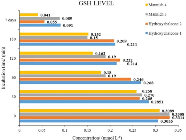
The mean GSH and GSSG values of the reactions with
compounds 1, 2, 3 and 4 are shown in Figures 12 and
13,respectively. As shown in Figure 12, the initial GSH
concentrations were between 0.3055 and 0.3314 mmol L-1,
while at the 180 min time point, the GSH levels were
found to be somewhat higher in the chalcone (1 and 2)
incubations: 0.211 and 0.2009 mmol L-1, respectively.
The results showed a considerable decreased amount of GSH after 7 days, possibly as consequence of an increase of the oxidized form GSSG. Accordingly, the measured GSSG levels substantially increased in the 7 day samples (Figure 13).
The reaction rate of α,β-unsatured ketones with
GSH was reported to be slower than their modified Mannich derivatives, possible as a result of decrease of the electron density of the carbonyl and/or stabilization
of the intermediate/conjugate by solvation.17,39,40 Electron
withdrawing effect of the aminoalkyl groups as well as hydrogen bond formation between the amino and carbonyl groups can decrease electron density of the carbonyl
and increase eletrophilicity of the β-carbon, however,
the nitrogen atoms of the Mannich bases 3 and 4 are
significantly protonated at the pH values of the present
experiments. Furthermore, the carbonyl 13C chemical shifts
of the Mannich derivatives appear at higher d values than
those of the 4’-hydroxychalcones (see above). However, there could be seen substantial difference in GSH-reactivity of the hydroxychalcones and their Mannich derivatives (see Figure 7). Therefore, we suggest the decreased electron density of the carbonyl is not an important contributing
factor for the high reactivity of 3 and 4 and stability of
the GS-3 and GS-4 conjugates. Besides, the hydrogen
bond (N−H...O−C) as well as the electron withdrawing
effect of Mannich moieties would also decrease the pKa
of α-hydrogenand the retro-Michael reaction would be
favored, which implies that these two factors do not support the relative stability of presented adducts.
In a recent study, the equilibrium and reaction kinetics of Michael addition reactions between chalcone analogues and thiol compounds, including GSH, were investigated
using density functional theory (DFT) calculations.41 While
the study in gas phase resulted in high deviated Gibbs free
energies (DG) from experimental values in water, the water
solvation model rendered well correlated measurements of
DG. Then, it is reasonable to expect that solvation effects
probably account to a significant extent for the reactivity
of the Mannich bases 3 and 4 with GSH in aqueous
environment.In addition, besides the solvent factor,
electrostatic interactions with local charges also is known
to strongly modulate cysteine sulfhydryl reactivity.42,43
Previous studies established that while conjugate
addition of the thiolate to the β-carbon to form enolate
intermediate is the rate limiting step of the Michael reaction, the rate of retro-Michael addition is proved to be limited by formation of the enolate anions in an elimination from
conjugated base mechanism (E1cB).14,41,44
The extent of ionization of GSH (pKa = 8.9)45 also
depends on the pH values of the solution. The increased rate of the Michael addition reactions of the investigated
bis Mannich chalcones (3 and 4) can be rationalized by
Figure 12. GSH levels in each time point (0, 30, 60, 120, 180 min, 7 days) of incubations of compounds 1, 2, 3, 4 and GSH. Determination of GSH levels were performed according to Rozmer et al.35 based on RP-HPLC method after derivatization with OPA. Incubations were performed in 0.2 mol L-1 phosphate buffer pH 7.4 at 37 °C.
Recent literature reveals that neighboring positive charges were shown to induce pKa shifts of thiols increasing its reaction with electrophiles and change the transition state energy affecting the reaction kinetics whereas the opposite effect was observed with nearby
negative charged groups.42,49,50 Indeed, positive charged
amino acids proximal to a Cys residue increased the Michael reaction rate as the protonation of the N atom in vinyl-substituted pyridine derivatives also promoted higher
reactivity towards Cys.42,49 Based on this findings and how
the effect of thiolate concentration overwhelms any other factor on Michael reaction kinetics, it is suggested that
protonated amino groups in compounds 3 and 4 can be
determinant for their high reactivity with GSH.49
Since E1cB reaction takes place by a base-induced
abstraction of an α-hydrogen unusually acidic by
resonance stabilization of the carbanion intermediate (conjugated base), a slower retro-Michael reaction would
be observed possibly if compounds 3 and 4 destabilize the
enol intermediate rendering more stable GS-3 and GS-4
conjugates.41,51
Conclusions
Present study showed bis Mannich bases 3 and 4 derived
from 4’-hydroxychalcones (1 and 2) had a high spontaneous
reactivity towards GSH, which demonstrated not to be significantly affected by the basicity of the morpholino and the dimethylamino groups. Considering how easily the compounds reacted with GSH, it would seem highly likely that they can also conjugate with another thiols in cellular environment. The reactions rendered stable monoadducts estimated in 85% of GSH conjugation of the starting material at 7-day time point. Taking into consideration the diverse biological effects of the investigated hydroxychalcones, it seems reasonable to suppose thiol-reactivity is not the only molecular mechanism that is involved in development of their biological effects. Application of HPLC analysis for this preliminary screening of the thiolalkylation reaction of Mannich bases proved to be an efficient and practical method to be used in further investigations. The findings could serve as useful structure-activity information for subsequent molecular modification of thiol-reactive 4’-hydroxychalcones.
Supplementary Information
Supplementary data [crystal data and refinement
parameters of the crystal structures of compounds 3
and 4 (Table S1); Fourier transform infrared (FTIR)
(Figures S1-S4) and NMR, spectra (Figures S5-S8)
of compounds 1-4] are available free of charge at
http://jbcs.sbq.org.br as PDF file.
Crystallographic data for the structures 3 and 4 were
deposited in the Cambridge Crystallographic Data Centre as supplementary publication numbers CCDC 1479638
and 1479615 for compounds 3 and 4, respectively. Copies
of the data can be obtained, free of charge, via www.ccdc. cam.ac.uk/conts/retrieving.html or from the Cambridge Crystallographic Data Centre, CCDC, 12 Union Road, Cambridge CB2 1EZ, UK; fax: +44 1223 336033. E-mail: [email protected].
Acknowledgments
The authors express their special thanks to Coordenadoria de Aperfeiçoamento de Pessoal de Ensino Superior (CAPES) (Grant No. BEX 6234/15-1) for a fellowship (to A. B.) for supporting this work. The authors express their sincere thanks to the Faculty of Medicine (University of Pécs) Research Fund (Grant No. AOK-KA-213/20) and the Conselho Nacional de Desenvolvimento Científico Tecnológico (CNPq) (Grant No. 478337/2013-2) for providing financial support. The authors express their thanks for Dr Monika Kuzma (Institute of Pharmaceutical Chemistry, University of Pécs) for her advice in the HPLC works.
References
1. Rozmer, Z.; Perjési, P.; Phytochem. Rev. 2016, 15, 87. 2. Dimmock, J. R.; Kandepu, N. M.; Hetherington, M.; Quail, J.
W.; Pugazhenthi, U.; Sudom, A. M.; Chamankhah, M.; Rose, P.; Pass, E.; Allen, T. M.; Halleran, S.; Szydlowski, J.; Mutus, B.; Tannous, M.; Manavathu, E. K.; Myers, T. G.; De Clercq, E.; Balzarini, J.; J. Med. Chem. 1998, 41, 1014.
3. Sabzevari, O.; Galati, G.; Moridani, M. Y.; Siraki, A.; O’Brien, P. J.; Chem.-Biol. Interact. 2004, 148, 57.
4. Calliste, C. A.; Le Bail, J. C.; Trouillas, P.; Pouget, C.; Habrioux, G.; Chulia, A. J.; Duroux, J. L.; Anticancer Res. 2000, 21, 3949. 5. Iwata, S.; Nishino, T.; Inoue, H.; Nagata, N.; Satomi, Y.;
Nishino, H.; Shibata, S.; Biol. Pharm. Bull. 1997, 20, 1266. 6. Xue, Y.; Zheng, Y.; An, L.; Zhang, L.; Qian, Y.; Yu, D.; Gong,
X.; Liu, Y.; Comput. Theor. Chem. 2012, 982, 74.
7. Elias, D. W.; Beazely, M. A.; Kandepu, N. M.; Curr. Med. Chem. 1999, 6, 1125.
8. Go, M. L.; Wu, X.; Liu, X. L.; Curr. Med. Chem. 2005, 12, 483. 9. Nowakowska, Z.; Eur.J. Med. Chem. 2007, 42, 125.
10. Benvenuto, J. A.; Connor, T. H.; Monteith, D. K.; Laidlaw, J. L.; Adams, S. C.; Matney, T. S.; Theiss, J. C.; J. Pharm. Sci. 1993, 82, 991.
12. Wilson, A. G. E. In New Horizons in Predictive Toxicology: Current Status and Application, 12th ed.; Wilson, A. G. E.; Thurston, D. E.; Rotella, D., eds.; Royal Society of Chemistry: Cambridge, UK, 2011.
13. Armstrong, R. N.; Chem. Res. Toxicol. 1991, 4, 131. 14. Baillie, T. A.; Slatter, J. G.; Acc. Chem. Res. 1991, 24, 264. 15. Dimmock, J. R.; Jha, A.; Zello, G. A.; Quail, J. W.; Oloo, E.
O.; Nienaber, K. H.; Kowalczyk, E. S.; Allen, T. M.; Santos, C. L.; De Clercq, E.; Balzarini, J.; Manavathu, E. K.; Stables, J. P.; Eur. J. Med. Chem. 2002, 37, 961.
16. Saab, A. N.; Sloan, K. B.; Beall, H. D.; Villanueva, R.; J. Pharm. Sci. 1990, 79, 1099.
17. Gul, H. I.; Yerdelen, K. O.; Das, U.; Gul, M.; Pandit, B.; Li, P. K.; Dimmock, J. R.; Chem. Pharm. Bull. 2008, 56, 1675. 18. Bala, S.; Sharma, N.; Kajal, A.; Kamboj, S.; Saini, V.; Int. J.
Med. Chem. 2014, 2014.
19. Cogan, P. S.; Fowler, C. R.; Post, G. C.; Koch, T. H.; Lett. Drug Des. Discovery 2004, 1, 247.
20. Gul, H. I.; Yerdelen, K. O.; Gul, M.; Das, U.; Pandit, B.; Li, P. K.; Secen, H.; Sahin, F.; Arch. Pharm. 2007, 340, 195. 21. Reddy, M. V. B.; Su, C. R.; Chiou, W. F.; Liu, Y. N.; Chen, R.
Y. H.; Bastow, K. F.; Lee, K. H.; Wu, T. S.; Bioorg. Med. Chem. 2008, 16, 7358.
22. Bilginer, S.; Gul, H. I.; Mete, E.; Das, U.; Sakagami, H.; Umemura, N.; Dimmock, J. R.; J. Enzyme Inhib. Med. Chem. 2013, 28, 974.
23. Reddy, M. V. B.; Chen, S. S.; Lin, M. L.; Chan, H. H.; Kuo, P. C.; Wu, T. S.; Chem. Pharm. Bull. 2011, 59, 1549.
24. Reddy, M. V. B.; Shen, Y. C.; Yang, J. S.; Hwang, T. L.; Bastow, K. F.; Qian, K.; Lee, K. H.; Wu, T. S.; Bioorg. Med. Chem. 2011, 19, 1895.
25. Modzelewska, A.; Pettit, C.; Achanta, G.; Davidson, N. E.; Huang, P.; Khan, S. R.; Bioorg. Med. Chem. 2006, 14, 3495. 26. Kumar, N.; Chauhan, L. S.; Sharma, C. S.; Dashora, N.; Bera,
R.; Med. Chem. Res. 2015, 24, 2580.
27. Jayapal, M. R.; Sreedhar, N. Y.; Int. J. Curr. Pharm. Res. 2010, 2, 60.
28. Garg, N.; Chandra, T.; Jain, A. B.; Kumar, A.; Eur. J. Med. Chem. 2010, 45, 1529.
29. Nguyen, V. S.; Shi, L.; Luan, F. Q.; Wang, Q. A.; Acta Biochim. Pol. 2015, 62, 547.
30. SADABS, APEX2 and SAINT, Bruker AXS Inc., Madison, WI, USA, 2009.
31. Sheldrick, G. M.; Acta Crystallogr., Sect. A 2008, 64, 112.
32. Macrae, C. F.; Bruno, I. J.; Chisholm, J. A.; Edgington, P. R.; Mccabe, P.; Pidcock, E.; Monge, L. R.; Taylor, R.; van de Streek J.; Wood, P. A.; J. Appl. Crystallogr. 2008, 41, 466.
33. Farrugia, L. J.; J. Appl. Crystallogr. 1997, 30, 565.
34. Perjési, P.; Maász, G.; Reisch, R.; Benkő, A.; Monatsh. Chem. 2012, 143, 1107.
35. Rozmer, Z.; Berki, T.; Perjési, P.; Toxicol. In Vitro2006, 20, 1354.
36. Jasinski, J. P.; Butcher, R. J.; Yathirajan, H. S.; Sarojini, B. K.; Musthafa Khaleel, V.; Acta Cryst. 2011, E67, o756.
37. van Iersel, M. L.; van Lipzig, M. M.; Rietjens, I. M.; Vervoort, J.; van Bladeren, P. J.; FEBS Lett. 1998, 441, 153.
38. Perjési, P.; Linnanto, J.; Kolehmainen, E.; Ősz, E.; Virtanen, E.; J. Mol. Struct. 2005, 740, 81.
39. Dimmock, J. R.; Smith, L. M.; Smith, P. J.; Can. J. Chem. 1980, 58, 984.
40. Jin, F.; Jin, X. Y.; Jin, Y. L.; Sohn, D. W.; Kim, S. A.; Sohn, D. H.; Kim, Y. C.; Kim, H. S.; Arch. Pharmacal Res. 2007, 30, 1359.
41. Chen, J.; Jiang, X.; Carroll, S. L.; Huang, J.; Wang, J.; Org. Lett. 2015, 17, 5978.
42. Lutolf, M. P.; Tirelli, N.; Cerritelli, S.; Cavalli, L.; Hubbell, J. A.; Bioconjugate Chem. 2001, 12, 1051.
43. Hou, L.; Honaker, M. T.; Shireman, L. M.; Balogh, L. M.; Roberts, A. G.; Ng, K. C.; Nath, A.; Atkins, W. M.; J. Biol. Chem. 2007, 282, 23264.
44. Kim, Y.; Mohrig, J. R.; Truhlar, D. G.; J. Am. Chem. Soc. 2010, 132, 11071.
45. Rabenstein, D. L.; J. Am. Chem. Soc. 1973, 95, 2797. 46. Rhile, I. J.; Markle, T. F.; Nagao, H.; Di Pasquale, A. G.; Lam,
O. P.; Lockwood, M. A.; Rotter, K.; Mayer, J. M.; J. Am. Chem. Soc. 2006, 128, 6075.
47. Costentin, C.; Robert, M.; Savéant, J. M.; Phys. Chem. Chem. Phys. 2010, 12, 11179.
48. Fang, Y.; Liu, L.; Feng, Y.; Li, X. S.; Guo, Q. X.; J. Phys. Chem. A 2002, 106, 4669.
49. Boutureira, O.; Bernardes, G. J.; Chem. Rev. 2015, 115, 2174. 50. Gitler, C.; Zarmi, B.; Kalef, E.; Methods Enzymol. 1995, 251,
366.
51. McMurry, J.; Organic Chemistry with Biological Applications, 3rd ed.; Cengage Learning: Stamford, USA, 2014.
Submitted: May 27, 2016