802
Rev Soc Bras Med Trop 49(6):802, November-December, 2016 doi: 10.1590/0037-8682-0110-2016
Images in Infectious Diseases
Corresponding author: Dr. Fred Luciano Neves Santos. e-mail: fred.santos@bahia.fi ocruz.br; fl [email protected] Received 23 March 2016
Accepted 9 May 2016
Meloidogyne
eggs in human stool in Northeastern Brazil
Fred Luciano Neves Santos
[1], Alina Maria Gonzaga Carlos de Souza
[2]and Filipe Dantas-Torres
[3][1]. Laboratório de Patologia e Biointervenção, Instituto Gonçalo Moniz, Fundação Oswaldo Cruz, Salvador, Bahia, Brasil. [2]. Laboratório Datalab, Grupo Promedica, Salvador, Bahia, Brasil. [3]. Laboratório de Imunoparasitologia, Departamento de Imunologia,
Centro de Pesquisas Aggeu Magalhães, Fundação Oswaldo Cruz, Recife, Pernambuco, Brasil.
Root-knot nematodes (Meloidogyne spp.) are among the most
economically damaging genera of plant-parasitic nematodes worldwide(1). Although they are not pathogenic to humans, their
eggs may eventually be found in human stools(2) and owing to their
similarity with eggs of pathogenic nematodes must be correctly identifi ed to avoid unnecessary treatments. Recently, researchers reported the presence of eggs similar to those of Trichostrongylus
spp. (identifi ed as Meloidogyne eggs) in three (0.5%) of 586 stool
samples collected from East Kwaio, Solomon Islands(2). From 2008
to 2014, 332,132 stool samples, referred to a private laboratory network (Datalab) in Salvador, Brazil, were examined using the Lutz method; 61 (0.02%) were positive for Meloidogyne
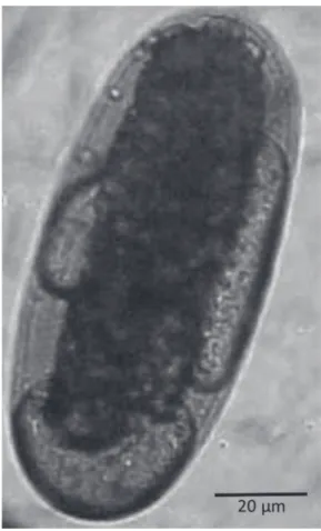
eggs (Figure 1).
Meloidogyne eggs may be identified based on their
shape, size, and absence or presence of characteristic internal
structures(3). Meloidogyne eggs have thin hyaline shells without
visible markings, elongate-ovoid with rounded ends. One of the sides can be concave or slightly fl attened. They measure 82-120µm in length × 24-43µm in width and can be seen inside a juvenile cell mass in the fi rst division phase or in a fully formed larva. They may present internal refractive corpuscles, located between the shell and the morula, which are important to distinguish them from eggs of Trichostrongylus spp. and
hookworms. The presence of corpuscles, resembling air-sacs,
on one of the poles, between the morula and the shell is very characteristic, although not always present. However, during its development, the concavity and the air-sacs may disappear, and
the egg becomes plano-convex or even biconvex.
Confl icts of Interest
The authors declare that there is no confl ict of interest.
FIGURE 1. A Meloidogyne egg showing a thin, hyaline shell and refractive
internal corpuscles located between the shell and the morula, resembling lipid droplets.
REFERENCES
1. Jones JT, Haegeman A, Danchin EGJ, Gaur HS, Helder J, Jones
MG, et al. Top 10 plant-parasitic nematodes in molecular plant pathology. Mol Plant Pathol 2013; 14:946-961.
2. Bradbury RS, Speare R. Passage of Meloidogyne eggs in human
stool: forgotten, but not gone. J Clin Microbiol 2015; 53:1458-1459.
3. Pardinti VC, Ferreira CJ, Moura DM, Mendonça AR. Ocorrência de ovos de Trichostrongylus sp. e Meloidogyne sp. em exames