Arq Neuropsiquiatr 2005;63(2-B):519-522
1Hospital das Clínicas, University of São Paulo School of Medicine, São Paulo SP Brazil;2Ludwig Institute for Cancer Research, São Paulo Branch, São Paulo SP, Brazil.
Received 15 September 2004, received in final form 6 December 2004. Accepted 4 February 2005.
Dr. Ricardo Nitrini - Rua Bartolomeu Feio 560 - 04580-001 São Paulo SP - Brasil.
ASYMMETRIC CORTICAL HIGH SIGNAL
ON DIFFUSION WEIGHTED-MRI IN A
CASE OF CREUTZFELDT-JAKOB DISEASE
Ricardo Nitrini
1, Renata Areza-Fegyveres
1, Vilma R. Martins
2,
Rosa Maria R.P.S. Castro
2, Michele C. Landemberger
2,
Nancy Huang
1, Luiz A. Bacheschi
1, Luiz E. Bacheschi
1,
Cláudia C. Leite
1, Carlos A. Buchpiguel
1, Sérgio Rosemberg
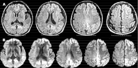
1ABSTRACT - High signal in the cerebral cortex and/or basal ganglia on diffusion-weighted magnetic reso-nance imaging (DW-MRI) has been described as a good diagnostic marker for sporadic Creutzfeldt-Jakob disease (sCJD). We report a case of sCJD with atypical clinical evolution and unusual DW-MRI findings. A 53-year-old man was seen with a 2-year history of a rapidly progressive dementia and cerebellar ataxia. Cerebrospinal fluid analysis, including the test for 14-3-3 protein, was normal. EEG did not show periodic activity. However, DW-MRI showed gyriform hyperintensity involving practically the entire cortical ribbon of the left hemisphere, whilst being limited to the posterior cingulate gyrus in the right hemisphere. DNA analysis showed no mutations or insertions in the prion protein gene, and homozigozity for methionine in codon 129. A subsequent brain biopsy confirmed the diagnosis of CJD. Thus, high signal on DW-MRI may be limited to the cerebral cortex and may present a very asymmetric distribution in sCJD.
KEY WORDS: Creutzfeldt-Jakob disease, prion disease, MRI, diffusion-weighted MRI.
Hipersinal cortical assimétrico na ressonância magnética na imagem em difusão em caso de doença de Creutzfeldt-Jakob
RESUMO - Hipersinal no cortex cerebral e/ou nos gânglios da base observado com a técnica de difusão da ressonância magnética (RMDIF) tem sido descrito como bom marcador diagnóstico da doença de Cre u t z f e l d t -Jakob esporádica (DCJe). Relatamos caso de DCJe com evolução clínica atípica e achados incomuns na RM-DIF. Homem de 53 anos foi examinado com história de dois anos de demência rapidamente progressiva e ataxia cere b e l a r. Exame do líquido cefalorraqueano, incluindo pesquisa da proteína 14-3-3, foi normal; EEG não revelou atividade periódica; RM-DIF mostrou hiperintensidade nos giros que afetava quase inteiramen-te o manto cortical do hemisfério cerebral esquerdo e que no hemisfério direito se limitava à parinteiramen-te posinteiramen-te- poste-rior do giro cíngulo. Análise do DNA revelou ausência de mutação ou de inserção no gene da proteína priôni-ca e a presença de homozigose para metionina no códon 129. Biópsia cerebral confirmou o diagnóstico de DCJ. Hipersinal na RM-DIF pode ser limitado ao córtex cerebral e pode distribuir-se de modo muito as-simétrico na DCJe.
PALAVRAS-CHAVE: doença de Creutzfeldt-Jakob, prion; ressonância magnética, difusão.
The clinical diagnosis of sporadic Creutzfeldt-Jakob disease (sCJD), particularly in its early man-ifestations, frequently presents a challenge to even the most experienced of neurologists. The clinical p i c t u re of most cases of sCJD is characterized by r a p-id progressive dementia, myoclonus, and multifo-cal neurologimultifo-cal dysfunction, with a fatal outcome in the first one or two years after the onset of the symptoms. The presence of periodic sharp-wave complexes on the electroencephalogram (EEG) has
been recognized as the most reliable non invasive test for the diagnosis of CJD1. However, this EEG a
b-n o rmality may be abseb-nt ib-n about ob-ne-third of the c a s e s2 , 3 . T h e re are cases of definite sCJD with a
m o rerelentless pro g ression and with atypical clini-cal features. Unfortunately, EEG periodic activity is usually absent in such cases2 , 3. Recently, two
520 Arq Neuropsiquiatr 2005;63(2-B)
intensity in the basal ganglia and/or in the cere-bral cortex on diffusion-weighted magnetic reso-nance imaging (DW-MRI)4,5.
We report a case of sCJD with atypical clinical evolution and unusual findings in DW-MRI.
CASE
A 53-year old Brazilian born from Japanese parents and an electronics technician, was seen for the first time in Febru a ry 2003 with a 2-year history of behavioral dis-turbances and cognitive decline. Since 1991 he had been working in Japan as a foreign worker, while his family continued to live in Brazil. He had kept in touch with his family by mail or telephone, sending money to his w i f e practically every month. He had been re t u rning once or twice a year to Brazil for vacations. In the beginning of 2001 he spent a vacation with his family, during which time he was considered to be a little more quiet than usual. In Febru a ry2001 he re t u rned to Japan, and for t h e following six months did not contact his family in Brazil. His wife and sons became very worried and tried to con-tact him unsuccessfully. In August 2001 he was found by a friend, living homeless and in a very confuse state. He was brought back to Brazil in October 2001, and on arr i v a l he was in a confused and apathetic state, refusing to w a s h or shave, but was partially independent for other daily activities, such as eating, still being able to control his sphincters. He was submitted to exhaustive medical and l a b o r a t o ry examinations which were inconclusive re g a rdi-ng the diagnosis. He was treated with paroxetine and b e-came agitated and aggressive. Haloperidol 5 mg bid as-sociated with biperiden 2mg daily reduced the agitat-ed and aggressive behavior, but he again became apa-thetic. The patient had been living in a nursing home since September 2002, since which time he had not be-en on medication. The subject’s condition had
continu-ed to deteriorate and in the few months prior to our first examination he had lost control of the sphincters. His past medical history was unremarkable whilst his mo-ther had died with Alzheimer disease at the age of 93. Physical examination was normal. On neurological examination the cranial nerves and muscle power were n o rmal, with brisk deep reflexes globally and flexor p l a n-tar reflexes bilaterally; muscle tone was slightly spastic in the legs. Gait was ataxic and a reduction of arm swin-ging was noticed on the left side. Ataxia was also pre sent in finger to nose and heel to knee tests bilaterally. He d i d not show concern over his condition and when questio-ned about his memory replied that he did not know if it was normal or not. Echolalia was also present. He sco-red 8 points in the MMSE (2 in spatial orientation, 1 i n registration, 2 in the verbal command, 2 in naming and 1 in repetition). Digit span was 3 in direct order and 0 in the reverse.
Routine laboratory tests, including haemogram, sed-imentation rate, serum glucose, urea, creatinine, sodiu m , potassium, calcium, phosphorus, transaminases, B12 and folate levels, were normal, except for a slightly re d u c e d t h y roid-stimulating hormone level (0.4 U/ml; norm a l levels: 0.5-5.0 U/ml) with a normal free-T4 level. CSF analysis, including VDRL, TPHA and protein electro p h o re-sis, were normal, and the test for 14-3-3 protein was neg-ative. EEG showed diffuse disorganization of the elec-trical activity and epileptic activity in the right tempo-ral region, without periodic activity.
Brain CT scan showed slight frontotemporal atro p h y. Brain single-photon emission computed tomography (SPECT) revealed mildly reduced regional cerebral blood flow in left frontal, temporal-parietal and superior o c c i p i-tal regions. Quantitative analysis using Statistical Para-metric Mapping (SPM-version 99) confirmed statistical-ly significant (p<0.001) hypoperfusion in left frontal re-gion and left parietal rere-gions. Additionally, SPM
Arq Neuropsiquiatr 2005;63(2-B) 521
sis showed decreased blood flow in posterior cingular cortex and left cerebellum. Mild abnormalities in right occipital-parietal lobes did not survive on statistical ana-lysis. Brain MRI study showed gyriform hyperintensity on fluid attenuated inversion re c o v e ry(FLAIR) and diff u-sion-weighted images involving almost entirely the cor-tical ribbon of the left hemisphere. In the right cere b r a l cortex, the hiperintensity was limited to the posterior cingulated gyrus (Fig 1).
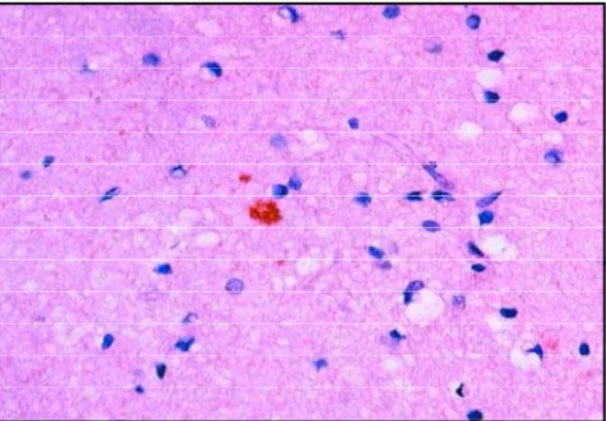
A brain biopsy taken from the right superior frontal g y rus revealed neuronal loss, mild gliosis and spongiform change in the cerebral cortex (Fig 2). Immunohistoche-m i s t rywith 3F4 antibody showed scattered focal granu-lar PrPResdeposits through the neuropil (Fig 3).
DNA analysis of peripheral blood cells using con-ventional sequencing or DHPLC 6showed no mutations or insertions in the prion protein gene, and the pre s e n-ce of 5 octarepeats, methionine in codon 129, asparagine in codon 171 and glutamic acid in codon 219 all in ho-mozigozity.
His condition continued to worsen, but he was able to walk with assistance until the beginning of 2004. A
year following the biopsy his condition had deteriorat-ed to akinetic mutism.
DISCUSSION
A c c o rding to the World Health Organization cri-teria, a typical EEG and/or a positive 14-3-3 CSF assay must be present for the diagnosis of pro b a b l e sCJD1. Approximately 60-80% of cases of sCJD
de-velop the characteristic EEG periodic activity1, and
although the sensitivity of the detection of 14-3-3 in the CSF is usually very high in the typical cases of sCJD7, it may be in low in cases with nonclassic
p re s e n t a t i o n8. In our case, a diagnosis of pro b a b l e
sCJD could not be assumed, a brain biopsy, how-ever, confirmed this diagnosis. MR FLAIR images and DW-MRI showed abnormal high intensity in the cortical ribbon with a very asymmetric distri-bution, involving mainly the left side, and absence of hyperintensity in the basal ganglia.
The abnormal high intensity on DW-MRI in sCJD usually involves the striatum and/or the cerebral cortex9. It is possible that high signal on DW-MRI
appears earlier in the cerebral cortex, while basal ganglia abnormalities appear later, when cortical high signals became less obvious5 , 1 0 , 1 1.The pre s e n c e
of high signal restricted to the cerebral cortex two years after the onset of the symptoms as seen in this case, is probably very uncommon. Asymmetric distribution had previously been re p o rt e d1 0 , 1 2, and
even unilateral cortical involvement has recently been described in a case of probable sCJD without pathological confirmation13.In several cases,
cor-relation was found between high intensity in the c e rebral cortex and clinical signs1 0 , 1 2, EEG abnorm a l
i-t i e s1 2 , 1 4or hypometabolism on PET- s c a n1 3 , 1 5. E x c e p t
for the echolalia, the clinical signs did not suggest a more severe involvement of the left cerebral
corFig 2. Cerebral cortex with moderate neuronal loss and spon -giform change (HE X 250).
522 Arq Neuropsiquiatr 2005;63(2-B)
tex in this case, while only brain SPECT showed cor-relation with the MRI findings.
The relatively long evolution raised the possi-bility of a familial form of CJD, which was exclud-ed by DNA analysis. The clinical characteristics of the cases of sCJD have been related to the poly-morphism in codon 129 and to the physicochemi-cal properties of the protease-resistant prion pro-tein (PrPSc) that is found in the brain16,17.There are
two types or strains of PrPS c, types 1 and 2, and thre e
possible genotypes at codon 129 [Methione homo-zigozity (MM), valine homohomo-zigozity (VV) and het-e rozigozity (MV)], which in conjunction arhet-e rhet-e s p o n-sible for six CJD subtypes. The majority of sporadic cases of CJD are MM1 or MV117. Given that we did
not ascertain the type of PrPSc, there are two
pos-sibilities for this case: MM1 or MM2. The clinical c h a-racteristics of MM1 cases are that of the typical sCJD, while MM2 cases may have a more protracted c o u r s e and absence of EEG periodic activity1 7. The most d i
s-tinctive pathological features of MM2 subtype are the type of spongiform degeneration, which is cha-racterized by large, confluent vacuoles, along with the pattern of PrPSc staining, which is coarse17. In
our case, the general pathological pattern was t h a t found in most cases of sCJD with MM1 subtype.
Attempts to correlate MRI findings with these six subtypes have been made. A recent study de-monstrated that DW-MRI abnormalities were pre s-ent in the first examination in 24 out of 26 patis-ents with CJD9. The second DW-MRI showed high
inten-sity lesions in the striatum in one of those patients, but repeated scans did not show abnormalities in the other patient, who was classified as MM2 (thal-amic form) at post-mortem examination9. In
anoth-er recent study, hypanoth-erintense signal changes in t h e basal ganglia were absent in the three patients clas-sified as MM21 8. In a re p o rted case of MV2 subtype
of sCJD, in which EEG periodic activity and 14-3-3 were negative, DW-MRI showed marked hyperin-tense signal abnormalities in the cerebral cortex and basal ganglia1 9. Recently, abnormal high
inten-sity on DW-MRI has been seen in the thalamus in two cases of VV2-type of sCJD20. It is well known
that hyperintense signal in the pulvinar is a typi-cal sign of the variant CJD, which in turn is associa-ted with bovine spongiform encephalopathy2 1.
Ta-ken together these data suggest that hyperinten-se signal changes in the brain on DW-MRI are fre-quent in all sCJD subtypes, except for the MM2 one. In conclusion, MRI is a useful tool for the diag-nosis of sCJD even when the presentation is
atypi-cal and asymmetric cortiatypi-cal high signal may be f o u n d in this disease.
A c k n o w l e d g e m e n t– We are indebted to Dr. Françoise
Gray for perf o rming the immunohistochemical analysis.
REFERENCES
1. WHO. Report of the consultation on the global surveillance, diagnosi-s and therapy of human trandiagnosi-smidiagnosi-sdiagnosi-sible diagnosi-spongiform encephalopathiediagnosi-s. Geneva: WHO, 1998.
2. Zerr I, Pocchiari M, Collins S, et al. Analysis of EEG and CSF 14-3-3 pro-tein as aids to the diagnosis of Creutzfeldt-Jakob disease. Neurology 2000;55:811-815.
3. Zeidler M, Green A. Advances in diagnosing Creutzfeldt-Jakob disease with MRI and CSF 14-3-3 analysis. Neurology 2004;63:410-411. 4. Hsich G, Kenney K, Gibbs CJ, Lee KH, Harrington MG. The 14-3-3 brain
p rotein in cere b rospinal fluid as a marker for transmissible spongiform encephalopathies. N Engl J Med 1996;335:924-930.
5. Demaerel P, Baert AL, Vanopdenbosch L, Robberecht W, Dom R. Dif-fusion-weighted magnetic resonance imaging in Creutzfeldt-Jakob dis-ease. Lancet 1997;349:847-848.
6. Castro RMRPS, Lamdenberger MC, Waltz R, et al. High capacity and low cost detection of prion protein gene variant alleles through dena-turing HPLC methodology. J Neurosc Meth 2004;62:751-755. 7. Zerr I, Bodemer M, Gefeller O, et al. Detection of 14-3-3 protein in the
c e re b rospinal fluid supports the diagnosis of Creutzfeldt-Jakob disease. Ann Neurol 1998;43:32-40.
8. Castellani, RJ, Colucci M, Xie Z, et al. Sensitivity of 14-3-3 protein test varies in subtypes of sporadic Creutzfeldt-Jakob disease. Neurology 2004;63:436-442.
9. Shiga Y, Miyazawa K, Sato S, et al. Diffusion-weighted MRI abnormali-ties as an early diagnostic marker for Creutzfeldt-Jakob disease. Neuro-logy 2004;63:443-449.
10. Yee AS, Simon JH, Anderson CA, Sze CI, Filley CM. Diffusion-weight-ed MRI of right-hemisphere dysfunction in Creutzfeldt-Jakob disease. Neurology 1999;52:1514-1515.
11. Tribl GG, Strasser G, Zeitlhofer J, et al. Sequential MRI in a case of Cre u t-zfeldt-Jakob disease. Neuroradiology 2002;44:223-226.
12. Cambier DM, Kantarci K, Worrell GA, Westmoreland BF, Aksamit AJ. Lateralized and focal clinical, EEG, and FLAIR MRI abnormalities in Creutzfeldt-Jakob disease. Clin Neurophysiol 2003;114:1724-1728. 13. Bavis J, Reynolds P, Tegeler C, Clark P. Asymmetric neuroimaging in
Creutzfeldt-Jakob disease. J Neuroimaging 2003;13:376-379. 14. Mao-Draayer Y, Braff SP, Nagle KJ, Pendlebury W, Penar PL, Shapiro
RE. Emerging patterns of diffusionweighted MR imaging in Cre u t z f e l d t -Jakob disease: case report and review of the literature. Am J Neuro r a d i o l 2002;23:550-556.
15. Tsuji Y, Kanamori H, Murakami G, et al. Heidenhain variant of Cre u t z-feldt-Jakob disease: diffusion-weighted MRI and PET characteristics. J Neuroimaging 2004;14:63-66.
16. Parchi P, Castellani R, Capellari S, et al. Molecular basis of phenotyp-ic variability in sporadphenotyp-ic Creutzfeldt-Jakob disease. Ann Neurol 1996; 39:767-778.
17. P a rchi P, Giese A, Capellari S, et al. Classification of sporadic Cre u t z f e l d t -Jakob disease based on molecular and phenotype analysis of 300 sub-jects. Ann Neurol 1999;46:224-233..
18. Meissner B, Köhler K, Körtner K, et al. Sporadic Creutzfeldt-Jakob dis-ease: magnetic resonance imaging and clinical findings. Neurology 2 0 0 4 ; 63:450-456.
19. Samman I, Schulz-Schaeffer WJ, Wohrle JC, Sommer A, Kretzschmar HA, Hennerici M. Clinical range and MRI in Creutzfeldt-Jakob disease with heterozygosity at codon 129 and prion protein type 2. J Neurol Neurosurg Psychiatry 1999;67:678-681.
20. Fukushima R, Shiga Y, Nakamura M, Fujimori J, Kitamoto T, Yoshida Y. MRI characteristics of sporadic CJD with valine homozygosity at codon 129 of the prion protein gene and PrPSc type 2 in Japan. J Neuro l Neurosurg Psychiatry 2004;75:485-487.