128 Arq Neuropsiquiatr 2013;71(2):127-132
Bilateral plexiform neurofibromas of the
brachial and lumbosacral plexuses
Neurofibroma plexiforme bilateral dos plexos braquial e lombossacral
Fernando H. Souza1, Carlos B. Dabdoub1, Silvya N. Bernardino2, Elisabeth Nascimento Silveira3, Silvio Litvin4,
Hildo Azevedo-Filho1
1Department of Neurosurgery, Restauração Hospital, Recife PE, Brazil; 2Department of Neurology, Hospital Getúlio Vargas, Recife PE, Brazil; 3Department of Nursing, Restauração Hospital, Recife PE, Brazil; 4Department of Radiology, Restauração Hospital, Recife PE, Brazil.
Correspondence: Fernando Henrique Souza; Departamento de Neurocirurgia, Hospital da Restauração; Avenida Agamenon Magalhães s/n; 52010-040 Recife PE - Brasil; E-mail: [email protected]
Conflict of interest: There is no conlict of interest to declare.
Received 24 May 2012; Received in inal form 11 July 2012; Accepted 18 July 2012
Neurofibromatosis type 1 (NF-1) is one the most com-mon autosomal dominant conditions affecting the ner-vous system1. Plexiform neurofibroma (PN) is a benign tumor that basically defines the diagnosis of NF-1. These entities consist of multiple, twisted masses that grow along the axis of a large nerve, infiltrating and separat-ing normal nerve fascicles. Malignant transformation is the main associated complication. Surgery is frequently reserved until PNs have progressed to the point of causing functional compromise, esthetic deformity or pain2. This paper describes a case of plexiform neurofibromas of the brachial and lumbosacral plexuses, with presentation of the clinical and radiological findings.
CASE REPORT
129
Arq Neuropsiquiatr 2013;71(2):127-132
Magnetic resonance imaging (MRI) of the brachial plexus was solicited and revealed the T2 weighted sequence, multi-ple, hyperintense, lobular, solid, expansive tumors in the dis-tribution of the brachial plexus bilaterally extending to the anterior mediastinum and invading the spinal canal (Fig 2). Lumbosacral plexus MRI showed impairment of both plexus-es and suggplexus-ested the prplexus-esence of a tumor toward the lumbo-sacral neural foramens (Fig 3) and its extension to the pelvic region (Fig 4).
An incisional biopsy was performed on the left cervical tumor and the histological indings were compatible with neuroibroma.
During the four-year follow-up involving imaging exams, the patient had been asymptomatic and free of neurologi-cal deicit. hus, the decision was to maintain conservative management.
DISCUSSION
PN is a common manifestation of NF-1. his entity may be classiied as a benign peripheral nerve sheath tumor that in-volves multiple nerve fascicles or branches of major nerves3.
MRI is the method of choice for the diagnosis and follow-up of neuroibromas. In T1 weighted image, a PN is isoin-tense in relation to muscle tissue. In T2 weighted sequence, a PN is seen as a hyperintense, homogeneous image with “tar-get sign”, owing to the central ibrous tissue surrounded by myxoid tissue4.
Malignant transformation is the leading complication of PN. The lifetime risk of this transformation is estimat-ed at 15 to 20% in patients with NF-15. Clinical signs of malignant degeneration include the emergence of consis-tent pain, neurological deficit and an increase in the tu-mor mass. In the present case, the patient did not present clinical signs of malignant transformation throughout the entire follow-up.
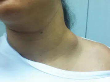
Fig 1. Color photograph showing left-sided neck mass.
Fig 2. (A) Coronal T2 weighted magnetic resonance imaging showing the brachial plexuses bilaterally irregular, enlarged and hyperintense, resembling a “bag of worms”. (B) Sagittal T2 sequence shows the extension of PNs into the spinal canal with indentation of the spinal cord at the C3–C6 level. (C) Axial T2 weighted magnetic resonance imaging showing many hyperintense nodules in the cervical paravertebral space.
he treatment of PNs in patients with NF-1 consists of regular follow-up examinations involving serial physical eval-uations and neuroimaging studies2. his was the conduct used in the present case.
B
A
C
Fig 4. (A) Axial T2 weighted MRI of the lumbar spine showing a large mass in the lumbar paravertebral space. (B) Axial T2 image showing deep, nodular neuroibroma of sciatic nerve in pelvis and gluteal region.
B
A
Fig 3. (A) Coronal T2 weighted MRI showing extensive PNs extending along the course of multiple nerve roots. (B) Sagittal T2 weighted MRI of the lumbar spine showing a large mass with multiple areas of low signal intensity centrally and high signal intensity peripherally. This inding is referred to as the target sign. (C) 3D CT scan reconstruction showing widening of the neural foramina at multiple levels of the lumbar spine.
B
1. Williams VC, Lucas J, Babcock MA, et al. Neuroibromatosis Type I Revisited. Pediatrics 2009;133:123-124
2. Serletis D, Parkin P, Bouffet E, et al. Massive plexiform neuroibromas in childhood: natural history and management issues. J Neurosurg 2007;106:363-367
3. Al-Otibi M, Rutka JT. Neurosurgical implications of neuroibromatosis type I in children. Neurosurg Focus 2006;19:E2
4. Krandsdorf MJ, Murphey MD. Neurogenic tumors. In: Krandsdorf MJ (Ed). Imaging of soft tissue tumors. 2nd ed. Philadelphia, USA: Lippincott Williams & Wilkins, 2006:335-348.
5. Anghileri M, Miceli R, Fiore M, et al. Malignant peripheral nerve sheath tumors: Prognostic factors and survival in a series of patients treated at a single institution. Cancer 2006;107: 1065-1074.