*e-mail: [email protected]; [email protected] Recebido: 27/01/2013 / Aceito: 28/05/2013
Web-based neuromuscular simulator applied to the teaching of
principles of neuroscience
Leonardo Abdala Elias*, André Fabio Kohn
Abstract Introduction: The learning of core concepts in neuroscience can be reinforced by a hands-on approach, either experimental or computer-based. In this work, we present a web-based multi-scale neuromuscular simulator that is being used as a teaching aid in a campus-wide course on the Principles of Neuroscience. Methods: The simulator has several built-in individual models based on cat and human biophysics, which are interconnected to represent part of the neuromuscular system that controls leg muscles. Examples of such elements are i) single neurons, representing either motor neurons or interneurons mediating reciprocal, recurrent and Ib inhibition; ii) afferent ibers that can be stimulated to generate spinal relexes; iii) muscle unit models, generating force and electromyogram; and iv) stochastic inputs, representing the descending volitional motor drive. Results: Several application examples are provided in the present report, ranging from studies of individual neuron responses to the collective action of many motor units controlling muscle force generation. A subset of them was included in an optional homework assignment for Neuroscience and Biomedical Engineering graduate students enrolled in the course cited above at our University. Almost all students rated the simulator as a good or an excellent learning tool, and approximately 90% declared that they would use the simulator in future projects. Conclusion: The results allow us to conclude that multi-scale neuromuscular simulator is an effective teaching tool. Special features of this free teaching resource are its direct usability from any browser (http://remoto.leb.usp.br/), its user-friendly graphical user interface (GUI) and the preset demonstrations.
purpose experimentation. Besides, experiments (even simple ones) may require highly specialized equipment and devices, which are frequently available in research labs only. At the same time, biologically plausible mathematical models have become largely accessible by means of public databases (Morse, 2008) including those that are able to represent
with idelity several aspects of the nervous and
musculoskeletal systems. This, allied to the increased power of personal computers, allows the student to reinforce his/her learning through active computer-based experimentation.
General-purpose simulators have been developed either with a focus on computational neuroscience or on motor control/biomechanics (e.g., (Bower and Beeman, 1998; Brette et al., 2007; Carnevale and Hines, 2006; Delp et al., 2007)). These packages can also be used for teaching or learning purposes; however, both the student and the instructor must have some familiarity with programming languages and a reasonable effort is needed to develop the structure to be studied. Moreover, the integration between different subsystems (primarily, the link between the nervous and the musculoskeletal systems) may be an arduous task, which may divert the instructor and student from the main focus.
Different multi-scale models have already been developed for the study of the neuromuscular system (Bashor, 1998; Fuglevand et al., 1993; Raphael et al., 2010; Winters, 1995). Nonetheless, these models were mainly intended to be used as tools for research labs and frequently their source-codes are not readily available or they are platform-dependent, limiting their applicability as educational tools. The large-scale neuromuscular system model developed by Maw et al. (1996) was primarily intended for education; however, the source code is not available and it is platform-dependent. Conversely, the simulator for neural networks described in (Ziv et al., 1994) is being used for research and educational purposes, but it’s focus is only on neural mechanisms. Therefore,
be used to explore the intricacies of the generation of
muscle force and muscle electrical activity. The irst
version of this neuromuscular system was intended for computational neuroscience research (Cisi and Kohn, 2008). The recently redesigned graphical user interface (GUI) and the inclusion of novel experimental protocols (called “Demonstrations” in the simulator) opened the way for its use for educational purposes. Several application examples will be provided after the presentation of the main details of the simulator, ranging from single cell behavior to motor behavior. Finally, the outcomes of a survey conducted on students from the Principles of Neuroscience course (see Course description in Methods) will be presented. Part of this work was previously presented as an abstract (Elias et al., 2012b).
Methods
Course description
The Principles of Neuroscience course is offered at the graduate entry level for students coming from different backgrounds (e.g., engineers, physical therapists,
biologists). The course is oficially offered in both the
Electrical/Biomedical Engineering Graduate Program and in the campus-wide Neuroscience Graduate Program at the University de São Paulo (USP, São Paulo, Brazil). The topics covered include: structure of the nervous system, structure of nerve cells and their membranes, ionic channels in the neuronal membrane, the ionic basis of the resting potential and the action potential, presynaptic and postsynaptic mechanisms in synaptic function, LTP and synaptic plasticity in memory and learning, sensory receptors in the skin and muscles, muscle contraction mechanisms, basics
of spinal cord neural circuits and their inluence on
the control of muscles acting around a joint, the inner ear (vestibular and cochlear functions), electrical activity of the cortex and sleep.
the students. They are required to come to class with a
pre-speciied topic of the textbook well studied at home.
The instructor pinpoints the most important concepts of that lecture and answers the questions posed by the students. At the end of randomly selected classes, there is an essay-based mini-exam. Therefore, the students come to every class with the subject matter well read
beforehand. At the end of the course there is a inal
exam. There are two optional computer simulation home-works, one based on the Hodgkin-Huxley model (Hodgkin and Huxley, 1952; Touretzky et al., 2003) and the other based on ReMoto. A student’s performance in these home-works contributes with
a small percentage to the inal grade.
Description of the neuromuscular model
General structure
A complete description of the mathematical models implemented in the simulator may be found elsewhere (Cisi and Kohn, 2008; Elias et al., 2012a). Briely, the simulator provides a detailed modeling of four spinal motor nuclei that command leg muscles responsible for ankle extension (Soleus – SOL; Gastrocnemius Medialis – GM; Gastrocnemius Lateralis – GL) and
ankle lexion (Tibialis Anterior – TA). Each nucleus
encompasses a motor neuron (MN) pool and spinal interneurons (INs) mediating recurrent inhibition (Renshaw cells – RC), reciprocal inhibition (Ia inhibitory INs that receive inputs from antagonist muscles), and Ib inhibition. Ia and Ib afferents are
present in ReMoto to allow studies of spinal relexes (e.g., H-relex) generated by electrical stimulation
applied to a nerve (Posterior Tibial Nerve for SOL, GL and GM; Common Peroneal Nerve for TA). This feature of nerve stimulation is useful for the understanding of some electrophysiological tests applied in the neurology clinic. Default numbers of spinal neurons and afferent axons were based on estimates from human and cat literature, but these values can be changed according to the application being studied (see examples in Results section). The MN pool drives muscle units, which generate both electrical (motor unit action potentials – MUAPs) and mechanical activity (force twitches). For each muscle, one output is the EMG, expressed as the sum of all MUAPs, and the force, which is the sum of all muscle twitches. Figure 1 summarizes the network structure of two motor nuclei (SOL and TA) and the pathways mediating monosynaptic Ia excitation, Ib inhibition, recurrent inhibition, and reciprocal inhibition. It is interesting to note that these modeled neural circuits
may inluence each other by means of the well-known
reciprocal inhibitory pathways.
Each type-speciied (i.e., S, FR and FF) MN
model is represented as two cylindrical compartments associated, respectively, with the soma and the dendritic tree. The somatic compartment encompasses Na+ and fast K+ conductances, responsible for the genesis of action potentials, and a slow K+ conductance yielding the afterhyperpolarization (AHP). Recently, an L-type Ca++ channel was added to the dendritic compartment to account for the persistent inward current (PIC), which is activated by the presence of neuromodulators (e.g., serotonin and norepinephrine) in the spinal cord (Elias and Kohn, 2013; Heckman et al., 2009). The level of neuromodulation can be adjusted to set the PIC magnitude during the simulation of different motor tasks (Elias and Kohn, 2013; Elias et al., 2012a). In addition, synaptically activated conductances (excitatory and inhibitory) are included in both somatic and dendritic compartments. INs are modeled as a single compartment (point models) with conductances similar to those included in the MN soma, but without PIC-generating conductances. The dynamic behavior
of each conductance is a simpliication of the
Hodgkin-Huxley formalism (Destexhe, 1997) that was adopted to speed up the simulations. Parameter values were chosen so that individual neuron models matched experimental data from cats (e.g., AHP time course;
iring rate versus injected current relation). The synaptic mechanisms are modeled following a kinetic model (Destexhe et al., 1994) with facilitation and depression dynamics, i.e., the amplitude of post-synaptic potentials can increase or decrease, respectively, after repetitive activations. A spike generated by a MN model reaches the MU end plate after a randomly chosen delay value, representing the effect of axonal conduction velocity. Similarly, there are delays in synaptic transmission
from descending commands and afferent ibers to
spinal neurons.
The MUAPs are modeled by Hermite-Rodrigez functions (Lo Conte et al., 1994; Zhou and Rymer, 2004), which are summed to generate the EMG. A
band-pass ilter is applied to the signal in order to
reproduce in the simulator what happens in a real bipolar surface EMG acquisition. The MU twitch is modeled as the impulse response of a critically damped second-order system which approximates the behavior obtained from human muscles (Milner-Brown et al., 1973).
In ReMoto, two nerves that supply the Triceps Surae (SOL, GM, and GL) and the TA can be electrically stimulated (short current pulses) as done in clinical neurophysiology, to obtain M-waves and
H-relexes. These electrical response signals are
(Pierrot-Deseilligny and Burke, 2012). In addition, electrical stimuli may be intracellularly applied to spinal neurons with different waveforms (step, pulse, sinusoid, ramp, and random signal) for studies of single neuron dynamics as done in invasive animal experiments. In order to mimic volitional force control, the MN pool may be driven by descending tracts whose spike trains are modeled as nonhomogeneous renewal point processes with Gamma-distributed interspike
intervals, which provide a lexible and realistic
stochastic source for the spinal neurons (Deger et al., 2012). Point process intensities may be modulated by different mathematical functions (e.g., sinusoid) in order to reproduce dynamic motor behaviors.
The simulator was developed in Java
(Oracle) programming language, as a web application (accessible at http://remoto.leb.usp.br/). It uses open
source technologies, which include i) Eclipse as the development environment ii) Tomcat as the servlet container iii) HSQLDB as the database manager system iv) Struts as the web framework v) and JFreeChart as the chart library. ReMoto is a multi-platform application that can be accessed from any web browser with support to the Java
virtual machine, including those embedded in mobile devices (e.g., smartphones and tablets). The user can also work in a logged-in
mode in which he/she could create speciic simulation
scenarios that remain available for his/her future studies. A series of default scenarios are provided to
assist irst-time users as well as instructors.
Graphical user interface (GUI)
The GUI was greatly improved in the new version of the simulator in comparison with the previous Figure 1. Schematic of the simulator. This igure shows only two of the motor nuclei, one for the Soleus (SOL) and the other for the Tibialis Anterior (TA) muscle. The descending commands, responsible for ankle extension and/or ankle lexion, activate the α-MN pools and spinal
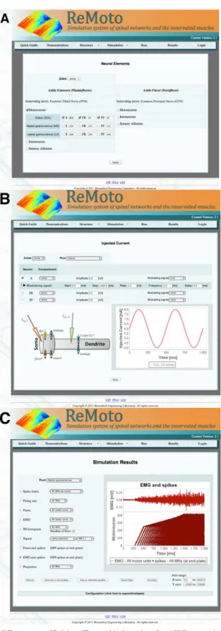
one, which was mainly intended for computational neuroscience research (see Figure 2 in (Cisi and Kohn, 2008)). The new GUI is designed with interactive menus representing more general structures (e.g., number of neuronal elements as shown in Figure 2A)
and internal menus permitting access to more speciic
elements. In addition, the user can now visualize the selected input waveforms and/or the statistical characteristics of these signals when setting up the
simulation coniguration (see Figure 2B), improving the control over what one wants to simulate. All these new features increased the potential use of ReMoto as a teaching tool.
Visualizing and exporting output data
Several output data can be visualized directly from the simulation environment (see Figure 2C). An interesting additional feature of the most recent version is the possibility to visualize the output data in an interactive graph (by means of an Applet, which is a Java application embedded in the HTML
code), with zooming and dragging capabilities.
However, if the user wants to perform a more speciic
analysis of the simulated data (e.g., peri-stimulus time histograms, EMG envelope, etc.) he/she may export the data in ASCII format, which is readable by most math programs (e.g., MatLab) or spreadsheets
(e.g., MS Excel).
Results
Application examples
Single neuron responses: action potential, synaptic facilitation, refractory period, and frequency-current relationship
The first set of examples will show how the neuromuscular simulator can be used to illustrate basic behaviors of a single neuron in response to different stimuli. Figure 3A shows simulation results from a single neuron (S-type MN from the SOL motor nucleus), which received synaptic inputs from Ia afferents of the PTN. Five electrical pulses stimulated this nerve so that the presynaptic input generated excitatory post-synaptic potentials (EPSPs) on the target neuron. The temporal summation of these EPSPs leads to membrane potential depolarization up to a given level (threshold) from which the action potential (AP) is generated by the ionic channel dynamics (see the inset in Figure 3A). The rising phase (or depolarization) of the AP occurs due to the opening of Na+ channels while its falling phase (or repolarization) is due to the opening of K+ channels,
as well as the closing of Na+ channels (inset of Figure 3A). This is the basic mechanism of AP
generation that was irstly described in the seminal
paper by Hodgkin and Huxley (1952). However, in a large variety of neurons (e.g., MNs) one can observe a hyperpolarization following the AP (referred to as the AHP), which is mainly due to a slow K+ channel, and
is functionally signiicant in regulating the neuronal iring rate during a sustained activity (Kernell, 2006). All these mechanisms can be easily explored using the simulator, and, in addition to the somatic membrane potential, one can monitor the dendritic membrane potential, which is the main location for synaptic inputs (see the red curve in Figure 3A).
A quite interesting topic covered in Neuroscience courses is the synaptic plasticity (Kandel et al., 2013). A kind of short-term synaptic plasticity is synaptic facilitation, which is an increase in EPSP amplitude during repetitive presynaptic activity. The main cause of this phenomenon is an excess (or residue) of Ca++ ions within the presynaptic membrane, leading to an increase in neurotransmitter release. Using the simulator, one can investigate how the synaptic facilitation time course (which may be easily changed)
inluences the genesis of an AP. Figure 3B shows an example of the effect of synaptic facilitation on the genesis of an AP. As the EPSP amplitude increases after each presynaptic spike, the AP threshold is reached earlier. The student/instructor can also simulate the opposite effect, i.e., synaptic depression (not shown here) that occurs during long-lasting presynaptic activity (Kandel et al., 2013; Kohn et al., 1997).
After the genesis of an AP, a second spike can only be generated after a certain minimal time interval referred to as the refractory period (Tr). This low-excitability period of the neuron is due to the AHP and to the Na+ channel inactivation (Kandel et al., 2013). The double pulse experiment illustrated in Figure 4 is frequently used to evaluate this property.
The irst stimulus pulse is injected into the neuron’s
soma so as to generate an AP. The second pulse is injected after a delay and having an amplitude which is barely supra-threshold. The minimal delay value that
results in the iring of an AP by the second stimulus pulse deines Tr (see the small blue and red bars in Figure 4 indicating the two stimulus pulses that
were able of iring the neuron once per pulse). If the
Figure 2. Screenshots taken from different parts of ReMoto. The graphical user interface (GUI) was entirely redesigned in comparison with
the irst version of the simulator (Cisi and Kohn, 2008). (A) Selection of neuronal elements: In this screen, the user can select the elements
that will be used in a speciic simulation. (B) Coniguration of the injected current: The graph illustrates the current waveform that is injected
A inal example concerning the basic mechanisms
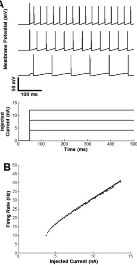
of single neurons is the response to steady currents injected through the cell’s membrane. Figure 5A shows qualitatively that the higher is the amplitude of the
injected current the higher is the neuron’s iring rate
(APs per seconds). This is valid for a great variety of neurons in the nervous system and the instructor may point out that many sensory receptors have their Figure 3. (A) Temporal summation of excitatory post-synaptic potentials (EPSPs) leading to action potential (AP) generation when the
somatic membrane crosses a iring threshold. The AP iring depends on the ionic conductances activated (inset). Depolarization depends
on the Na+ conductance (g
Na) and repolarization on both gNa and fast K
+ (g
Kf). A slow K
+ conductance (g
Ks) yields the afterhyperpolarization
(AHP). All these variables are readily available as simulation outputs. (B) Somatic membrane time courses in two conditions: without and with synaptic facilitation. Note short-term increases in EPSP amplitudes during facilitation, which makes the membrane potential reach
functioning based on a monotonic relation between
iring rate and input intensity, as shown in Figure 5B. For more advanced students, the instructor may point out that in some cases the relation between
the neuron’s iring rate (f) and the injected current (I) – referred to as f-I curve – may be approximately linear (Figure 5B), as in anaesthetized cats (Kernell, 2006), and at other times it will be nonlinear as in decerebrate non-anaesthetized cats or in different animals after administration of neuromodulators (e.g., serotonin or norepinephrine) (Elias and Kohn, 2013; Kernell, 2006)).
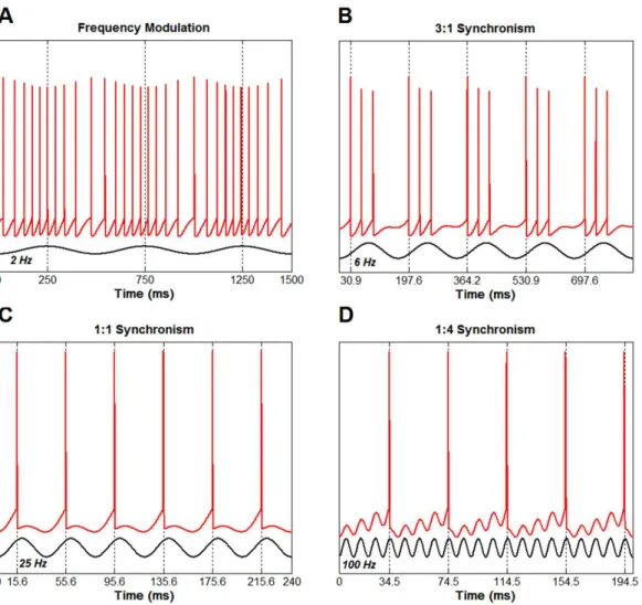
Frequency modulation and phase locking
This application, illustrated using a single neuron, is more challenging since it evidences the highly nonlinear behavior of the neuron as an information-processing unit. Initially, a steady current is injected into the neuron so as to maintain an approximately
constant iring rate (~20 Hz). Next, a sinusoidal
modulation is superimposed to the steady current and will be called simply the input to the neuron.
For very low input frequencies (2 Hz in Figure 6A)
the neuron’s iring rate is sinusoidally modulated, i.e.,
the output rate follows the input signal. This relation
between the neuron’s output iring rate and the neuron’s
input amplitude is the so-called rate coding principle of sensory neurophysiology (Kandel et al., 2013). As the input frequency increases, a nonlinear behavior starts to emerge. For instance, for a 6 Hz input frequency (Figure 6B) the neuron output synchronizes with the input in a 3:1 fashion, i.e., the neuron discharges three APs at every cycle of the sinusoidal input. It is worth noting that the discharge burst occurs at the
same phase of the sinusoid, characterizing the phase locking phenomenon. For modulation frequencies in a range around the natural frequency of the neuron (i.e., 20 Hz), a 1:1 synchronism is observed (note that one AP is generated at each cycle of the sinusoid in Figure 6C). Finally, for frequencies well above the natural frequency, a 1:M (with M representing the number of input cycles) synchronism is observed. For example, if the modulation frequency is set at 100 Hz (Figure 6D) for the neuron used in this application, a 1:4 synchronism is achieved. This intricate phenomenon, whose mechanisms are out of the scope of this paper, has been described in many neural systems, such as in invertebrate sensory neurons (Martinez et al., 1983) and neurons of the auditory system (cochlear nerve Figure 4. Double pulse paradigm used to evaluate the refractory
period of a neuron. When the interpulse interval (IPI) is about or higher than 55 ms (small gray vertical bars at the bottom) the neuron discharges a second spike; however, for lower IPIs the second pulse (barely supra-threshold) does not generate a spike (see EPSP following the black vertical bar) due to the refractory period of the neuron.
Figure 5. (A) A steady current injected into the neuron’s soma evokes
repetitive discharges with a certain frequency (iring rate). The higher the injected current amplitude, the higher the neuron’s iring rate. (B) Relationship between injected current and iring rate for a
these ibers will respond to weak stimuli, generating a relexive wave that can be recorded from the muscle
EMG. Starting at some higher stimulus intensity, a few motor axons will also be activated generating a direct motor response (termed M-wave and occurring
at a smaller latency) while the H-relex will attain its
maximal amplitude. For stimulus intensities higher than
this causing the maximum H-relex, the antidromic
APs generated in the motor axons will annihilate the
orthodromic APs generated by the relex. This will lead to a decrease in H-relex amplitude. These mechanisms
lead to a recruitment curve of the simulated responses as shown in Figure 7A, where the ordinate is the
H-relex (or M-wave) normalized amplitude and the
abscissa is the stimulus intensity. The data points in
ibers) that code the incoming sound information
(Kandel et al., 2013).
H-relex and reciprocal inhibition
The H-relex is a common electrical response used
in clinical neurophysiology and in human research to estimate the excitability of the spinal cord (Daube and Rubin, 2009; Pierrot-Deseilligny and Burke, 2012).
Basically, the H-relex is generated by activating
the monosynaptic pathway encompassing the Ia afferents, spinal MNs and the muscle. In this technique, the peripheral nerve is electrically stimulated by a rectangular pulse (1 ms duration), which evokes a
synchronous discharge in the ibers. As Ia afferents
have larger diameters than motor axons, a fraction of
Figure 6. Frequency modulation and phase locking between the input and output of a single neuron model. (A) For a low-frequency sinusoidal
input, the neuron’s iring rate approximately follows the input signal imposed on the neuron. (B) 3:1 synchronism is shown when the input
effect of the RI on the H-relex by applying a test
stimulus to the PTN and a conditioning stimulus to the CPN (antagonist nerve; see Figure 1) 2-3 ms before the test. Gray curves in Figure 7B represent
valuable (and unavailable from human experiments) to illustrate what happens to the neuronal population
during studies of spinal relexes.
Recruitment and rate coding in force generation
During a given motor task, two fundamental mechanisms are involved to increase the force level (Heckman and Enoka, 2012): i) the recruitment of new motor units, and ii) previously recruited motor units
increase their iring rate, called rate coding in motor
control. Both mechanisms may be easily studied using the neuromuscular simulator presented in this work.
Figure 8 shows the effects of a linearly rising command (1500 ms duration) imposed on the GM muscle and the resulting force increase in the upper panel. The shadowed box represents the recruitment range, i.e., the force range when additional (higher threshold) motor units are being recruited as the contraction increases. After the end of the recruitment range, the increase in force is due solely to the increase
in motor unit iring rate. It is worth noting that for this muscle new motor units are recruited up to ~80%
Figure 7. (A) H-relex recruitment curve. Black circles represent
the peak-to-peak amplitude of the direct motor M-wave, whereas empty circles correspond to peak-to-peak amplitude (in percentage
of the maximum M-wave) of the H-relex. (B) Effect of reciprocal inhibition (RI) on H-relex amplitude. Top panel shows simulated
EMGs (n = 5) evidencing small M-waves and the H-relexes. Gray
curves represent control conditions in which the RI pathway was not
activated. Black curves show that the H-relexes were reduced due
to the conditioned stimulus applied to the Common Peroneal Nerve (CPN). Bottom panel shows two graphs with raster plots for one control and one RI condition (a raster plot shows for any MN of the pool, numbered in the ordinate, when it discharges in the time axis, as indicated by a single dot). Note the lower number of recruited MNs in RI condition.
Figure 8. Recruitment and rate coding in force control. During a
slowly rising contraction, new motor units are recruited up to ~80% of the maximum voluntary contraction (MVC). Gray box deines the
recruitment range in this case. The increase in muscle force observed above the full motor unit recruitment (to the right of the box) is due
of the maximum “voluntary” contraction (MVC). The graph in the middle panel of Figure 8 shows the simulated EMG. Note the increase in EMG intensity as the contraction progresses.
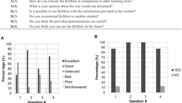
Student feedback
A survey was conducted on a subset of eight graduate students that took the Principles of Neuroscience course in 2012 at the USP. The questionnaire (Table 1) asked them about their opinion about ReMoto used in the second optional homework which explored several
concepts, including H-relex and force control (similar to the examples presented above). Identiication ield
was not provided to avoid bias in the responses. Figure 9 shows the survey outcomes. Almost all students rated the simulator as a good or an excellent learning tool, even when comparing with other simulators frequently used. They considered the graphical user interface adequate to be used by any student, but some (3) recommended a tutorial on the use of the simulator or onscreen tips so as to
help the user in coniguring a simulation. It should
be noted that all the surveyed students completed successfully the proposed homework. All the students recommended the use of ReMoto as a learning tool
for other students, and approximately 90% wrote
that they would use the simulator in future projects.
Discussion
In this paper, we presented an open-source web-based neuromuscular simulator, ReMoto, which may be a helpful aid to the teaching of principles of neuroscience. The multi-scale structure of the simulator allows one to explore concepts ranging from single cell dynamics to whole muscle control, generating force
and EMG. Due to its GUI, simulation coniguration
and data visualization are more straightforward than the previous version reported elsewhere (Cisi and Kohn, 2008). Its availability as a web application is a distinguishing characteristic in comparison to other packages (e.g., (Bower and Beeman, 1998; Brette et al., 2007; Carnevale and Hines, 2006; Delp et al., 2007)), mainly when viewed from the didactic perspective.
One of the guiding lights for the construction of this software was its usage by persons without a background on mathematical modeling and/or programming. The advantage of this approach is that all the structure needed for the simulation of a given neurophysiological experiment is already implemented. The user should only select the appropriate elements encompassing
Table 1. Questionnaire used in the survey. Questions were separated in two blocks: In block A the students rated the simulator as Excellent, Good, Irrelevant, Bad, and Poor; Block B comprised Yes/No questions.
Block(#) Question
A(1) What is your opinion about the graphical user interface (GUI) of the simulator?
A(2) What is your opinion about the ReMoto as a learning tool?
A(3) How do you classify the ReMoto in comparison to other learning tools?
A(4) What is your opinion about the way results are presented?
B(1) Is it possible to use ReMoto with the information provided in the website?
B(2) Do you recommend ReMoto to another student?
B(3) Do you think the provided demonstrations are useful?
B(4) Do you think you can use the ReMoto in the future?
its inherent power, the simulator could be used also in courses on Neural Engineering, Motor Control or Computational Neuroscience, as well as in research (Cisi and Kohn, 2008; Elias et al., 2012a). The instructor may use ReMoto in real time during classes
to illustrate speciic topics that are being taught (this
has been done in previous years in the Principles of Neuroscience course mentioned before). At the same time, for the student, ReMoto may serve as an adjunct tool in the learning both at home or inside the classroom. It may act as a virtual laboratory in which the tutor (instructor) provides a challenging
experiment and the student tries to igure out the
best way to solve and interpret the problem using the knowledge acquired from the theory. In fact, several disciplines, including neurosciences, are being taught using a problem-based (or hands-on) approach (Deslauriers et al., 2011; Hosseinzadeh and Hesamzadeh, 2012; Wiznia et al., 2012). Technologies such as that presented here are in accordance with this teaching/learning method and may be used to improve education in neuroscience, neural engineering
and related ields.
The outcomes of the survey conducted on graduate students from the Principles of Neuroscience course suggest that ReMoto was an effective resource. The (few) criticisms received are being used to improve future versions of the simulator, as well as in the development of additional documentation and video tutorials. A detailed quantitative evaluation of the
eficacy of ReMoto as a learning tool may be a topic
for a future study. Nonetheless, this preliminary result from a relatively small number of students was relevant to highlight the strengths and weaknesses of the simulator and was useful as a guide for future improvements.
The examples shown here (see Application examples in Results section) are just a small fraction of a multitude of possible scenarios that can be simulated in ReMoto. Several other examples frequently covered in principles of neuroscience courses can be also evaluated, for instance: i) drug effects on ionic
which is important to represent the spatial location of dendritic ionic channels (ElBasiouny et al., 2005) as well as synaptic dispersion (Segev et al., 1990); ii) the muscle force has no inluence from intrinsic viscoelastic properties (Zajac, 1989); and iii) the simulator operates in open-loop, i.e., there is no proprioceptive feedback from peripheral receptors. Nevertheless, the system is being continuously incremented, and several new features are under way. A recent study from our lab (Chaud et al., 2012) used a beta version (not yet available as a web-application) to evaluate the short-latency component
of the stretch relex of the SO muscle (Cronin et al., 2008). This new system included: muscle spindle model (Mileusnic et al., 2006) providing the proprioceptive feedback and a Hill-type muscle model (Zajac, 1989),
with viscoelastic properties of muscle ibers and tendon.
In the future, several of these new elements will be made available on the web application (as part of the ReMoto simulator) and several new examples may emerge for the teaching of fundamental mechanisms regarding the neuromusculoskeletal system.
Acknowledgements
This study was supported by grants from FAPESP and CNPq (Brazilian Funding Agencies). LAE holds a scholarship from FAPESP (proc. no. 2009/15802-0). The authors are grateful to V. M. Chaud for his invaluable work in improving the simulator’s graphical user interface (GUI).
References
Bashor DP. A large-scale model of some spinal relex circuits. Biological Cybernetics. 1998; 78(2):147-57. PMid:9525039. http://dx.doi.org/10.1007/s004220050421
Bower JM, Beeman D. The book of Genesis: Exploring realistic neural models with the general neural simulation system. New York: Springer; 1998.
B, Djurfeldt M, Lansner A, Rochel O, Vieville T, Muller E, Davison AP, El Boustani S, Destexhe A. Simulation of networks of spiking neurons: A review of tools and strategies. Journal of Computational Neuroscience. 2007; 23(3):349-98. PMid:17629781 PMCid:2638500. http://dx.doi.org/10.1007/ s10827-007-0038-6
Cannon SC. Pathomechanisms in channelopathies of skeletal muscle and brain. Annual Review of Neuroscience. 2006; 29: 387-415. PMid:16776591. http:// dx.doi.org/10.1146/annurev.neuro.29.051605.112815
Carnevale NT, Hines ML. The Neuron book. New York: Cambridge University Press; 2006. http://dx.doi.org/10.1017/ CBO9780511541612
Chaud VM, Elias LA, Watanabe RN, Kohn AF. A simulation study of the effects of activation-dependent muscle stiffness on proprioceptive feedback and short-latency relex. In: 4th IEEE RAS/EMBS International Conference on Biomedical Robotics and Biomechatronics: Proceedings of the International Conference on Biomedical Robotics and Biomechatronics; 2012; Rome. Rome: IEEE; 2012. p. 133-8.
Cisi RRL, Kohn AF. Simulation system of spinal cord motor nuclei and associated nerves and muscles, in a Web-based architecture. Journal of Computational Neuroscience. 2008; 25(3):520-42. PMid:18506610. http:// dx.doi.org/10.1007/s10827-008-0092-8
Cronin NJ, Peltonen J, Ishikawa M, Komi PV, Avela J, Sinkjaer T, Voigt M. Effects of contraction intensity on muscle fascicle and stretch relex behavior in the human triceps surae. Journal Applied Physiology. 2008; 105(1):226-32. PMid:18483163. http://dx.doi.org/10.1152/japplphysiol.90432.2008
Daube JR, Rubin DI. Clinical Neurophysiology. 3rd. Oxford: Oxford University Press; 2009.
Deger M, Helias M, Boucsein C, Rotter S. Statistical properties of superimposed stationary spike trains. Journal of Computational Neuroscience. 2012; 32(3):443-63. PMid:21964584 PMCid:3343236. http://dx.doi.org/10.1007/ s10827-011-0362-8
Delp SL, Anderson FC, Arnold AS, Loan P, Habib A, John CT, Guendelman E, Thelen DG. OpenSim: open-source software to create and analyze dynamic Simulations of movement. IEEE Transactions on Biomedical Engineering. 2007; 54(11):1940-50. PMid:18018689. http:// dx.doi.org/10.1109/TBME.2007.901024
Deslauriers L, Schelew E, Wieman C. Improved learning in a large-enrollment physics class. Science. 2011; 332(6031):862-4. PMid:21566198. http://dx.doi.org/10.1126/science.1201783 Destexhe A. Conductance-based integrate-and-ire models. Neural Computation. 1997; 9(3):503-14. PMid:9097470. http://dx.doi.org/10.1162/neco.1997.9.3.503
Destexhe A, Mainen ZF, Sejnowski TJ. An eficient method for computing synaptic conductances based on a kinetic-model of receptor-binding. Neural Computation. 1994; 6(1):14-8. http://dx.doi.org/10.1162/neco.1994.6.1.14
Dolecek GJ. MATLAB-Based Program for teaching autocorrelation function and noise concepts. IEEE Transactions on Education. 2012; 55(3):349-56. http:// dx.doi.org/10.1109/TE.2011.2176736
ElBasiouny SM, Bennett DJ, Mushahwar VK. Simulation of dendritic Ca(V)1.3 channels in cat lumbar motoneurons: Spatial distribution. Journal of Neurophysiology. 2005; 94(6):3961-74. PMid:16120667. http://dx.doi.org/10.1152/jn.00391.2005
Elias LA, Chaud VM, Kohn AF. Models of passive and active dendrite motoneuron pools and their differences in muscle force control. Journal of Computational Neuroscience. 2012a; 33(3):515-31. PMid:22562305. http://dx.doi.org/10.1007/s10827-012-0398-4
Elias LA, Chaud VM, Watanabe RN, Kohn AF. A web-based neuromuscular simulator applied to the teaching of the basics of neuroscience. In: 42nd Annual Meeting of the Society for Neuroscience: Proceedings of the Annual Meeting of the Society for Neuroscience; 2012; New Orleans. New Orleans; 2012b. p. 23.06SA.
Elias LA, Kohn AF. Individual and collective properties of computationally eficient motoneuron models of types S and F with active dendrites. Neurocomputing. 2013; 99: 521-33. http://dx.doi.org/10.1016/j.neucom.2012.06.038
Fuglevand AJ, Winter DA, Patla AE. Models of recruitment and rate coding organization in motor-unit pools. Journal of Neurophysiology. 1993; 70(6):2470-88. PMid:8120594.
Heckman CJ, Enoka RM. Motor unit. Comprehensive Physiology. 2012; 2:2629-82. PMid:23720261.
Heckman CJ, Mottram C, Quinlan K, Theiss R, Schuster J. Motoneuron excitability: The importance of neuromodulatory inputs. Clinical Neurophysiology. 2009; 120(12):2040-54. PMid:19783207. http://dx.doi.org/10.1016/j. clinph.2009.08.009
Hodgkin AL, Huxley AF. A quantitative description of membrane current and its application to conduction and excitation in nerve. Journal of Physiology-London. 1952; 117(4):500-44.
Hosseinzadeh N, Hesamzadeh MR. Application of project-based learning (PBL) to the teaching of electrical power systems engineering. IEEE Transactions on Education. 2012; 55(4):495-501. http://dx.doi.org/10.1109/ TE.2012.2191588
Illert M, Wiese H, Wolfram U. Electroencephalography and evoked potentials: a PC-based analysis program for laboratory courses in physiology. Advances in Physiology Education. 1992; 263(6 Pt 3):S16-22.
Kandel ER, Schwartz JH, Jessell TM, Siegelbaum SA, Hudspeth AJ. Principles of neural science. 5th ed. New York: McGraw-Hill; 2013.
Kernell D. The motoneuron and its muscle ibres. New York: Oxford University Press; 2006. PMid:17054451. http:// dx.doi.org/10.1093/acprof:oso/9780198526551.001.0001
Kohn AF, Floeter MK, Hallett M. Presynaptic inhibition compared with homosynaptic depression as an explanation for soleus H-relex depression in humans. Experimental Brain Research. 1997; 116(2):375-80. http://dx.doi.org/10.1007/ PL00005765
Education. 2003; 27(3):120-9. PMid:12928321. http:// dx.doi.org/10.1152/advan.00055.2002
Mileusnic MP, Brown IE, Lan N, Loeb GE. Mathematical models of proprioceptors. I. Control and transduction in the muscle spindle. Journal of Neurophysiology. 2006; 96(4):1772-88. PMid:16672301. http://dx.doi.org/10.1152/jn.00868.2005
Milner-Brown HS, Stein RB, Yemm R. The contractile properties of human motor units during voluntary isometric contractions. Journal of Physiology. 1973; 228(2):285-306. PMid:4687100 PMCid:1331298.
Morita H, Petersen N, Christensen LO, Sinkjaer T, Nielsen J. Sensitivity of H-reflexes and stretch relexes to presynaptic inhibition in humans. Journal of Neurophysiology. 1998; 80(2):610-20. PMid:9705454.
Morse TM. Neuroinformatics: from bioinformatics to databasing the brain. Bioinformatics and Biology Insights. 2008; 2:253-64. PMid:19812780 PMCid:2735961.
Pierrot-Deseilligny E, Burke D. The circuitry of the human spinal cord: Spinal and corticospinal mechanisms of movement. Cambridge: Cambridge University Press; 2012. http://dx.doi.org/10.1017/CBO9781139026727
Raphael G, Tsianos GA, Loeb GE. Spinal-like regulator facilitates control of a two-degree-of-freedom wrist. Journal of Neuroscience. 2010; 30(28):9431-44. PMid:20631172.
Segev I, Fleshman JW, Burke RE. Computer-simulation of group Ia EPSPs using morphologically
Vincent A. Autoimmune channelopathies: well-established and emerging immunotherapy-responsive diseases of the peripheral and central nervous systems. Journal of Clinical Immunology. 2010; 30 Suppl 1:S97-102. PMid:20387102. http://dx.doi.org/10.1007/s10875-010-9401-x
Winters JM. An improved muscle-relex actuator for use in large-scale neuromusculoskeletal models. Annals of Biomedical Engineering. 1995; 23(4):359-74. PMid:7486344. http://dx.doi.org/10.1007/BF02584437
Wiznia D, Korom R, Marzuk P, Safdieh J, Grafstein B. PBL 2.0: enhancing problem-based learning through increased student participation. Medical Education Online. 2012; 17: 17375. PMid:22778540 PMCid:3374499. http://dx.doi.org/10.3402/meo.v17i0.17375
Zajac FE. Muscle and tendon: Properties, models, scaling, and application to biomechanics and motor control. Critical Reviews in Biomedical Engineering. 1989; 17(4):359-411. PMid:2676342.
Zhou P, Rymer WZ. MUAP number estimates in surface EMG: Template-matching methods and their performance boundaries. Annals of Biomedical Engineering. 2004; 32(7):1007-15. http://dx.doi.org/10.1023/ B:ABME.0000032463.26331.b3
Ziv I, Baxter DA and Byrne JH. Simulator for neural networks and action potentials: description and application. Journal of Neurophysiology. 1994; 71(1):294-308. PMid:7512628.
Authors
Leonardo Abdala Elias*, André Fabio Kohn