Blood gas analysis, anion gap, and strong ion difference in horses treated with
polyethylene glycol balanced solution (PEG 3350) or enteral and
parenteral electrolyte solutions
Hemogasometria, anion gap e diferença de íons fortes em equinos tratados com solução balanceada contendo polietilenoglicol (PEG 3350) ou soluções eletrolíticas enteral e parenteral
Cláudio Luís Nina GomesI José Dantas Ribeiro FilhoII* Rafael Resende FaleirosIII Fernanda Timbó D’el Rey DantasII Lincoln da Silva AmorimII
Waleska de Melo Ferreira DantasII ISSN 0103-8478
ABSTRACT
Large volumes of different electrolytes solutions are commonly used for ingesta hydration in horses with large colon impaction, but little is known about their consequences to blood acid-base balance. To evaluate the effects of PEG 3350 or enteral and parenteral electrolyte solutions on the blood gas analysis, anion gap and strong ion difference, fi ve adult female horses were used in a 5x5 latin square design. The animals were divided in fi ve groups and distributed to each of the following treatments: NaCl (0.9% sodium chloride solution); EES (enteral electrolyte solution), EES+LR (EES plus lactated Ringer’s solution); PEG (balanced solution with PEG 3350) and PEG+LR (PEG plus lactated Ringer’s solution). Treatments PEG or PEG + LR did not change or promoted minimal changes, while the EES caused a slight decrease in pH, but its association with lactated Ringer’s solution induced increase in AG and SID values, as well as caused hypernatremia. In turn, the treatment NaCl generated metabolic acidosis. PEG 3350 did not alter the acid-base balance. Despite it’s slight acidifying effect, the enteral electrolyte solution (EES) did not cause clinically relevant changes.
Key words: Acid-base balance, cathartic, fl uid therapy.
RESUMO
Grandes volumes de diferentes soluções eletrolíticas são comumente usados na hidratação da ingesta em equinos com compactação, mas pouco se sabe sobre suas consequências sobre o equilíbrio ácido base sanguíneo. Para avaliar os efeitos do PEG 3350 e soluções eletrolíticas enterais e parenterais sobre a hemogasometria, anion gap e diferença de íons fortes, foram utilizadas cinco fêmeas adultas em um quadrado latino 5x5. Os animais foram distribuídos em cinco grupos e submetidos a cada um dos seguintes tratamentos: solução NaCl (cloreto de sódio 0,9%); EES (solução eletrolítica enteral); EES + RL (solução eletrolítica enteral mais Ringer lactato); PEG 3350 (solução
balanceada com PEG 3350) e PEG + RL (PEG 3350 mais Ringer lactato). Os tratamentos PEG ou PEG + RL não alteraram ou promoveram alterações mínimas, enquanto a EES ocasionou discreta diminuição no pH, mas sua associação com Ringer lactato induziu o aumento nos valores do AG e DIF, além de ocasionar hipernatremia. Por sua vez, o tratamento NaCl resultou em acidose metabólica hiperclorêmica. O PEG 3350 não alterou o equilíbrio ácido base. Apesar do seu discreto efeito acidifi cante, a solução eletrolítica enteral (EES) não promoveu alteração clínica relevante.
Palavras-chave: Equilíbrio ácido base, catártico, hidratação.
INTRODUCTION
Large intestine impaction is one of the most frequent causes of colic in horse (DABAREINER & WHITE, 1995; RIBEIRO FILHO et al., 2012). Traditionally, treatment has been based on the administration of large volume of intravenous electrolyte solution in order to promote hydration of ingesta and faeces. Among the solutions for parenteral use in horses, lactated Ringer’s and 0.9% sodium chloride are probably the most common solutions used worldwide. Lactated Ringer’s solution (LR) has a polyionic composition similar to plasma, and is therefore commonly administered for general use (ROSE, 1981). LR has a slight alkalinizing effect in blood, which has already been described in horses (RIBEIRO FILHO et al., 2007) and dogs (RIBEIRO FILHO et al., 2008). On the other hand, the 0.9%
IDepartamento de Clínicas Veterinárias, Universidade Estadual do Maranhão (UEMA), Maranhã, MA, Brasil.
IIDepartamento de Veterinária, Universidade Federal de Viçosa (UFV), Avenida Peter Henry Holfs s/n, 36570-000, Campus Universitário,
Viçosa, MG, Brasil. E-mail: [email protected]. *Autor para correspondência.
sodium chloride solution can lead to hyperchloremic metabolic acidosis and has been indicated to most animals suffering from dehydration associated with sodium and chloride loss (ROSE, 1981; MATHEWS, 1998).
More recently, enteral electrolyte solution (EES) therapy has gain popularity as a direct and effective way to promote hydration of the ingesta (LOPES et al., 2002; RIBEIRO FILHO et al., 2012). Enteral fl uids are also quickly absorbed through intestine and expand blood volume, rehydrate the tissues and correct electrolytes imbalances (RIBEIRO FILHO et al., 2007). Another advantage is the possibility of the enteral solution to be formulated as needed by the patient, as well as it’s low cost. Despite its advantages, the enteral technique might also cause electrolyte and acid-base imbalances, depending on its composition (RIBEIRO FILHO et al., 2009).
Laxatives are substances that increase defecation frequency, fecal bulk, or alter the fecal consistency (CLARK & BECHT, 1987), and can also be used to treat large intestine impactions. Polyethylene glycol (PEG 3350) is an effective osmotic laxative used in humans (CLEVELAND et al., 2001). It has a high molecular weight, does not suffer tissue or bacterial degradation, and acts as a pure osmotic agent, being releasedquantitatively in the colon (SCHILLER, 1999). In Brazil, was launched recently the MunvilaxR, a laxative of polyethylene glycol 3350. In commercially available products for laxative use, PEG 3350 is combined with balanced amounts of sodium bicarbonate, sodium chloride and potassium in such way that the exchange of water and electrolytes with the blood is minimal (ATTAR et al., 1999), reducing the risk of dehydration that can occur with other osmotic laxatives. To our knowledge, there are currently no studies evaluating the use of this substance in veterinary medicine, with the exception of an experiment using cats (TAM et al., 2011). The aim of this study was to evaluate the effects of a standard PEG 3350 solution compared with other electrolyte solutions given by intravenous and enteral routes, on the acid-base balance, anion gap and strong ion difference.
MATERIAL AND METHODS
Five female crossbred horses aged between fi ve and nineteen years (mean = 12.5 years), with good body condition 392 ± 18kg (mean ± sd) were used. Mares had no history of intestinal disease in the last six months and were considered healthy based on physical examination, blood biochemistry and cell counts, and urinalysis. Ten days prior to the study, horses were dewormed (Ivermectin + Praziquantela,
PO) and received a deltamethrin spray bath (0.025%) for ticks and fl ies control. During all the research period horses were kept in individual stalls and were fed with commercial concentratedb (1% of bwt daily, divided into two meals), Tifton 85 hay (2% of bwt daily, two meals), 50g /day of a minerals mixturec and water “ad libitum”. During the treatment period horses were fasted (T0h to T12h).
Each mare was submitted to 5 different treatments in a 5x5 Latin square design, considering the effects period, treatment and time with the animal effect being random. The treatments were as follow: NaCl – received 0.9% sodium chloride solutiond administered intravenously in a dose of 15mL kg-1 h-1 for 12 hours under continuous fl ux. Osmolarity and pH of the solution: 309.80mMol L-1 and 5.60, respectively. EES – received enteral electrolyte solution containing 6g NaCl; 0.5g KCl; 1g calcium gluconate; 0.3g magnesium pidolatee; 5g maltodextrin in 1.000mL of water, via small-caliber nasogastric tube in a dose of 15mL kg-1 h-1 for 12 hours under continuous fl ux. Osmolarity and pH of the solution: 321.14mMol L-1 and 7.2, respectively. EES+LR – received enteral electrolyte solution (same composition of EES treatment), via small-caliber nasogastric tube in a dose of 7.5mL kg-1 h-1 for 12 hours under continuous fl ux, plus 7.5mL kg-1 h-1 of lactated Ringer’s solution administered intravenously for 12 hours under continuous fl ux. PEG – a single dose of PEG 3350f (1.5g kg-1) diluted in fi ve liters of water given via nasogastric tube. Osmolarity and pH of the solution: 252.66mMol L-1 and 8.65, respectively. PEG+LR – a single dose of PEG 3350 (1.5g kg-1) diluted in fi ve liters of water, given via nasogastric tube, plus 15mL kg-1 h-1 of lactated Ringer’s solution administered intravenously for 12 hours under continuous fl ux. Osmolarity and pH of the solution of lactated Ringer’s: 272.22mMol L-1 and 6.75, respectively. The interval between treatment periods was 14 days.
Baseline blood samples for blood gas and biochemical tests were taken immediately before the start of the treatment (T0h) and 6 (T6h), 12 (T12h), 24 (T24h) and 48 (T48h) hours after treatment beginning. For biochemistry analysis, samples were collected aseptically via jugular venipuncture, and stored in vials with sodium fl uoride or without anticoagulantg. Sodium and potassium serum concentrations were measure using fl ame photometryh while a multi-biochemical analyzerj was used to measure serum chloride and plasma L-lactate concentrations.
using previously heparinizedk 3mL disposable plastic syringes and 30x7 needles. A blood gas analyzerl was used to obtain the following variables: pH(v), oxygen partial pressure - pO2(v), carbon dioxide partial pressure - pCO2(v), carbon dioxide total concentration - tCO2(v), plasma bicarbonate concentration - cHCO
-3(v) and base excess - BE(v).The strong ion difference (SID) and anion gap (AG) were calculated using the respective formulas: SID (mMol) = (Na+ + K+) - (Cl- + lactate-); AG (mMol) = (Na+ + K+) - (Cl- + HCO
3
-) (CONSTABLE, 1999).
Data was analyzed using specifi c statistical software (SAEG UFV-01.09.2007). ANOVA was used to compare the effect of treatments within each time and the effect of times within each treatment. Post-hoc comparisons were made using the Tukey test. The nonparametric Kruskal–Wallis test was used when data did not meet the assumptions of normality or equality of variance. The null hypothesis was rejected when P<0.05.
RESULTS AND DISCUSSION
Our results showed that therapeutic protocols commonly used for ingesta hydration using intravenous or enteral routes can produce signifi cant changes in the blood acid-base and electrolyte balances in normal horses. On the other hand, the proposed protocol using PEG 3350 solution alone administered in a single dose did not alter any of the studied variables.
An important change in the physiologic balance occurred in NaCl group, where hyperchloremia (Table 1) with a concomitant decrease in blood pH were detected at the end of electrolyte solution administration (T12h). Normal saline (0.9% NaCl solution) has higher levels of chloride compared to plasma (MATHEUS, 1998) and administration of large volumes can cause changes in acid-base balance, i.e. hyperchloremic metabolic acidosis (CONSTABLE, 1999; PROUGH & BIDANI, 1999).Metabolic acidosis is traditionally divided into hyperchloremic (normal anion gap) and normochloremic (high anion gap) based on the anion gap and chloride (DeMORAIS & BIONDO, 2012). Chloride is the most prevalent strong anion in the ECF. At a constant [Na+], an increase in [Cl-] decreases SID causing hyperchloremic acidosis. The effects of increasing [Cl-] without changing [Na+] can be understood when considering a fl uid with an SID = 0 (e.g., 0.9% NaCl where [Na+] = [Cl-] and thus SID = [Na+] - [Cl-] = 0) (CONSTABLE, 2003), producing the occurrence of the acidifying effect of 0.9% NaCl, which can be seen in tables 1 and 2.
The traditional approaches for assessing blood gas analysis also demonstrated by the presence of metabolic acidosis in the animals of NaCl group, ie a decrease in pH, tCO2, HCO3 and base excess values compared with baseline was seen in animals of that group at T12h (Table 1 and 2), which was also coincident with a similar increase in the chloride levels (Table 3). The decrease in the concentration of HCO3, and thus in the base excess, occurred due to the compensation mechanism, when there is an increase of the chloride anion, the body increases the excretion of bicarbonate anion. Increases in serum chloride concentrations in horses after the enteral administration of electrolyte solutions were recorded by others (ECKE et al., 1998; ALVES et al., 2005). For other treatments of this research, despite the decrease in pH values , tCO2, HCO
-3 and base excess did not detect the presence of metabolic acidosis as can be seen in tables 1 and 2.
In T12h, pCO2 values declined (P<0.05) in treatments EES, compared to T6h, and NaCl, compared to T0h, with the lowest average for animals treated with NaCl (Table 2). According to CARLSON & BRUSS (2008), pCO2 decreases as a compensatory response of the respiratory component to metabolic acidosis. Therefore, in the same way that EES treatment caused a decrease in pH, tCO2, HCO3 and base excess, it also reduced pCO2, which recovered quickly after treatment completion. Emphasizing that despite the decrease, all variables remained within the reference, even in animals of NaCl treatment. In turn, there was no difference between treatments either in treatments over time nor in the values of pO2.
When using enteral electrolyte solutions containing carbohydrates as an energy source, there is the possibility of these substances to predispose to the onset of acidosis. This is because sugars and starches undergo bacterial metabolism in the gastrointestinal tract, generating organic acids (ZHANG et al., 2003). Among these acids are the isomers L-lactate and D-lactate (FALL & SZERLIP, 2005). Once absorbed in the digestive tract, the D-lactate metabolism is carried out at a very slow rate (FALL & SZERLIP, 2005), because mammals have little specifi c mechanisms for metabolizing D-lactate and the accumulation in the blood can cause metabolic acidosis (EWASCHUK et al., 2005). But the results of this experimental study show that the maltodextrin (10g L-1) present in treatments ESS and ESS+LR did not cause the appearance of this adverse effect.
T6h and T12h and in treatments throughout the experimental phase, making them without clinical signifi cance. The D-lactate was not measured in this study, however its level can be estimate through the anion gap (AG). The AG reference values for horses range from 5 to 16.2 mMol L-1 (CARLSON & BRUSS, 2008). It is used primarily to identify metabolic acidosis, confi rm the mixed disturbances and prognosis (GOSSETT et al., 1987). Higher rates to the reference range indicate metabolic acidosis due to an increase in unmeasured anions, especially the D-lactate, as occurs in lactic acidosis, determining the bicarbonate decrease (DeMORAIS & DiBARTOLA, 1993). As signifi cant changes in the AG values were not detected in the EES group, corroborates the assertion that the maltodextrin
administered at a dose of 10g L-1 does not cause the onset of metabolic acidosis (Table 1).
The animals receiving EES+RL treatment showed an important increase in sodium concentration at T12h (P<0.05). Hypernatremia is characterized by a value of serum sodium exceeding 147 mEq L-1 (STEWART, 1998). Although water loss through dehydration is considered the main cause of hypernatremia, it can also be caused by excessive sodium intake through hydration with the electrolyte solutions, such as sodium bicarbonate administered intravenously and also with 0.9% NaCl solution given enterally (BARBOSA & SZTAJNBOK, 1999). The increase of sodium concentration in the EES+LR at T12h (P<0.05) can be attributed to the fact that animals received sodium from three sources:
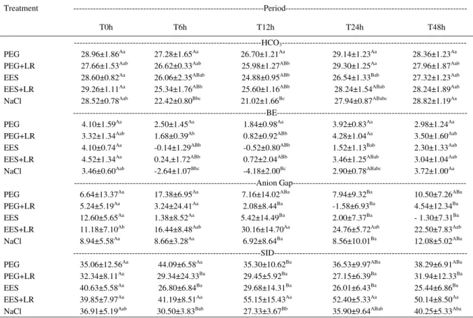
Table 1 - Means and standard deviations values of cHCO3- (mMol L-1), BE (mMol L-1), Anion Gap (mMol L-1) and SID (mMol L-1), in
venous blood (v) of five horses treated with PEG, PEG+LR, EES, EES+LR, and NaCl in pretreatment (T0h), treatment (T6h and T12h) and post-treatment (24h and T48h) periods.
Treatment
---Period---T0h T6h T12h T24h T48h
---HCO3
---PEG 28.96±1.86Aa 27.28±1.65Aa 26.70±1.21Aa 29.14±1.23Aa 28.36±1.23Aa
PEG+LR 27.66±1.53Aab 26.62±0.33Aab 25.98±1.27ABb 29.30±1.25Aa 27.96±1.87Aab
EES 28.60±0.82Aa 26.06±2.35ABab 24.88±0.95ABb 26.54±1.33Bab 27.32±1.23Aab
EES+LR 29.26±1.11Aa 25.34±1.76ABb 25.60±1.16ABb 28.24±1.54ABab 28.24±1.89Aab
NaCl 28.52±0.78Aab 22.42±0.80Bbc 21.02±1.66Bc 27.94±0.87ABabc 28.82±1.19Aa
---BE---PEG 4.10±1.59Aa 2.50±1.45Aa 1.84±0.98Aa 3.92±0.83Aa 2.98±1.24Aa
PEG+LR 3.32±1.34Aab 1.68±0.39Ab 0.82±0.92ABb 4.28±1.04Aa 3.50±1.60Aab
EES 4.10±0.74Aa -0.14±1.29ABb -0.52±0.80ABb 1.52±1.13Bab 2.30±1.33Aab
EES+LR 4.52±1.34Aa 0.24,±1.72ABb 0.72±2.04ABb 3.46±1.25ABab 3.04±1.04Aab
NaCl 3.46±0.60Aab -2.64±1.07Bbc -4.18±2.00Bc 2.90±0.78ABabc 3.72±1.00Aa
---Anion
Gap---PEG 6.64±13.37Aa 17.38±6.95Aa 7.16±14.02ABa 7.94±9.32Ba 10.50±7.26ABa
PEG+LR 5.24±5.19Aa 3.24±24.41Aa 2.08±8.44Ba -1.58±6.93Ba 4.54±12.34Ba
EES 12.60±5.65Aa 1.38±8.52Aa 5.42±14.49Ba 2.00±7.37Ba - 1.30±7.31Ba
EES+LR 11.18±7.10Ab 16.44±8.48Aab 30.16±14.70Aa 24.76±5.72Aab 22.50±7.83Aab
NaCl 8.94±5.58Aa 8.66±3.28Aa 6.92±8.64Ba 8.56±10.01Ba 12.08±5.02ABa
---SID---PEG 35.06±12.56Aa 44.09±6.58Aa 35.30±10.62Ba 36.53±9.97ABa 38.29±6.91ABa
PEG+LR 32.34±8.11Aa 29.34±24.33Ba 29.45±5.92Ba 27.15±6.39Ba 31.94±12.33Ba
EES 40.63±5.58Aa 26.80±6.84Ba 29.68±14.31Ba 26.01±6.43Ba 25.44±6.86Ba
EES+LR 39.85±7.97Aa 41.19±8.51Aa 55.15±15.43Aa 52.40±5.33Aa 50.14±8.50Aa
NaCl 36.91±5.19Aab 30.50±3.83Bab 27.33±3.67Bb 35.90±9.64ABab 40.25±5.33Aba
Means in the same column followed by equal capital letters and in the same row followed by equal lowercases letters do not differ at 5% level of probability by Tukey test.
PEG (Polyethylene glycol)
PEG+LR (PEG plus lactated Ringer' s solution) EES (enteral electrolyte solution)
the EES with 6g L-1 of sodium chloride, lactated Ringer’s solution containing 6g of sodium chloride and 3g of sodium lactate. The sum of them yielded in sodium concentration of 232.2 mMol L-1, which is higher than plasma’s (139±3.5 mMol L-1), resulting hypernatremia after their administration. As horses had access to water and food after the hydration phase (T24h and T48h), there was a gradual decrease in serum sodium during this time in animals of this group. Other study using isotonic enteral solution also reported hypernatremia due to the use of more than one sodium source (ALVES et al., 2005).
The hypernatremia observed in animals in the EES+RL at T12h was concomitant with the increase in the values of AG and SID (P<0.05) at time 12 hours (Table 1). This increase was caused
by the presence of high serum sodium concentration due to the composition of electrolyte solutions used in such treatment. As the AG and SID were calculated by the equations: AG = (Na+ + K+) - (Cl- + HC0
-3) and SID = (Na +
+ K+) - (Cl- + lactate), the increase in the concentration of serum sodium, explained above, determined the elevation of both values.Since the change in AG value was not commensurate with bicarbonate’s (Table 1) in the same period (T12h), one should suspect the presence of mixed disorders as quoted CARLSON & BRUSS (2012).
The treatments had no effect on serum potassium values that have remained unchanged throughout the experimental phase and within the reference range (KANEKO et al., 2008).
Although the protocol using PEG 3350 balanced solution (PEG) alone did not alter any of the
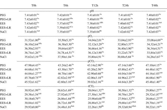
Table 2 - Means and standard deviations values of pH, pO2 (mmHg), pCO2 (mmHg) and tCO2 (mMol L-1) in venous blood (v) of five horses
treated with PEG, PEG+LR, EES, EES+LR, and NaCl in pretreatment (T0h), treatment (T6h and T12h) and post-treatment (24h and T48h) periods.
Treatment
---Period---T0h T6h T12h T24h T48h
---pH---PEG 7.41±0.01Aa 7.42±0.01Aa 7.41±0.01Aa 7.41±0.02Aa 7.40±0.03Aa
PEG+LR 7.42±0.02Aa 7.41±0.02ABa 7.40±0.01ABa 7.41±0.01Aa 7.40±0.02Aa
EES 7.43±0.02Aa 7.37±0.02CBb 7.38±0.01ABb 7.40±0.02Aab 7.41±0.01Aab
EES+LR 7.42±0.02Aa 7.39±0.02ACa 7.40±0.02Aa 7.42±0.02Aa 7.40±0.02Aa
NaCl 7.41±0.01Aab 7.35±0.03Cb 7.35±0.04Bb 7.42±0.02Aab 7.42±0.07Aa
---pO2
---PEG 31,52±1,80Ba 35,50±5,35Aa 34,04±2,01Aa 33,04±2,93Aa 35,08±4,69Aa
PEG+LR 36,10±2,64ABa 36,38±5,30Aa 32,12±3,29Aa 32,80±3,37Aa 34,32±4,21Aa
EES 36,50±2,93Aa 39,64±4,85Aa 36,66±4,16Aa 36,40±3,90Aa 36,34±4,51Aa
EES+LR 36,74±1,26Aa 34,78,±4,51Aa 36,12±3,90Aa 33,22±5,78Aa 33,84±3,03Aa
NaCl 35,62±3,19 ABa 37,50±1,84 Aa 35,80±4,91 Aa 30,88±5,68 Aa 34,26±3,67 Aa
---pCO2
---PEG 47.98±4.43Aa 43.24±2.46Aa 43.14±2.90Aa 47.14±3.60Aa 47.10±4.15Aa
PEG+LR 44.68±1.91Aa 43.30±1.70Aa 43.20±2.50Aa 45.88±2.43Aa 44.48±1.82Aa
EES 44.04±1.27Aab 46.70±1.66Aa 42.90±0.68Ab 44.04±3.04Aab 44.16±1.03Aab
EES+LR 45.76±0.75Aab 42.82±2.95Aab 42.06±3.18Ab 44.96±2.27Aab 46.68±1.90Aa
NaCl 45.52±1.62Aa 42.60±3.10Aab 39.02±2.16Ab 44.64±3.20Aa 45.48±1.40Aa
---tCO2
---PEG 30,92±1,99Aa 28,62±1,69Aa 28,04±1,32Aa 30,58±1,32Aa 29,80±1,27Aa
PEG+LR 29,38±1,54Aab 27,92±0,37Aab 27,30±1,34ABb 30,70±1,29Aa 29,32±1,87Aab
EES 30,18±0,79Aa 26,86±1,68ABb 25,98±0,68ABb 27,94±1,32Bab 28,66±1,27Aab
EES+LR 30,84±1.02Aa 26,72±1,88ABb 28,88±5,21Aab 29,68±1,67ABab 29,70±1,19Aab
NaCl 29,92±0,80Aa 24,48±1,81Bab 22,26±1,59Bb 29,32±0,94ABab 30,22±1,19Aa
Means in the same column followed by equal capital letters and in the same row followed by equal lowercases letters do not differ at 5% level of probability by Tukey test.
PEG (Polyethylene glycol)
PEG+LR (PEG plus lactated Ringer' s solution) EES (enteral electrolyte solution)
studied variables, the combination with lactate Ringer’s solution (PEG+RL) induced minor changes like decreases of cHCO3- in T12h, compared to T24h, and of BE in T6h and T12h, compared to T24h. Emphasizing that despite the decrease had been signifi cant, the values remained within the reference range (KANEKO et al., 2008), making them of no clinical signifi cance. Moreover, the values promptly returned to baseline levels in T24h (Table 1).
CONCLUSION
The PEG 3350 electrolyte solution did not interfere with the acid-base balance, and should be consider as therapeutic option for horses that need laxatives. Despite the slight acidifying effect, the proposed enteral electrolyte solution showed to be clinically safe to be used in normal horses. The association of intravenous
Ringer lactate with the used enteral electrolyte solutions or the infusion of intravenous 0.9% NaCl solution using doses and infusions rates presented in this study can cause important electrolytes and acid-base imbalances and should be used with caution in clinical cases.
ACKNOWLEDGEMENTS
ToFAPEMIG (Fundação de Amparo à Pesquisa do Estado de Minas Gerais).
SOURCES AND MANUFACTURES
a Padock NFgel® - Lab Vetbrands
b Equisul Special Ration 15 - Total Foods, Três Corações, MG c Hiposal 80% - Total Foods, Três Corações, MG
d Sodium chloride 0.9% - Texon e Pidomag® - Baldacci Laboratory, Brazil f Muvinlax - Libbs
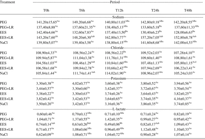
Table 3.- Means and standard deviations values of sodium (mMol L-1), chloride (mMol L-1), potassium (mMol L-1) and L-lactate (mMol L-1)
serum of five horses treated with PEG, PEG+LR, EES, EES+LR and NaClin pretreatment (T0h), treatment (T6h and T12h) and
post-treatment (24h and T48h) periods.
Treatment
---Period---T0h T6h T12h T24h T48h
---Sodium---PEG 141,20±15,65Aa 149,20±6,68Aa 140,80±11,03ABa 142,80±9,18ABa 142,20±8,55ABa
PEG+LR 137,40±8,88Aa 137,60±21,35Aa 138,40±5,13ABa 133,60±5,18Ba 137,60±11,33ABa
EES 142,40±4,66Aa 132,60±7,83Aa 137,40±13,50Ba 130,40±6,23Ba 128,00±6,63Ba
EES+LR 143,20±7,60Ab 148,20±6,30Aab 162,80±3,77Aa 157,20±7,05Aab 152,80±8,78Aab
NaCl 139,80±5,07Aa 139,40±3,58Aa 138,80±4,15ABa 141,60±8,68ABa 142,00±4,53ABa
---Chloride---PEG 108,90±4,33Aa 108,56±2,24Aa 108,56±2,22Ba 109,52±2,03Aa 107,28±4,10Aa
PEG+LR 109.94±5,87Aa 111,04±3,38Aa 111,78±1,31ABa 109,60±1,40Aa 108,80±1,61Aa
EES 104,58±1,03Ad 108,46±1,29Aab 110,84±1,60ABa 107,48±1,13Abc 105,80±1,15Acd
EES+LR 106,58±1,68Aa 109,84±2,78Aa 110,68±2,42ABa 107,94±2,69Aa 106,22±3,41Aa
NaCl 105,84±1,44Ac 111,74±1,41Aab 114,02±1,90Aa 108,96±2,07Abc 105,24±3,03Ac
---Potassium---PEG 3,30±0,38Aa 4,02±0,77Aa 3,60±0,38Aa 3,80±0,52Aa 3,94±0,56Aa
PEG+LR 3,44±0,57Aa 3,30±0,60Aa 3,42±0,37Aa 3,72±0,67Aa 3,70±0,54Aa
EES 3,38±0,22Aa 3,30±0,63Aa 3,74±0,26Aa 3,64±0,43Aa 3,82±0,25Aa
EES+LR 3,82±0,42Aa 3,42±0,53Aa 3,64±0,65Aa 3,74±0,35Aa 4,16±0,37Aa
NaCl 3,50±0,20Aa 3,42±0,37Aa 3,16±0,36Aa 3,86±0,35Aa 3,74±0,85Aa
---L-lactate---PEG 0,60±0,46Aa 0,79±0,12ABa 0,71±0,10ABa 0,71±0,24Aa 0,82±0,19Aa
PEG+LR 1,04±0,71Aa 1,37±0,55Aa 1,42±0,35Aa 0,99±0,27Aa 0,95±0,42Aa
EES 0,74±0,14Aab 0,63±0,26Bab 0,49±0,06Bb 0,82±0,17Aab 1,09±0,43Aa
EES+LR 0,71±0,17Aa 1,08±0,66ABa 0,96±0,49ABa 1,12±0,48Aa 1,10±0,33Aa
NaCl 0,62±0,09Aa 1,08±0,71ABa 1,04±0,72ABa 0,90±0,28Aa 1,07±0,14Aa
Means in the same column followed by equal capital letters and in the same row followed by equal lower cases letters do not differ at 5% level of probability by Tukey test.
PEG (Polyethylene glycol)
PEG+LR (PEG plus lactated Ringer' s solution) EES (enteral electrolyte solution)
g Frasco Vacuum II – Bacton & Dickinson Ind. Cirúrgica Ltda., Brasil h Single Channel Flame Photometer - FC 180 Model: Celm i Automatic device Alizé - Clinine 150
k Parinex – Hipolabor Laboratory
l Venous blood gas i-STAT - i-STAT Corporation, USA
BIOETHICS AND COMMITTEE APPROVAL
The experimental design was submitted to Ethics Committee of the institution of origin, and was approved by the protocol number 050/2007.
REFERENCES
ALVES, G.E.S. et al. Tratamento da compactação experimental do cólon maior em equinos: resultados de laboratório e exames bioquímicos. Arquivo Brasileiro de Medicina Veterinária e Zootecnia, v.57, n.3, p.281-287, 2005. Disponível em: <http:// www.scielo.br/scielo.php?pid=S0102...script>. Acesso em 21 jun. 2012. doi: 10.1590/S0102-09352005000300001.
ATTAR, A. et al. Comparison of a low dose polyethylene glycol electrolyte solution with lactulose for treatment of chronic constipation.
Gut, v.44, n.2, p.226-230, 1999. doi:10.1136/gut.44.2.226.
BARBOSA, P.A.; SZTAJNBOK. J. Electrolyte disturbances. Review Article. Journal Pediatrics, v.75, n.2, p.223-233, 1999.
BLACK, D.A.K. Body fl uid depletion. Lancet,v.21, p.305-311, 1953. doi:10.1016/S0140-6736(53)91039-3.
CARLSON, G.P.; BRUSS, M. Fluid, electrolyte, and acid-base balance. In: KANEKO, et al. Clinical biochemistry of domestic animal. 6.ed. San Diego : Academic, 2008. Cap.17, p.529-559.
CLARCK, E.S.; BECHT, J.L. Clinical pharmacology of the gastrointestinal tract. Veterinary Clinical North America Equine Practice, v.3, n.1, p.101-122, 1987.
CLEVELAND, M.V. et al. New polyethylene glicol laxative for treatment of constipation in adults: a randomized, double-blind, placebo-controlled study. South Med J, v.94, p.478-481, 2001.
CONSTABLE, P.D. Clinical assessment of acid-base status: strong ion difference theory. Veterinary Clinical North America Food Animal Practice, v.15, n.3, p.447-472, 1999.
CONSTABLE, P.D. Hyperchloremic acidosis: the classic example of strong ion acidosis. Anesth Analg, v.96, p.919–922, 2003.
DABAREINER, R. M.; WHITE, N. A. Large colon impaction: 147 cases (1985-1991). Journal of the American Veterinary Medical Association, v.206, p.679-685, 1995.
DeMORAIS, H.A.; BIONDO, A.W. Disorders of chloride: Hyperchloremia and hypochloremia. In: DiBARTOLLA, S.P.
Fluid, electrolyte and acid-base disorders in small animal practice. 4.ed. St. Louis : Elsevier, 2012. Cap.4, p.80-91.
DeMORAIS, H.A.; DiBARTOLA, S.P. Mixed acid-base disorders. Part I: Clinical approach. Compendium Continuing Education Practicing Veterinarian, v.15, n.12, p.1619-1626, 1993.
ECKE, P. et al. Induced diarrhea in horses. Part 2: response to administration of an oral rehydration solution. Veterinary Journal, v.155, n.2, p.161-170, 1998.
EWASCHUK, J.B. et al. D-lactate in human and ruminant metabolism. Critical Review. J American Society Nutrition Science, v.135, n.7, p.1619-1625, 2005.
FALL, P.J.; SZERLIP, H.M. Lactic acidosis: from sour milk o septic shock. Analytic Reviews. Journal Intensive Care Medicine,v.20, n.5, p.254-271, 2005. doi: 10.1177/0885066605278644.
KANEKO, J.J. et al. Clinical biochemistry of domesticanimal. 6.ed. San Diego : Academic, 2008. 904p.
LOPES, M. A. F. et al. Treatments to promote colonic hydration: enteral fl uid therapy versus intravenous fl uid therapy and magnesium sulphate. Equine Veterinary Journal, v.34, p.505-509, 2002.
MATHEWS, K.A. The various types of parenteral fl uids and their indication. Veterinary Clinical North America SmallAnimal Practice, v.28, n.3, p.483-513, 1998.
PROUGH, D.S.; BIDANI, A. Hiperchloremic metabolic acidosis is a predictable consequence of intraoperative infusion of 0,9% saline. Anesthesiology, v.90, n.5, p.1247-1249, 1999.
RIBEIRO FILHO, J.D. et al. Hemogasometria em equinos com compactação experimental do cólon maior tratados com sene, fl uidoterapia enteral e parenteral. Ciência Rural, v.37, n.3, p.755-761, 2007. Disponível em: <http://dx.doi.org/10.1590/S0103-84782007000300024>.
RIBEIRO FILHO, J. D. et al. Tratamentos da compactação experimental do cólon maior de equinos com hidratação enteral, intravenosa e sene (Cassia augustifolia Vahl). Revista Ceres, v.59, p.32-38, 2012. Dísponível em: <http://dx.doi.org/10.1590/ S0034-737X2012000100005>.
RIBEIRO FILHO, J.D. et al. Hemogasometria em cães com desidratação experimental tratados com soluções eletrolíticas comerciais administradas por via intravenosa. Ciência Rural, v.38, n.7, p.1914-1919, 2008. Disponível em: <http://dx.doi. org/10.1590/S0103-84782008000700017>.
RIBEIRO FILHO, J.D. et al. Hidratação enteral em ruminantes e equídeos. Efi ciência e baixo custo. Revista Conselho Medicina Veterinária, v.48, p.63-67, 2009.
ROSE, R.J. A physiological approach to fl uid and electrolyte therapy in the horse. Equine Veterinary Journal, v.13, n.1, p.7-14, 1981. doi: 10.1111/j.2042-3306.1981.tb03439.
SAEG. Versão 9.1. Viçosa: UFV, Fundação Arthur Bernardes, 2007. 301p.
SCHILLER, L.R. Clinical pharmacology and use of laxatives and lavage solutions. Journal Clinical Gastroenterology,v.28, n.1, p.11-18, 1999.
STEWART, R.H. Considerations in fl uid and electrolyte therapy. In: REED, S.M.; BAYLY, W.M. Equine internal medicine. Philadelphia: Saunders, 1998. p.192-198.
TAM, F.M. et al. Safety and palatability of polyethylene glycol 3350 as an oral laxative in cats. Journal Feline Medicine Surgery, v.13, n.10, p.694-7, 2011. doi: 10.1016/j.jfms.2011.05.017.