*e-mail: [email protected]
Investigation of Sintered Cobalt-zinc Ferrite Synthesized by Coprecipitation
at Different Temperatures: A Relation between
Microstructure and Hysteresis Curves
Ana Maria Rangel de Figueiredo Teixeiraa*, Tsuneharu Ogasawarab, Maria Cecília de Souza Nóbregab a
Department of the Analytical Chemistry, Fluminense Federal University, Niteroi, RJ, Brazil
b
Department of Metallurgical and Materials Engineering of COPPE,
Federal University of Rio de Janeiro, RJ, Brazil
Received: August 1, 2005; Revised: August 16, 2006
The magnetic properties of sintered samples of cobalt-zinc ferrites produced from the corresponding coprecipitate were calculated based on hysteresis curves (Hc). The Hc values confirmed that soft ferrites were obtained by the procedure. A possible relation between the magnetic hysteresis curves and the microstructure of the sintered samples was investigated. X ray diffraction, thermal analysis and scanning electron microscopy were used to characterize the microstructure and the phases present in the sintered ceramic pieces, as well as those of their coprecipitated tri-metallic hydroxide precursor powders. It was found that sintering of Co0.5Zn0.5Fe2O4 at 1400 °C led to “honeycombing” of the ferrite grains and that there was no single phase in the microstructure of a sample sintered at 1400 °C. Thus, a more complete study was made of the behavior of the microstructure at lower sintering temperatures, i.e., in the 1100-1350 °C range.
Keywords: cobalt-zinc ferrite, hysteresis curve, coprecipitation
1. Introduction
Studies of spinel ferrites are highly relevant to modern technolo-gies, so the synthesis and sintering of ferrites have become an impor-tant part of modern ceramic research1-3. Cobalt-zinc ferrite is one of the soft ferrites used in electronic devices such as transformer cores, electric motors and generators. In its applications, it is subjected to alternating magnetic fields, and should therefore present low energy losses. Co(1-x)ZnxFe2O4 ferrites with x ranging from 0.1 to 0.5 prepared
by the coprecipitation method have not yet been as extensively studied as Mn-Zn and Ni-Zn substituted ferrites2,4,5 have.
The distribution of the cations in cobalt-zinc ferrite can be rep-resented by Zn2+
xFe3+1-x[Co2+1-xFe3+1+x]O46-8. The substitution of Zn2+ for Fe3+ reduces the Curie temperature of the ferrite6,7,9. On the other hand, increasing the zinc content of cobalt-zinc ferrites increases their lattice parameter10-12 while decreasing the saturation magnetization due to augmented B-B interaction followed by reduced A-B interac-tion. Also, the presence of Co2+ ion in the cobalt-zinc ferrite hastens the Co2+ + Fe3+⇔ Co3+ + Fe2+ exchange reaction in octahedral sites, while tetrahedral sites are preferentially occupied by zinc cations. This exchange reaction supports the electronic conduction mechanism in cobalt-zinc ferrites12,13; the octahedral sites become enlarged when they are occupied by Fe2+ ions instead of Co2+ ions6,8,14.
In this study, the cobalt-zinc ferrite precursor was prepared by coprecipitation from an aqueous solution of the pure salts of the cations, a method that offers the advantage of easy and homogene-ous mixing of the main components. The purpose of this study was to investigate the possible relation between the magnetic hysteresis curves and the microstructure of sintered Co0.5Zn0.5Fe2O4.
2. Experimental Procedure
The starting material was an aqueous solution of pure cobalt, zinc and ferric nitrates in a molar ratio of 1: 1: 4. Titration by NH4OH or NaOH yielded a triple hydroxide precipitate at pH 10. Filtration,
care-ful drying in a furnace and calcination at 400 °C for 5 hours resulted in anhydrous triple-oxide with minimum particle aggregation.
The heat-treated samples were characterized by X ray diffraction to ascertain the effective formation of the desired cobalt-zinc ferrite. All the samples were analyzed with a Phillips X ray diffractometer using CoKα (λ = 1.79 A°) radiation.
The thermal behavior of the ferrite powder was monitored by TG and DTA, with the sample heated to a maximum of 1400 °C under synthetic air (Neztsch, Luxx 409 STA). The Co0.5Zn0.5Fe2O4 powder containing 2 wt. (%) of polyvynil alcohol (PVA) was pressed into ring-shaped compacts (rectangular cross section, 1.7 cm outer diameter and 0.6 cm inner diameter) under 1.5 ton/cm2 for 1 minute in a steel die. The compacts weighed about 2.50 g before sintering and were sintered at preset temperatures (950, 1100, 1200, 1300, 1350 and 1400 °C) for 5 hours. The heating rate applied was 4 °C/min up to 600 °C, where the compacts were held for 1 hour to eliminate PVA, after which this rate was increased to 8 °C/min up to the sintering temperature. The sintered compacts were analyzed by atomic absorption spectrometry, X ray diffraction (XRD), and scanning electron microscopy (SEM). One sintered compact from each sintering temperature was equipped with varnished copper wire winding for use as a solenoid, which was then subjected to magnetic hysteresisgraphy using a Walker Scientific model AMH-20 hysteresisgraph at a frequency of 60 Hz.
3. Results and Discussion
3.1. Characterization of the powder
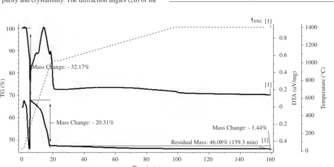
An exothermic reaction between 575 and 925 °C, corresponding on the TGA graph to a 16.75% weight loss, suggests that the ferrite still contains hydroxyl groups, which is congruent with the exother-mic band depicted in the DTA graph. According to the literature5, hydroxyl groups are retained in Co0.5Zn0.5Fe2O4 even after sintering at 700 °C and are completely removed after sintering at 925 °C. A similar behavior was observed5 for Co
0.2Zn0.8Fe2O4 prepared by coprecipitation, but the exothermic band ended at 550 °C. The pres-ence of hydroxyl groups has also been reported3 in nanocrystalline ferrite powders prepared by hydrothermal synthesis. The weight loss occurring between 300 and 600 °C is not significant but, in this range, the DTA graph shows an exothermic band, which may be due to the formation of spinel ferrite.
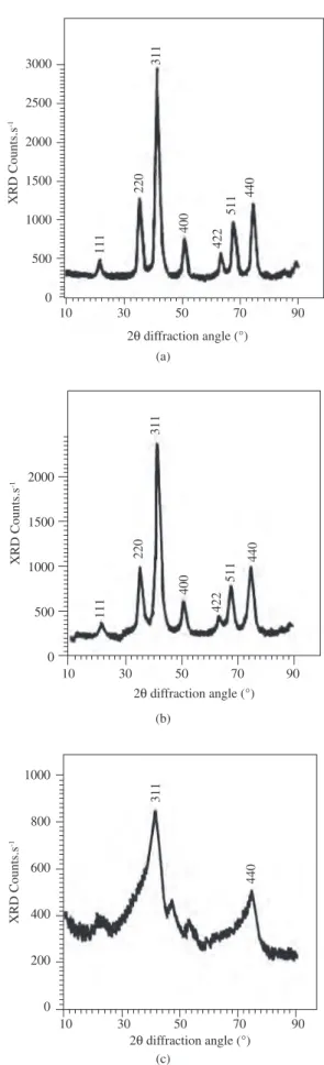
Figure 2 shows the X ray diffraction patterns of the powder samples precipitated by NaOH and NH4OH in the same time period but at different temperatures. The peaks observed here were found to correspond to those of standard diffraction patterns of a spinel ferrite15 with no extra peaks, confirming that the cubic ferrite phase was formed in all the samples.
An X ray diffraction pattern similar to that shown in Figures 2a and 2b indicated that both precipitants can be used. However, when ammonium hydroxide is used as the precipitating agent, the pH must be strictly controlled because complexes with zinc17 can be formed and zinc ions may remain in the solution, influencing the ferrite’s molar ratio. Therefore, sodium hydroxide was chosen as the precipitant in this study. The XRD pattern also showed that the ferrite phase was dependent on the reaction temperature. The sample heated to 200 °C presented mostly an amorphous phase (Figure 2c), but peaks (311) and (440) attributed to the spinel phase began to appear. The powders treated at 400 °C presented a high level of crystallization. The literature1,5,17 cites similar results for several spinel ferrites with high levels of crystallization.
The lattice parameter for the ferrite powder prepared at 400 °C was calculated as 8.38 Å based on the XRD pattern. This result was confirmed by the literature1,6 (which cites values of 8.37 and 8.36 Å).
3.2. Characterization of the sintered material
The sintered compacts were characterized by XRD to confirm the phase purity and crystallinity. The diffraction angles (2θ) of the
Table 1. Data from X ray diffraction patterns of Co-Zn ferrites sintered at
different temperatures.
950-1350 °C 1400 °C
2θ I(%) (h,k,l) 2θ I(%)
35.3 10 2,2,0 28.9 11
41.3 100 3,3,1 30.5 25
50.4 20 4,0,0 48.9 23
65.7 18 4,2,2 68.8 55
69.1 40 5,1,1 71.8 100
73.2 50 4,4,0 84.4 10
0.8
0.6
0.4
0.2
0
- 0.2
- 0.4
1400
1200
1000
800
600
400
200
0
T
emperature (°C)
DT
A (uV/mg)
0 20 40 60 80
Time (min)
100 120 140 160
100
90
80
70
60
50
TG (%)
Mass Change: - 32.17%
Mass Change: - 20.31%
Mass Change: - 1.44%
Residual Mass: 46.08% (159.3 min) [1] [1] [1] exc
Figure 1. TGA-DTA graph of a sample of coprecipitated triple hydroxide (--- temperature, DTA, TGA).
cobalt-zinc ferrite were calculated at different sintering temperatures (Table 1), based on the X ray diffraction pattern. The values remained essentially constant in the range of 950-1350 °C, but clearly differed at 1400 °C, indicating the onset of a phase transformation just above 1350 °C, which means that Co0.5Zn0.5Fe2O4 cannot be produced above that temperature. The strongest reflection came from the (3,1,1) plane for all ferrites sintered from 950 to 1350 °C, while the strongest reflec-tion for the ferrite sintered at 1400 °C came from the (4,4,4) plane. Table 2 presents the true density (Dx) of sintered Co0.5Zn0.5Fe2O4 obtained in this work, which was calculated based on the XRD as a function of the sintering temperature. These values range from 5.29 to 5.35 g/cm3 and are congruent with the reported9 value of 5.30 g/cm3 for Co-Zn ferrite also prepared by coprecipitation. The true density of the material sintered at 1400 °C was not calculated because the diffraction peaks could not be assigned to the Co0.5Zn0.5Fe2O4. This is evidence of the presence of extra phases.
Figure 2. X ray diffraction patterns of cobalt-zinc ferrites prepared by co-precipitation using as co-precipitation agent: a) NaOH and calcined at 400 °C; b) NH4OH and calcined at 400 °C; and c) NaOH and calcined at 200 °C. (calcination time in all cases: 5 hours).
(a)
111
220
311
400
422
511
440
3000
2500
2000
1500
1000
500
0
XRD Counts.s
-1
10 30 50 70 90
2Q diffraction angle (°)
(b)
111
220
311
400
422
511
440
10 30 50 70 90
2Q diffraction angle (°)
XRD Counts.s
-1
(c)
10 30 50 70 90
2Q diffraction angle (°)
XRD Counts.s
-1
311
440
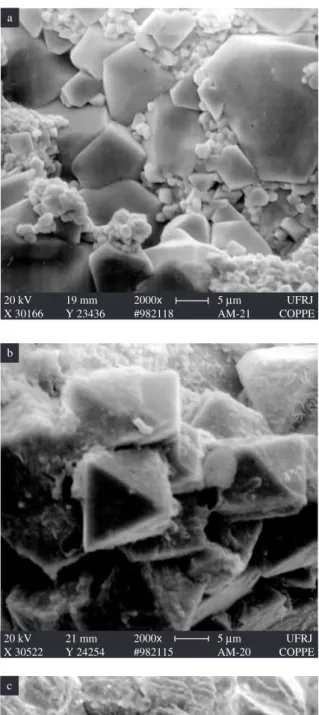
grains welded into a continuous mass dispersed in the honeycomb holes. Honeycombing in the material sintered at 1400 °C (Figure 3f) was so intense that numerous large channels emerged to the sample’s surface. This phenomenon is likely related with the decomposition of cobalt-zinc ferrite and loss of zinc by evaporation18-20.
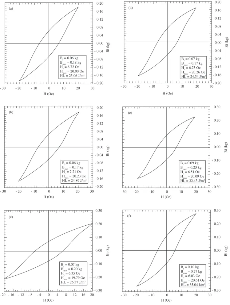
The magnetic behavior of Co0.5Zn0.5Fe2O4 was examined. Figure 4 presents the hysteresis curves of the sintered Co0.5Zn0.5Fe2O4 com-pacts.
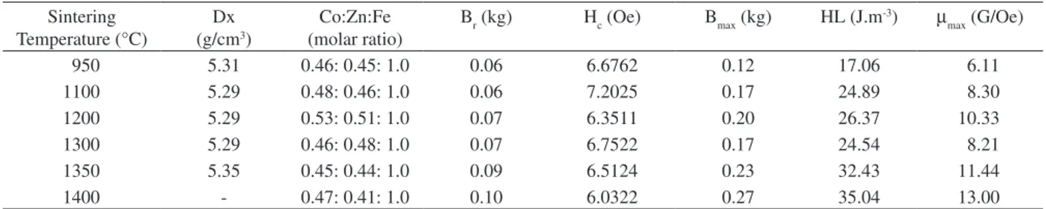
Table 2 summarizes the main findings of the material’s magnetic behavior.
It can be considered that Br (remanescent magnetic induction), Bmax (maximum magnetic induction), HL (magnetic hysteresis loss) and µmax (maximum magnetic permeability) increase as the tempera-ture rises from 950 °C to 1400 °C, while the Hc (magnetic coercive force) shows the opposite trend, decreasing as the temperature in-creases in the same range.
The difference in the Hc values is understandable because of the substantial increase in grain size in response to the increase in sinter-ing temperature from 950 to 1350 °C. In other words, above 1100 °C the mean grain size far exceeded 5 µm, facilitating the movement of the magnetic domain. However, a comparison of Figures 3a and 3d leaves room for doubt concerning this substantial grain growth. The low coercive force, which confirms that the cobalt-zinc ferrite produced here is a soft ferrite, is congruent with the values reported in the literature1,21 for some substituted cobalt-zinc-ferrite powders prepared by coprecipitation.
On the other hand, the values of the other magnetic properties were more difficult to interpret because of the influence of many vari-ables, including the zinc substitution. Tetrahedral sites in the spinel structure are suitable for cationic radii in the range of 0.58 Å to 0.67 Å, while octahedral sites can accept cations with radii in the range of 0.70 Å to 0.75 Å22. Therefore, in the unit cell structure, Co2+ (0.72 Å) and Fe2+(0.75 Å) may replace Zn2+(0.74 Å), while Co3+(0.63 Å) can exchange sites with Fe3+(0.64 Å)12,13. This cation exchange depends on the sintering conditions, since the oxygen partial pressure affects cobalt and iron oxidation states and, hence, influences the magnetic properties of ferrite.
4. Conclusions
a) The powder coprecipitation method produced well crystallized cobalt-zinc ferrite powder upon heating the coprecipitated triple hydroxide to 400 °C for 5 hours, as confirmed by X ray diffraction;
b) The coprecipitation method provided the densest compact when the green compact was sintered for 5 hours at 1350 °C. Furthermore, the method required relatively low temperatures and short calcination times to produce well crystallized cobalt-zinc ferrite powders;
c) Coprecipitated Co0.5Zn0.5Fe2O4 ceramics should be sintered at temperatures up to 1350 °C, as indicated by “honeycombing” degradation of the ceramic sample above this temperature, and by the sharp change in the X ray diffraction pattern in response to the increase in the sintering temperature from 1350 °C to 1400 °C; and
d) In spite of the change in the microstructure caused by the sintering process, all the Hc values were sufficiently low to confirm that the ferrite was a soft ferrite.
Acknowledgments
Figure 3. Micrographs of cobalt-zinc ferrite rings sintered for 5 hours at: a) 950 °C; b) 1100 °C; c) 1200 °C; d) 1300 °C; e) 1350 °C; and f) 1400 °C.
20 kV 19 mm 2000x 5 Mm UFRJ
X 30166 Y 23436 #982118 AM-21 COPPE
a
20 kV 21 mm 2000x 5 Mm UFRJ
X 30522 Y 24254 #982115 AM-20 COPPE
b
20 kV 22 mm 1000x 10 Mm UFRJ
#981712 AM-14 COPPE
c
20 kV 16 mm 500x 20Mm UFRJ
X 39280 Y 40650 007 #981565 AM-11 COPPE
d
20 kV 20 mm 1000x 10 Mm UFRJ
#981710 AM-13 COPPE
e
20 kV 23 mm 500x 20Mm UFRJ
X 29453 Y 21674 #982119 AM-22 COPPE
Figure 4. Magnetic hysteresisgraphs of cobalt-zinc ferrite rings sintered for 5 hours at: a) 950 °C; b) 1100 °C; c) 1200 °C; d) 1300 °C; e) 1350 °C; and
f) 1400 °C. Frequency = 60 Hz.
0.30
0.20
0.10
0.00
- 0.10
- 0.20
- 0.30
Bi (kg)
- 30 - 20 - 10 0 10 20 30
H (Oe)
Br = 0.10 kg Bmax = 0.27 kg Hc = 6.03 Oe Hmax = 20.61 Oe HL = 35.04 J/m3 (f)
Bi (kg)
0.30
0.20
0.10
0.00
- 0.10
- 0.20
- 0.30
- 20 - 16 - 12 - 8 - 4 0 4 8 12 16 20
H (Oe)
Br = 0.07 kg Bmax = 0.20 kg Hc = 6.35 Oe Hmax = 19.79 Oe HL = 26.37 J/m3 (c)
Br = 0.09 kg Bmax = 0.23 kg Hc = 6.51 Oe Hmax = 20.09 Oe HL = 32.43 J/m3
0.30
0.20
0.10
0.00
- 0.10
- 0.20
- 0.30
Bi (kg)
- 30 - 20 - 10 0 10 20 30
H (Oe) (e)
Br = 0.06 kg Bmax = 0.17 kg Hc = 7.21 Oe Hmax = 20.23 Oe HL = 24.89 J/m3
0.20
0.16
0.12
0.08
0.04
0.00
- 0.04
- 0.08
- 0.12
- 0.16
- 0.20
Bi (kg)
- 30 - 20 - 10 0 10 20 30
H (Oe) (b)
- 30 - 20 - 10 0 10 20 30
H (Oe)
Br = 0.07 kg Bmax = 0.17 kg Hc = 6.75 Oe Hmax = 20.26 Oe HL = 24.54 J/m3
0.20
0.16
0.12
0.08
0.04
0.00
- 0.04
- 0.08
- 0.12
- 0.16
- 0.20
Bi (kg)
(d)
- 30 - 20 - 10 0 10 20 30
0.20
0.16
0.12
0.08
0.04
0.00
- 0.04
- 0.08
- 0.12
- 0.16
- 0.20
Bi (kg)
H (Oe)
References
1. Arulmurugan R, Jeyadevan B, Vaidyanathan G, Sendhilnathan S. Effect of zinc substitution on Co–Zn and Mn–Zn ferrite nanoparticles prepared by co-precipitation. J Magn Magn Mater. 2005; 288:470-7.
2. Virden AE, O’Grady K. Structure and magnetic properties of NiZn ferrite nanoparticles. J Magn Magn Mater. 2005; 290-291: 868-70.
3. Verma S, Joy PA, Khollam YB, Potdar HS, Deshpande SB. Synthesis of nanosized MgFe2O4 powders by microwave hydrothermal method. Mater Lett. 2004; 58(6):1092-5.
4. Costa ACF, Tortella E, Neto EF, Morelli MR, Kiminami RHGA. Sintering of Ni-Zn ferrite nanopowders by the constant heating rate (CHR) method. Mater Res. 2004; 7(4):523-8.
5. Dey S, Ghose J. Synthesis, characterisation and magnetic studies on nanocrystalline Co0.2Zn0.8Fe2O4. Mater Res Bull. 2003; 38(11-12):1653-60.
6. Pandya PB, Joshi HH, Kulkarni RG. Bulk magnetic properties of Co-Zn ferrites prepared by the coprecipitation method. J Mater Sci. 1991; 26(20):5509-12.
7. Suzuki K.Preparation of zinc and aluminium substituted Co-ferrite thin films and their faraday rotation. Jpn J Appl Phys. 1988; 27(3):361-5. 8. Ahmed MA. Electrical properties of Co-Zn ferrites. Phys Stat Sol A.
1989; 111: 567-72.
9. Tawfik A. Effect of magnetic order on the conductivity in Co-Zn ferrites. J Therm Anal. 1989; 35:141-5.
10. Murthy SR, Seshagiri Rao T. Effect of magnetic field and temperature on the elastic behaviour of Co-Zn ferrites. J Less Common Met. 1979; 65:19-26.
11. Murthy SR. Dielectric behaviour of Co-Zn ferrites. J Mater Sci Lett. 1984; 3:1049-51.
12. Abd El-Ati MI, Kafafy MA, Tawfik A. Magnetic properties of zinc doped ferrites. Acta Phys Pol A. 1991; 79(6):889-94.
13. Gaballa GA. Effect of Mn addition on physical properties of Co0.6 Zn0.4Mnx Fe2-xO4 system. Phase Transitions. 1994; 46:66-67.
14. Darwish NZ, Hemeda OM, Abd El-Ati MI. Thermal properties of gama-ray irradiated Co-Zn ferrite. Appl Radiat Isot. 45(4):445-8.
15. JCPDS International Centre for Diffraction Data. 2nd ed.,USA; 1979. 16. Abd El-Ati MI. Thermal conductivity of Zn doped CoFe2O4 ferrites.
Phase Transitions. 1994; 46(4):209-15.
17. Alexéev V. Análise Qualitativa. Porto (Portugal): Editora Lopes da Silva; 1982. p. 302.
18. Drofenik M, Rozman M. Sintering of nanosized MnZn ferrite powders. J Amer Ceram Soc. 1998; 81(7):1757-64.
19. Inaba H, Matsui T. Vaporization and diffusion of manganese–zinc ferrite. J Solid State Chem. 1996; 121(1):143-8.
20. Sainamthip P, Amarakoon VRW. Role of zinc volatilization on the micro-structure development of manganese zinc ferrites. J Amer Ceram Soc. 1988; 71(8):644-8.
21. Brownlow JM. Preferential volatilization of cations from ferrites during sintering. J Appl Phys. 1958; 29(3):373-5.
22. Chen CW. Magnetism and Metallurgy of Soft Materials. Netherlands: North-Holland Publishing Co.; 1977.
23. Buchanam RC, Ceramic Materials for Electronics: Processing, Properties and Applications. N. York (USA): Marcel Dekker Inc.; 1986.
Table 2. Summary of properties of cobalt-zinc ferrites sintered at different temperatures.
Sintering Temperature (°C)
Dx (g/cm3)
Co:Zn:Fe (molar ratio)
Br (kg) Hc (Oe) Bmax (kg) HL (J.m-3) µ
max (G/Oe)
950 5.31 0.46: 0.45: 1.0 0.06 6.6762 0.12 17.06 6.11
1100 5.29 0.48: 0.46: 1.0 0.06 7.2025 0.17 24.89 8.30
1200 5.29 0.53: 0.51: 1.0 0.07 6.3511 0.20 26.37 10.33
1300 5.29 0.46: 0.48: 1.0 0.07 6.7522 0.17 24.54 8.21
1350 5.35 0.45: 0.44: 1.0 0.09 6.5124 0.23 32.43 11.44