Outward potassium current oscillations
in macrophage polykaryons:
extracellular calcium entry and
calcium-induced calcium release
Instituto de Biofísica Carlos Chagas Filho, Universidade Federal do Rio de Janeiro, Rio de Janeiro, RJ, Brasil
R.M. Saraiva, M.O. Masuda and G.M. Oliveira-Castro
Abstract
Outward current oscillations associated with transient membrane hyperpolarizations were induced in murine macrophage polykaryons by membrane depolarization in the absence of external Na+.
Oscilla-tions corresponded to a cyclic activation of Ca2+-dependent K+
cur-rents (IKCa) probably correlated with variations in intracellular Ca2+
concentration. Addition of external Na+ (8 mM) immediately
abol-ished the outward current oscillations, suggesting that the absence of the cation is necessary not only for their induction but also for their maintenance. Oscillations were completely blocked by nisoldipine. Ruthenium red and ryanodine reduced the number of outward current cycles in each episode, whereas quercetin prolonged the hyperpolar-ization 2- to 15-fold. Neither low molecular weight heparin nor the absence of a Na+ gradient across the membrane had any influence on
oscillations. The evidence suggests that Ca2+ entry through a pathway
sensitive to Ca2+ channel blockers is elicited by membrane
depolariza-tion in Na+-free medium and is essential to initiate oscillations, which
are also dependent on the cyclic release of Ca2+ from intracellular
Ca2+-sensitive stores; Ca2+ ATPase acts by reducing intracellular Ca2+,
thus allowing slow deactivation of IKCa. Evidence is presented that
neither a Na+/Ca2+ antiporter nor Ca2+ release from IP
3-sensitive Ca2+
stores participate directly in the mechanism of oscillation. Correspondence
R.M. Saraiva
Laboratório de Eletrofisiologia Cardíaca
Instituto de Biofísica Carlos Chagas Filho, UFRJ Bloco G, CCS, Ilha do Fundão 21949-900 Rio de Janeiro, RJ Brasil
Fax: 55 (021) 212808193 E-mail: [email protected]
The present address of G.M. Oliveira-Castro is Universidade Estácio de Sá, Rua do Bispo, 71, Reitoria, 20261-061 Rio de Janeiro, RJ, Brasil. Research supported by CNPq, FINEP and FAPERJ.
Received March 14, 1997 Accepted August 19, 1997
Key words
•Macrophage
•Macrophage polykaryon
•Ca2+-dependent K+ current
•Membrane potential oscillations
•Calcium-induced calcium release
•Ca2+ ATPase
Introduction
Transmembrane potential oscillations have been encountered in many cell types, including both excitable and non-excitable cells. They are often associated with oscilla-tions in intracellular Ca2+ concentrations that
activate Ca2+-dependent K+ currents (IK Ca)
(1-3) and can be useful as an alternative to
direct measurement of intracellular Ca2+
con-centration (4,5). This correlation has been established by simultaneous recordings of membrane potential and intracellular Ca2+ in
fibroblasts (2) and endothelial cells (3). In non-excitable cells, Ca2+ oscillations are
gen-erated by different mechanisms: cyclic Ca2+
oo-cytes and megakaryooo-cytes (6-8), or by cyto-plasmic Ca2+-activated mechanisms as in
hamster eggs (9), or Ca2+ entry-dependent
oscillations as in endothelial cells (3). These different mechanisms may also interact to modulate Ca2+ oscillations, as is the case for
IP3- and Ca2+-sensitive mechanisms in
pan-creatic acinar cells (10), hepatocytes and endothelial cells (1). The presence of extra-cellular Ca2+ is also important, since
oscilla-tions cannot be sustained when Ca2+ is
re-moved from the extracellular medium. Ex-tracellular Ca2+ allows replenishment of
in-tracellular stores via Ca2+ influx pathways
that are not yet fully understood (1). In non-excitable cells, the oscillation patterns vary widely (1). In a single cell type, the pattern of oscillations can vary depending on the stimu-lus and on the individual cell. However, the response of the same cell to repeated expo-sure to the same agonist can be remarkably similar (1).
In macrophages, spontaneous membrane potential oscillations due to a Ca2+
-depend-ent increase in K+ conductance have been
described (11). Because of their episodic nature, these spontaneous oscillations are virtually inaccessible experimentally. How-ever, qualitatively similar oscillations can be induced reproducibly in macrophage poly-karyons when the cells are depolarized in Na+-free medium. These oscillations consist
of cyclic hyperpolarizations of membrane potential of progressively decreasing ampli-tude. Each hyperpolarization lasts 3 to 5 s. Each oscillation event, on average, is com-posed of 5 cycles. These oscillations have been shown to depend on extracellular Ca2+,
and the underlying mechanism is a cyclic opening of Ca2+-dependent K+ channels (12).
Thus, these oscillations may serve as a mo-del for studying the mechanisms underlying spontaneous oscillations.
However, there is no agreement with re-spect to the role of Ca2+ oscillations or the
physiological relevance of electrical activity in macrophages. While Ca2+ is a known
sec-ond messenger, it is not clear how oscillatory changes in internal Ca2+ would have an
ad-vantage over step changes (4). Thus, there are few well-established links between Ca2+
oscillations and cellular responses. A role for Ca2+ oscillations has been proposed for
fluid secretion in pancreatic acinar cells (13) and in neutrophil migration (14) and in terms of allowing replenishment of intracellular calcium stores (4). Calcium oscillations are also detected during frustrated phagocytosis and adherence in macrophages (15). None-theless, the fact that an oscillatory signal can vary in amplitude and frequency, as well as in position (calcium waves (16)), suggests that Ca2+ oscillations have an unusual
poten-tial for encoding cellular functions (1,4,16). There are also indications of a link be-tween electrical activity and macrophage functions: ionic currents are activated by the Fc fraction of immunoglobulin (17) and by extracellular ATP (18-20); oscillations of potassium currents are induced in mouse peritoneal macrophages by C5a (21) and activation of a Ca2+-dependent,
non-selec-tive current is associated with superoxide release subsequent to phagocytosis (22).
In the present study, we examined the genesis and modulation of the depolariza-tion-induced outward K+ current oscillations
in macrophage polykaryons and evaluated the role of the various mechanisms involved in intracellular Ca2+ homeostasis, including
calcium channels, Na+/Ca2+ antiporter, Ca2+
ATPase and intracellular Ca2+ stores.
Knowl-edge of these mechanisms can help elucidate the physiological role of electrical activity and Ca2+ oscillations in macrophages.
Material and Methods
Cells
15 days, coverslips were removed from the animal and washed in RPMI-1640 (Gibco, Grand Island, NY) buffered with 6 mM HEPES, pH 7.3, and containing 5% fetal calf serum, penicillin (100 U/ml) and streptomy-cin (100 µg/ml) at 37oC. Coverslips were
transferred to a 5-ml culture chamber on the stage of a phase-contrast Leitz microscope equipped with a Heineke condenser and a UMK 50/0.60 objective. The large macro-phage polykaryons were easily identified on the surface of the coverslips.
Solutions
Unless otherwise stated, experiments were performed in standard extracellular saline con-taining 5 mM KCl, 2 mM CaCl2, 1 mM MgCl2,
6 mM HEPES and 140 mM Tris-HCl, pH 7.3. In whole-cell patch-clamp recordings the in-ternal solution contained 140 mM KCl, 5 mM NaCl, 0.2 mM CaCl2, 2 mM MgCl2, 10 mM
HEPES-KOH, 0.6 mM K2EGTA, pH 7.2, and
pCa 7.04 (calculated according to Fabiato and Fabiato (24)). When sodium concentration was increased (both in the external and inter-nal saline) osmolarity was corrected by de-creasing the major cation Tris in the external saline and potassium in the internal saline. No osmolarity correction was made for the so-dium-free internal saline.
Reagents
Nisoldipine (Bayer, Munich, Germany) and ryanodine (K & K Laboratories, Califor-nia) were dissolved in dimethyl sulfoxide (DMSO), quercetin (Sigma, St. Louis, MO) was dissolved in absolute ethanol, low mo-lecular weight heparin (Sigma) was dissolved in internal saline, and ruthenium red (British Drug Houses, Poole, England) in water.
Electrophysiological technique
Experiments were carried out at room temperature (23-28oC). Whole-cell
patch-clamping was performed using an EPC-7 amplifier (List Electronic, Darmstadt, Ger-many) according to standard procedures (25). A giga-ohm seal was formed using a heat-polished micropipette back-filled with the internal saline solution. Standard electro-physiological recordings were performed using glass microelectrodes (30 to 60 MΩ tip resistance). The electrodes were back-filled with a 2.5 M KCl solution and connected to a high-input-impedance preamplifier with an active bridge circuit (M4A Electromer, WP Instruments, New Haven, CT) allowing simultaneous recording of the transmem-brane potential and of the injected currents. Iontophoretic intracellular calcium injec-tions were performed through a second glass microelectrode back-filled with 0.5 M CaCl2,
as previously described (23).
The statistical tests used are specified together with the corresponding data in the Results section. In all cases differences were considered to be significant when P<0.05.
Results
Membrane potential oscillations and sodium dependence
Oscillations of membrane potential and current were obtained reproducibly when macrophages were depolarized in the ab-sence of external Na+ (Figure 1). Both
The presence of 8 mM NaCl in the exter-nal saline was sufficient to prevent mem-brane potential oscillations (N = 8). Addi-tion of Na+ to the external medium during an
oscillation abolished both hyperpolarizing voltage (Figure 2A) and outward current
oscillations (Figure 2B) in 88% of the cells tested (N = 8).
Participation of the Na+/Ca2+ antiporter
in the Ca2+ influx may explain the
depend-ence on the absdepend-ence of external Na+ (26).
The inverted Na+ gradient could result in
reversal of this antiporter, leading to Ca2+
influx. This hypothesis was tested using Na+
-free internal saline to eliminate any Na+
gradient across the membrane. An increase in the amplitude of the resting membrane potential was observed under these experi-mental conditions (Table 1). However, the same percentage of cells exhibited depolar-ization-induced oscillations with character-istics similar to those observed for cells main-tained under control conditions (Figure 2 and Table 1). An increase in the internal Na+
concentration to 15 mM did not significantly change resting potential or the pattern of the oscillatory response (Table 1).
Extracellular calcium
The possible participation of an L-type Ca2+ channel in the genesis of the
oscilla-tions was investigated. Nisoldipine, a blocker specific for L-type Ca2+ channels (27),
com-pletely blocked the oscillations when added to the external medium (N = 6) (Figure 3C). This effect persisted even when the depolar-izing pulse amplitude was increased and was reversed by washing out the nisoldipine (data not shown). Addition of the solvent (DMSO) did not affect membrane potential oscilla-tion (data not shown). The calcium-activated potassium efflux in human erythrocytes (28) and N-type potassium channels (Kn
chan-nels) in human and murine T lymphocytes (29,30) is blocked by dihydropyridine and other Ca2+ channel blockers. In order to test
whether nisoldipine acts by blocking IKCa,
calcium was injected iontophoretically be-fore (Figure 3B) and after (Figure 3D) the addition of the blocking agent. Calcium in-jection induced hyperpolarization in both situations.
40 mV 0
100 pA 0
10 s
40 mV 0
100 pA 0
10 s
A
B
40 mV 0
100 pA
0
10 s
40 mV 0
100 pA
0
10 s
A
B
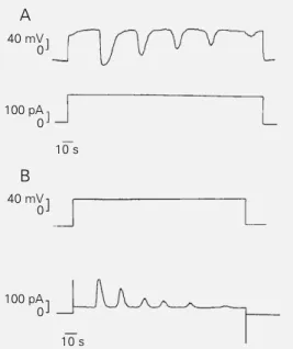
Figure 1 - Voltage and current oscillations induced by depolar-ization in Na+-free medium. A,
Depolarization by constant out-ward current injection induced membrane potential oscilla-tions in macrophage polykary-ons bathed in Na+-free saline
solution. Resting membrane potential = -48 mV. B, Outward current oscillation elicited when the transmembrane po-tential is clamped at +40 mV. Holding potential = -50mV. Note that current and voltage oscillations have similar pat-terns. Records were obtained by means of whole-cell patch-clamp under current patch-clamp (A) and voltage clamp (B) condi-tions in two different cells. Cali-bration bars in all figures were positioned in such a way that the current and voltage values presented correspond to actual values in the tracings.
Figure 2 - Effect of external Na+. Both voltage (A) and
cur-rent (B) oscillations were inter-rupted by the addition of NaCl to the external medium to a final concentration of 8 mM (ar-rowheads). Intracellular saline was Na+ free in both cases.
Calcium ATPase
Quercetin is a flavone of plant origin that blocks Ca2+ ATPase in plasma membranes
as well as in sarcoplasmic reticulum (31,32). Figure 4 illustrates one experiment where quercetin, added to the external medium af-ter membrane potential oscillation had been induced, generated a long-lasting hyperpo-larization (~30 s). An increase in duration of hyperpolarization ranging from 2- to 15-fold was seen in 48% of the cells tested (N = 21). Hyperpolarizing cycles lasted 9.5 ± 5.0 s (mean ± SD; N = 21) before quercetin addi-tion and increased to 25.2 ± 28.9 s in the presence of quercetin (paired t-test after log-transformation, P<0.05). A single, long-last-ing hyperpolarization similar to that shown in Figure 4 was observed in 60% of the cells that responded to quercetin addition. Quer-cetin seems to prolong the time during which intracellular Ca2+ concentration is high
enough to activate IKCa, thus maintaining a
hyperpolarized membrane potential.
Intracellular calcium stores
There are two mechanisms of Ca2+
re-lease from the endoplasmic reticulum, one sensitive to IP3 and the other to Ca2+ itself
(33). Both mechanisms were tested using specific inhibitors.
Low molecular weight heparin (200 µg/ ml), which blocks Ca2+ release from IP
3
-sensitive intracellular Ca2+ stores (33), did
not significantly alter the membrane poten-tial, the percentage of cells responding to depolarization or the characteristics of the membrane potential oscillations (Table 2).
The possible involvement of Ca2+-induced
Ca2+ release was tested by using ruthenium
red in the patch-clamp pipette, or ryanodine in the external medium (6,33). A compari-son of cells from the same population indi-cated a striking decrease in the number of hyperpolarizing cycles in the presence of ruthenium red (50 µM, Table 2). Ryanodine
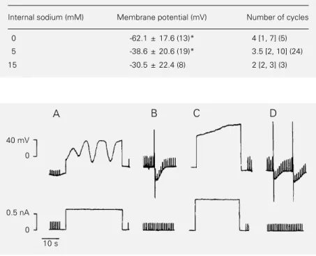
Table 1 - Effect of internal sodium on membrane potential and oscillations.
Data are reported as mean ± SD or median [min, max] for the number of cases given in parentheses. *P<0.05 between the two groups (ANOVA). No statistically significant differences in the number of cycles were observed among groups (Kruskal-Wallis test).
Internal sodium (mM) Membrane potential (mV) Number of cycles
0 -62.1 ± 17.6 (13)* 4 [1, 7] (5)
5 -38.6 ± 20.6 (19)* 3.5 [2, 10] (24)
15 -30.5 ± 22.4 (8) 2 [2, 3] (3)
40 mV
0
0.5 nA
0
A B C D
10 s
Table 2 - Effect of intracellular calcium release blockers on oscillations.
Data are reported as median [min, max] for the number of cells given in parentheses. *P<0.05 (Mann-Whitney test); +P<0.05 (Wilcoxon test; data
from the same cells before (control) and during ryanodine treatment (experimental)).
Number of cycles
Control Experimental
Ruthenium red (50 µM) 3 [0, 7] (13) 1 [0, 3] (17)* Heparin (200 µg/ml) 3 [0, 7] (13) 3 [1, 13] (8)
Ryanodine (1 µM) 3 [2, 4] (9) 0 [0, 1] (9)+
Figure 3 - Inhibition of membrane potential oscillation by nisoldipine. Control situation: depolarization-induced membrane potential oscillation (A) and hyperpolarization induced by iontophoretic intracellular calcium injection (B). Transmembrane potential oscillation was blocked by nisoldipine (20 µM) in extracellular saline (C). Hyperpolarizations induced by iontophoretic intracellular calcium injection were not blocked by nisoldipine (D). Records (A-D) were obtained from a single cell using standard intracellular microelectrodes. In B and D, iontophoretic injections of calcium were performed through a second microelectrode containing CaCl2. Current pulses of 0.20 nA were injected in order to monitor membrane
(1 µM) also induced a significant decrease in the number of oscillations in the same cell (Figure 5, Table 2). Ryanodine opens the Ca2+-sensitive Ca2+ channel of the internal
stores at low concentrations (nanomolar) but blocks them at the concentration used here (micromolar) (6). The results obtained with heparin, ruthenium red and ryanodine indi-cate that oscillations depend on the release of intracellular Ca2+ from Ca2+-sensitive
stores but not from IP3-sensitive stores.
Discussion
In the present study, we investigated the participation of various processes related to cytoplasmic Ca2+ homeostasis in the
regula-tion of the outward potassium current oscil-lations activated by depolarization in Na+
-free saline. Our working hypothesis pro-poses that the cyclic activation of the IKCa
would depend on cyclic increases in
cyto-plasmic free Ca2+ activity produced by an
influx through the plasma membrane, by release from intracellular stores, or by both events.
Here, we showed that nisoldipine, an L-type Ca2+ channel blocker, abolished
mem-brane potential oscillations and that this ef-fect was not due to the direct inhibition of IKCa by nisoldipine (28,30), since
intracellu-lar Ca2+ injection restored membrane
hyper-polarization in the presence of the drug (Fig-ure 3D). Other evidence for the participation of Ca2+ influx in the genesis of these
mem-brane potential oscillations has been pre-sented previously, and includes their de-pendence on extracellular Ca2+ and
block-ade by D-600 (an L-type Ca2+ channel
blocker) (12).
These findings suggest that Ca2+ influx
through a dihydropyridine-sensitive, depo-larization-activated pathway is essential for the oscillations. However, a voltage-depend-ent Ca2+ channel, such as the L-type Ca2+
channel, has not yet been found in the mac-rophage membrane (29,34) or in the major-ity of non-excitable cells (1). Calcium influx pathways in non-excitable cells include chan-nels activated by second messengers such as Ca2+, inositol 1,3,4,5-tetrakisphosphate (IP
4)
(16) and IP3 (29), channels activated by
de-pletion of intracellular Ca2+ stores (1), and
non-selective cation channels (35). In mac-rophages, a Ca2+-selective current activated
by depletion of intracellular Ca2+ stores (36)
and an ATP-activated Ca2+ channel (18) have
been described. Voltage-sensitive, non-se-lective cation channels (37) and another ATP-activated cation channel (19) are also pres-ent. Thus, one possible pathway for Ca2+
influx in macrophage polykaryons would be a voltage-dependent cation channel which, in a Na+-free environment, allows Ca2+
per-meation. An analogous situation has been reported for the potassium channel in guard cell membranes of Vicia faba, where Ca2+
enters the cell in the absence of potassium (38). However, the fact that we did not detect 40 mV
0
0.5 nA 0
10 s Figure 4 - Effect of quercetin on
membrane potential oscillation. Quercetin (0.1 mM) applied dur-ing the oscillation (arrowhead) prolonged the subsequent hy-perpolarizing cycle to almost three times the control value. The record was obtained using a standard electrophysiologic re-cording technique. Resting membrane potential = -20 mV.
40 mV 0
10 s 100 pA
0 100 pA 0
40 mV 0
10 s
Ryanodine Figure 5 - Effect of ryanodine
on membrane potential and outward current oscillations. The number of hyperpolarizing cycles of transmembrane poten-tial (A) and outward current (B) oscillations was reduced by the addition of ryanodine (1 µM) to the extracellular saline. Records were obtained using a whole-cell patch-clamp under current clamp (A) and voltage clamp conditions (B). Resting membrane potential = -12 mV. Holding potential = -40 mV(B).
A
an inward current associated with depolar-ization is puzzling. Either this current is too small to be detected under our experimental conditions or some outward current is acti-vated simultaneously.
The reversal of the Na+/Ca2+ antiporter
was another possible Ca2+ influx pathway
studied. Reversal of this antiporter in the absence of extracellular Na+ has been
dem-onstrated in aortic smooth muscle cells (26). However, the use of Na+-free internal
solu-tion in the patch-clamp experiments in order to minimize the Na+ gradient had no
influ-ence on the induction of oscillations by de-polarization (Figure 2). Nevertheless, indi-rect participation of this exchanger cannot be ruled out, since reversal or blockade would raise the background cytoplasmic Ca2+
con-centration.
When macrophage polykaryons were ex-posed to the sodium-free saline pipette in patch-clamp experiments, a significant in-crease in the resting membrane potential was observed. This increase cannot be attributed to the expected decrease in the electrochemi-cal sodium gradient or to the inhibition of the sodium potassium pump activity since both conditions would depolarize the cell. One possibility is that the decrease in internal sodium would somehow increase resting potassium conductance and thus the resting potential. Actually, a blocking effect of in-tracellular sodium on a calcium-dependent potassium current has been reported in bo-vine adrenal chromaffin cells (39).
The intracellular Ca2+ stores may
partici-pate in the genesis of oscillations through Ca2+- and/or IP
3-sensitive mechanisms, both
known to be functional in macrophages (40). Ruthenium red and ryanodine, which block Ca2+-induced Ca2+ release at the
concentra-tions used (6,33), significantly decreased the number of oscillation cycles in macrophage polykaryons, thus indicating the participa-tion of this mechanism in the genesis of the oscillations. This observation reinforces the importance of Ca2+ influx for increasing
cy-toplasmic Ca2+ and triggering Ca2+ release
from this store. On the other hand, low mo-lecular weight heparin, a blocker of IP3
-induced Ca2+ release (16), did not have a
significant effect on the oscillations, exclud-ing the participation of this Ca2+ pool in its
genesis.
The prolongation of the duration of the hyperpolarization of the first oscillation cycle following quercetin addition (Figure 4) sug-gests that each hyperpolarizing cycle is ter-minated normally by a decrease in cytoplas-mic free Ca2+. Such a decrease, mediated by
Ca2+ ATPase activity, would deactivate IK Ca
and allow membrane potential repolariza-tion.
The fact that the amplitudes of the hyper-polarizing cycles usually decrease progres-sively and finally subside after a mean of 5 cycles may be the result of several factors, including voltage-dependent inactivation of the Ca2+ influx pathway, depletion of
intra-cellular Ca2+ stores, inactivation of IK Ca,or
desensitization of the intracellular Ca2+
-re-lease mechanism. At present, we do not know which of these processes contribute to the termination of the oscillations. The fact that new oscillations can be induced by a further increase in depolarization (data not shown) rules out a voltage-dependent inacti-vation of the Ca2+ influx pathway and of
IKCa. Inactivation of IKCa is also unlikely
because intracellular calcium injection re-stored one hyperpolarization cycle (data not shown). Considering that intracellular Ca2+
stores are only partially repleted after each hyperpolarizing cycle, it is conceivable that a sustained depolarization leads to a progres-sive decrease in the Ca2+ content of those
stores, with the end of oscillations coincid-ing with their depletion.
Thus, we propose the following hypoth-esis for the genhypoth-esis of the depolarization-induced membrane potential oscillations in macrophage polykaryons in Na+-free
medi-um. Upon depolarization, Ca2+ enters the
path-way. The resulting increase in cytoplasmic Ca2+ activity induces the release of Ca2+
from Ca2+-sensitive intracellular stores. This
increase in Ca2+ activates IK
Ca which
hyper-polarizes the cell membrane. The Ca2+
ATPase pumps Ca2+ out of the cell and/or
into intracellular stores, decreasing cytoplas-mic Ca2+ and deactivating IK
Ca.
Addition-ally, the Ca2+-sensitive intracellular stores
are modulated by cytoplasmic calcium itself (41): above a certain intracellular calcium concentration, the open state probability of the calcium-sensitive calcium channels is reduced (42), further decreasing calcium re-lease and consequently IKCa, allowing the
membrane potential to return to its previous level. If the depolarizing pulse is sustained, intracellular Ca2+ may rise again, so that new
cycles occur until one or more of the mecha-nisms discussed above terminates the oscil-lation.
The present study demonstrates that the
depolarization-induced oscillation of mem-brane potential in macrophage polykaryons depends on events occurring at both the plasma membrane and the endoplasmic reticulum levels. In addition, we propose that a similar mechanism may account for the spontaneous oscillations that occur in macrophages and macrophage polykaryons. The reproducible oscillatory phenomena pre-sented here can be very useful as a model for the study of the mechanisms for generation of oscillations and their role in cell responses, including those of macrophages.
Acknowledgments
We are grateful to Drs. Pedro Muanis Persechini, Luiz Anastácio Alves, Cristóvão de Albuquerque and Martha Sorenson for helpful discussions and a critical review of the manuscript, and to Dr. Doris Rosenthal for help with the statistical analysis.
References
1. Fewtrell C (1993). Ca2+ oscillations in
non-excitable cells. Annual Review of Physiol-ogy, 55: 427-454.
2. Ueda S, Oiki S & Okada Y (1986). Oscilla-tions of cytoplasmic concentraOscilla-tions of Ca2+ and K+ in fused L cells. Journal of
Membrane Biology, 91: 65-72.
3. Laskey RE, Adams DJ, Cannel M & van Breemen C (1992). Calcium entry-depend-ent oscillations of cytoplasmic calcium concentration in cultured endothelial cell monolayers. Proceedings of the National Academy of Sciences, USA, 89: 1690-1694.
4. Hille B, Tse A, Tse FW & Almers W (1994). Calcium oscillations and exocytosis in pi-tuitary gonadotropes. Annals of the New York Academy of Sciences, 710: 261-270. 5. Nathanson MH (1994). Cellular and sub-cellular calcium signaling in gastrointesti-nal epithelium. Gastroenterology, 106: 1349-1364.
6. Berridge MJ (1993). Inositol triphosphate and calcium signaling. Nature, 361: 315-325.
7. DeLisle S, Krause K, Demming G, Potter BVL & Welsh MJ (1990). Effect of inositol triphosphate and calcium on oscillation el-evations of intracellular calcium in Xeno-pus oocytes. Journal of Biological Chem-istry, 265: 11726-11730.
8. Uneyama C, Uneyama H, Takashi M & Akaike N (1993). Cytoplasmic pH regu-lates ATP-induced Ca2+-dependent K+
-current in rat megakaryocytes. Biochemi-cal Journal, 295: 317-320.
9. Berridge MJ & Galione A (1988). Cytoso-lic calcium oscillators. FASEB Journal, 2: 3074-3082.
10. Wakui M, Osipchuk YV & Petersen OH (1990). Receptor-activated cytoplasmic Ca2+ spiking mediated by inositol
triphos-phate is due to Ca2+-induced Ca2+
re-lease. Cell, 63: 1025-1032.
11. Oliveira-Castro GM (1983). Ca2+-sensitive
K+ channels in phagocytic cell
mem-branes. Cell Calcium, 4: 475-492. 12. Soldati L & Persechini PM (1988).
Depo-larization of macrophage polykaryons in the absence of external sodium induces a cyclic stimulation of a calcium-activated potassium conductance. Biochimica et Bi-ophysica Acta, 972: 283-292.
13. Kasai H & Augustine GJ (1990). Cytosolic Ca2+ gradients triggering unidirectional
fluid secretion from exocrine pancreas. Nature, 348: 735-738.
14. Marks PW & Maxfield FR (1990). Tran-sient increases in cytosolic free calcium appear to be required for the migration of adherent human neutrophils. Journal of Cell Biology, 110: 43-52.
15. Kruskal BA & Maxfield FR (1987). Cytoso-lic-free calcium increases before and os-cillates during frustrated phagocytosis in macrophages. Journal of Cell Biology, 105: 2685-2693.
16. Amundson J & Clapham D (1993). Cal-cium waves. Current Opinion in Neurobi-ology, 3: 375-382.
17. Young JDE, Unkeless JC, Young TM, Mauro A & Cohn ZA (1983). Role for mouse macrophage IgG Fc receptor as ligand-dependent ion channel. Nature, 306: 186-189.
19. Albuquerque C, Oliveira SMC, Coutinho-Silva R, Oliveira-Castro GM & Persechini PM (1993). ATP- and UTP-induced cur-rents in macrophages and macrophage polykaryons. American Journal of Physiol-ogy, 265: C1663-C1673.
20. Hara N, Ichinose M, Sawada M, Imai K & Maeno T (1990). Activation of single Ca2+
-dependent K+ channel by external ATP in
mouse macrophages. FEBS Letters, 267: 281-284.
21. Ichinose M, Hara N, Sawada M & Maeno T (1992). Induction of two K+ currents by
complement component C5a in mouse macrophages. Biochimica et Biophysica Acta, 1111: 165-170.
22. Holevinsky KO & Nelson DJ (1995). Si-multaneous detection of free radical re-lease and membrane current during phag-ocytosis. Journal of Biological Chemistry, 270: 8328-8336.
23. Persechini PM, Araujo EG & Oliveira-Castro GM (1981). Electrophysiology of phagocytic membranes: induction of slow membrane hyperpolarizations in macro-phages and macrophage polykaryons by intracellular calcium injection. Journal of Membrane Biology, 61: 81-90.
24. Fabiato A & Fabiato F (1979). Calculator programs for computing the composition of the solutions containing multiple met-als and ligands used for experiments in skinned muscle cell. Journal de Physiolo-gie, 75: 463-505.
25. Hamil OP, Marty E, Neher B, Sackmann B & Sigworth FJ (1981). Improved patch-clamp techniques for high-resolution cur-rent recording from cells and cell-free membrane patches. Pflügers Archives. European Journal of Physiology, 391: 85-100.
26. Smith JB, Zheng T & Smith L (1989). Rela-tionship between cytosolic free Ca2+ and
Na+-Ca2+ exchange in aortic muscle cells.
American Journal of Physiology, 256 (Cell Physiology, 25): C147-C154.
27. Fleckenstein A (1985). Calcium antago-nists and calcium agoantago-nists: fundamental criteria and classification. In: Fleckenstein A, van Breemen C, Groβ R & Hoffmeister F (Editors), Cardiovascular Effects of Di-hydropyridine-type Calcium Antagonist and Agonists. Springer-Verlag, Berlin, Heidelberg.
28. Kaji DM (1990). Nifedipine inhibits cal-cium-activated K transport in human eryth-rocytes. American Journal of Physiology, 259: C332-C339.
29. Gallin EK (1991). Ion channels in leuko-cytes. Physiological Reviews, 71: 775-811.
30. Randriamampita C, Bismuth G, Debre P & Trautmann A (1991). Nitrendipine-induced inhibition of calcium influx in a human T-cell clone: role of T-cell depolarization. Cell Calcium, 12: 313-323.
31. Fewtrell CMS & Gompters BD (1977). Ef-fect of flavone inhibitors of transport ATPases on histamine secretion from rat mast cells. Nature, 265: 635-636. 32. Shoshan V & MacLennan DH (1981).
Quercetin interaction with the (Ca2+
+Mg2+)-ATPase of sarcoplasmic
reticu-lum. Journal of Biological Chemistry, 256: 887-892.
33. Petersen OH & Wakui M (1990). Oscillat-ing intracellular Ca2+ signals evoked by
activation of receptors linked to inositol lipid hydrolysis: mechanism of generation. Journal of Membrane Biology, 118: 93-105.
34. Randriamampita C & Trautmann A (1987). Ionic channels in murine macrophages. Journal of Cell Biology, 105: 761-769. 35. Von Tscharner V, Prod‘hom B, Baggiolini
M & Reuter H (1986). Ion channels in human neutrophils activated by a rise in free cytosolic calcium concentration. Na-ture, 324: 369-372.
36. Malayev A & Nelson DJ (1995). Extracel-lular pH modulates the Ca2+ current
acti-vated by depletion of intracellular Ca2+
stores in human macrophages. Journal of Membrane Biology, 146: 101-111. 37. Nelson DJ, Jow B & Jow F (1990).
Whole-cell currents in macrophages: I. Human monocyte-derived macrophages. Journal of Membrane Biology, 117: 29-44. 38. Fairley-Grenot KA & Assmann SM (1992).
Permeation of Ca2+ through K+ channels
in the plasma membrane of Vicia faba guard cells. Journal of Membrane Biol-ogy, 128: 103-113.
39. Marty A (1983). Blocking of large unitary calcium-dependent potassium currents by internal sodium ions. Pflügers Archives, 396: 179-181.
40. Randriamampita C, Bismuth G & Trautmann A (1991). Ca2+-induced Ca2+
release amplifies the Ca2+ response
elic-ited by inositol triphosphate in macro-phages. Cell Regulation, 2: 513-522. 41. Tepikin AV & Petersen OH (1992).
Mecha-nisms of cellular calcium oscillations in secretory cells. Biochimica et Biophysica Acta, 1137: 197-207.