1Department of Anatomic Pathology, Faculty of Medical Sciences, State University of Campinas (UNICAMP), Campinas SP, Brazil:
This study was supported by grant 1078/96 from FAEP (Fundação de Apoio ao Ensino e à Pesquisa) UNICAMP. Received 27 October 2003, received in final form 22 January 2004. Accepted 5 March 2004.
Dr. Luciano Queiroz - Department of Anatomic Pathology - Faculty of Medical Sciences - UNICAMP - PO Box 6111 - 13081-970 Campinas SP - Brazil. E-mail: [email protected]
PAPILLOMAS AND CARCINOMAS
OF THE CHOROID PLEXUS
Histological and immunohistochemical studies
and comparison with normal fetal choroid plexus
Ana Silvia Carvalho de Menezes Barreto
1, José Vassallo
1, Luciano de Souza Queiroz
1ABSTRACT - Background: Choroid plexus tumors are rare. Results on immunohistochemical features are scanty and controversial even regarding normal plexus. Method: Thirteen cases of choroid plexus tumors and five samples of normal fetal choroid plexus were submitted to immunohistochemical study using a panel of epithelial, neuronal and stromal markers. Results/Conclusions: Relevant histological findings were presence of clear cells in 3/5 papillomas (PP) and 7/8 carcinomas (CA) and all 5 fetal plexuses; rhabdoid cells, desmoplasia and vascular proliferation were found respectively in 3, 4 and 5 cases out of 6 poorly differ-entiated CA and were absent in PP and well differdiffer-entiated CA. Pancytokeratin AE1/AE3 was strongly pos-itive in all 13 cases, even in the undifferentiated component of poorly differentiated CA, where reactivity was focal in 3 and diffuse in 3 cases. Low molecular weight cytokeratin (35βH11) was not expressed in any of the 8 CA, but was present in all 5 PP. In 4 of 6 poorly differentiated CA there was reactivity for smooth muscle actin (1A4) in 10 to 30% of the cells. This was true also for one case lacking rhabdoid cells. Laminin was undetectable in all 6 cases of poorly differentiated CA but was present in 4 PP and 2 well differenti-ated CA. All 5 fetal plexuses expressed GFAP.
KEY WORDS: choroid plexus tumors, normal fetal choroid plexus, immunohistochemistry, central nervous system.
Papilomas e carcinomas do plexo coróide: estudo histológico e imuno-histoquímico e compara-ção com plexo coróide fetal normal
RESUMO - Contexto: Os tumores do plexo coróide são raros. Os resultados de dados imuno-histoquímicos são escassos e controversos, o mesmo valendo para o plexo coróide normal. Método: Treze casos de tumores do plexo coróide e cinco exemplares de plexo coróide fetal normal foram submetidos a estudo imuno-his-toquímico, utilizando-se marcadores para antígenos epiteliais, neurais e estromais. Resultados/Conclusão: Os achados histológicos mais relevantes foram células claras em 3/5 papilomas (PP) e 7/8 carcinomas (CA) e em todos os 5 plexos fetais; células rabdóides, desmoplasia e proliferação vascular foram encontradas, respectivamente, em 3, 4 e 5 casos de 6 CA pouco diferenciados, mas não nos PP e CA bem diferenciados. A pancitoqueratina AE1/AE3 foi fortemente positiva em todos os 13 casos, mesmo no componente indiferen-ciado do CA pouco diferenindiferen-ciado, em que a reatividade foi focal em 3 casos e difusa em outros 3. A citoque-ratina de baixo peso molecular (35βH11) não foi expressa em nenhum dos 8 CA, mas estava presente em todos os 5 PP. Em 4/6 CA pouco diferenciados houve reatividade para actina de músculo liso (1A4) em 10-30% das células. Este achado ocorreu também em um caso sem células rabdóides. Laminina não foi detec-tada em nenhum dos 6 CA pouco diferenciados, mas estava presente em 4 PP e em 2 CA bem diferencia-dos. Todos os 5 plexos fetais expressaram GFAP.
PALAVRAS-CHAVE: tumores do plexo coróide, plexo coróide fetal normal, imuno-histoquímica, sistema ner-voso central.
Choroid plexus tumors are infrequent (0.4-1% of central nervous system (CNS) tumors)1-5 and
their immunohistochemical pattern is still contro-versial, in part due to the paucity of cases available
Arq Neuropsiquiatr 2004;62(3-A) 601
reactivity to cytokeratins (CK), especially of low mo-lecular weight, vimentin, epithelial membrane anti-gen (EMA), transthyretin (TTR), S100 protein, glial fibrillary acidic protein (GFAP) and neuron specific enolase (NSE), but the frequency of reaction varies greatly6,9-11,13,14,16-21. In contrast, normal choroid
plexus, fetal or adult, has been described as cons-tantly negative to GFAP, with rare exceptions7-15.
Poorly differentiated choroid plexus CA, especial-ly in pediatric patients with posterior fossa neo-plasms, must be distinguished from other anaplas-tic tumors with solid diffuse pattern and undiffe-rentiated cells, such as the rare and controversial aty-pical teratoid / rhabdoid tumor (AT/RT)22-25. In these
situations an immunohistochemical panel may be helpful.
The variation of results in the literature concern-ing the immunohistochemical patterns in choroid plexus tumors and in the normal choroid plexus prompted us to study their immunoreactivity using a panel of epithelial, neuronal and stromal mark-ers. It was also intended to compare these findings with the normal fetal choroid plexus to evaluate whether neoplastic cells may show immunohisto-chemical features of fetal cells.
METHOD
Cases of choroid plexus tumors occurring in patients up to the age of 25 years between 1966 and 1999 were selected from the files of the Department of Anatomic Pathology, State University of Campinas, São Paulo, Brazil. This study was approved by the Ethics Committee of the Faculty of Medical Sciences of our institution. Only those cases in which paraffin embedded tissue was avail-able for immunohistochemical study and the amount of tissue was large enough (at least 1.5 cm in largest diam-eter) were included. Age and sex of patients and topog-raphy of the tumors were recorded. Archival slides sta-ined with H&E were reviewed for diagnosis. Cases we-re classified according to the WHO nomenclatuwe-re26as pa-pilloma (PP) and carcinoma (CA). We further divided the carcinomas into well [WCA] or poorly differentiated [PCA], similarly to what was done by others21,27. Five ca-ses of normal fetal choroid plexus (NFCP) (between 16 and 40 gestational weeks) were also studied to compare their immunohistochemical pattern with those of tumors. New sections were cut for immunohistochemical stu-dies, placed on silanized slides, dewaxed and hydrated. Antigen retrieval was achieved by immersing slides in citrate buffer, pH 6.0, 10 mM, for 25 minutes in steam-er (95oC). Sections were incubated with the primary anti-bodies at 4oC overnight (Table 1). Revelation of the reac-tion was made using the streptavidin-biotin-peroxidase complex (Dakopatts, Carpenteria, USA), stained with 3,3-diaminobenzidine, and counterstained with hema-toxylin.
Table 1. Antibodies used in the present study.
Antibody to Dilution
Cytokeratin, AE1/AE3 1:50
Cytokeratin, 35βH11 1:50
Cytokeratin, 34βE12 1:50
Epithelial membrane antigen (EMA) 1:80 Carcinoembrionic antigen polyclonal (CEA) 1:1000
Vimentin, V9 1:100
S100 protein 1:1000
Neurofilament 1:200
Synaptophysin 1:50
Neuron specific enolase, monoclonal (NSE) 1:100 Glial fibrillary acid protein (GFAP) 1:100 Transthyretin protein (TTR) 1:100
Desmin, D33 1:20
Smooth muscle actin, 1A4 1:25
Laminin 1:1000
P53 protein, DO7 1:100
Source of antibodies: Dakopatts, Carpenteria, USA.
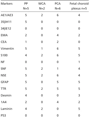
Table 2. Immunodetection of proteins in choroid plexus tumors and fetal normal choroid plexus.
Markers PP WCA PCA Fetal choroid
N=5 N=2 N=6 plexus n=5
AE1/AE3 5 2 6 4
35βH11 5 0 0 0
34βE12 0 0 0 0
EMA 2 0 4 2
CEA 1 0 2 0
Vimentin 5 1 6 5
S100 4 2 6 5
NF 0 0 0 1
SNF 5 2 1 4
NSE 5 2 6 4
GFAP 5 0 5 5
TTR 5 2 5 5
Desmin 4 0 0 3
1A4 2 0 4 2
Laminin 4 2 0 5
P53 0 0 0 0
Cases were considered positive when at least 10% of the cells showed the characteristic brown staining, either in the nuclei, cytoplasm or membrane, according to each antibody pattern. The frequency of antigen im-munodetection was studied comparatively in each tumor group and in normal fetal choroid plexus.
RESULTS
Between 1966 and 1999, 184 cases of CNS tumor were diagnosed in patients younger than 25 years. Thirteen (7%) corresponded to choroid plexus tu-mors: 5 PP and 8 CA (2 WCA and 6 PCA). Among the PP 3 patients were females and 2 males; age
Arq Neuropsiquiatr 2004;62(3-A) 603
ranged between 1 month and 25 years, with medi-an age of 1y 6mo. In 2 cases tumors were intraven-tricular without specification, one was in the pos-terior fossa / IV ventricle, another in the left later-al ventricle and in one case no information was ava-ilable. Among the CA 6 patients were females and 2 males; age ranged between 5 months and 3 years, with median of 11 months. In one case tu-mor was intraventricular without specification, 5 were in the posterior fossa / IV ventricle and in 2 no information was available.
The cases of choroid plexus papillomas recalled the normal architecture of the choroid plexus: cu-boidal or columnar epithelial cells formed a mono-layer on papillary vascular connective tissue stro-ma. The nuclei were ovoid, with regular, well distri-buted chromatin and the luminal surface of the neoplastic epithelium was smooth and straight, as opposed to the hobnail appearance of normal cho-roid epithelium. The PP tended to have higher cel-lularity than normal choroid plexus, although no quantitation was attempted. Choroid plexus
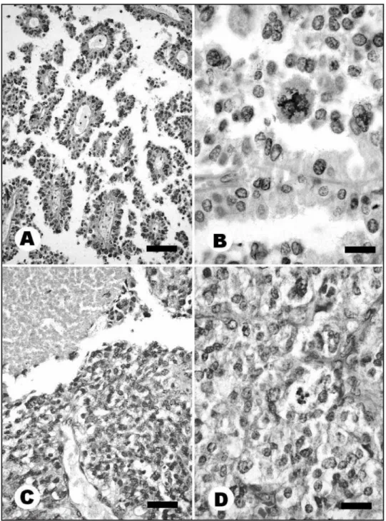
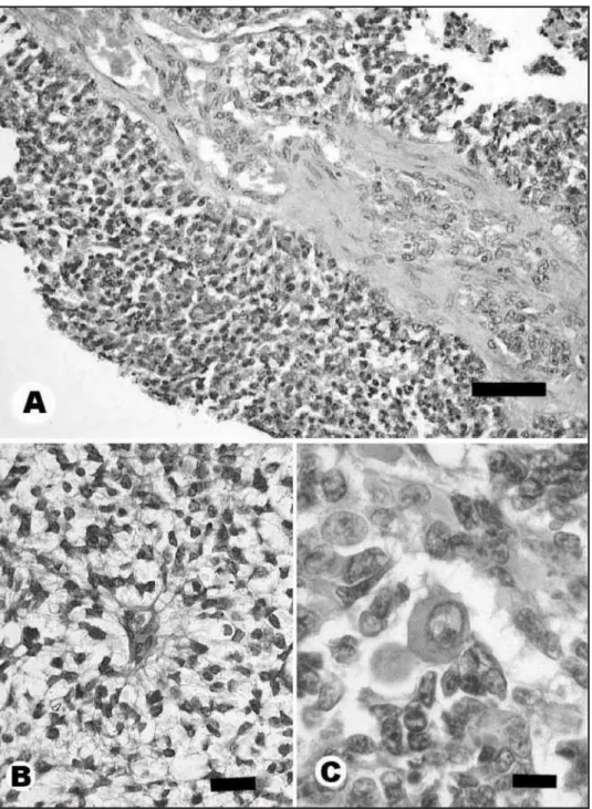
nomas were characterized by unequivocal malignant features, such as cytological atypia, necrosis, mitot-ic activity, brain invasion and/or loss of papillary architecture. Carcinomas were subclassified into well differentiated (WCA) and poorly differentiat-ed (PCA) basdifferentiat-ed on the prdifferentiat-edominance of papillary vs solid areas. There were 2 cases of WCA, with at least 50% of papillary areas and 6 PCA (Fig 1). Of these, only one showed a single focus of papillary structures. The other 5 consisted of solid tissue only. Besides these characteristic findings, some note-worthy observations were found in our cases. Vas-cular endothelial proliferation was found in 3 of 6 PCA (Fig 2A). In the same 3 cases, stromal prolif-eration formed thick irregular septa (Fig 2A).
Pe-rivascular pseudorosettes were present in the 2 cas-es of WCA and in 5 out of 6 cascas-es of PCA. Clear cells were a common finding (3/5 PP, 2/2 WCA and 5/6 PCA), mostly in focal areas (Fig 2B). In one case of PCA larger areas of clear cells were seen. These cells were a major finding in all examples of fetal cho-roid plexus examined. In every case, PAS reaction was negative in clear cells. Rhabdoid cells were found in 3 out of 6 PCA (Fig 2C). They were obser-ved mostly in perivascular distribution and were characterized by clear vesicular nuclei, with single prominent nucleolus and abundant acidophilic cytoplasm devoid of the classical hyaline body usu-al in rhabdoid cells. These cells showed immunore-activity for smooth muscle actin 1A4.
Arq Neuropsiquiatr 2004;62(3-A) 605
Immunohistochemical findings are summarized in Table 2 and shown in Figure 3, for AE1/AE3, GFAP, 1A4 and laminin.
DISCUSSION
Case selection was limited to ages from birth to 25 years because of the high incidence of choroid plexus tumors in the pediatric population with significant decrement after the second decade.
Some noteworthy points regarding frequency, histopathological features and immunohistochem-istry of choroid plexus tumors in childhood arose from our study. Choroid plexus tumors account for 1.5 to 3.9% of CNS tumors in children, PP being at least four times more common than CA1,2. There is
slight male predominance and the lateral ventricles are the most common site at this age. In the pres-ent series, the frequency of choroid plexus tumors was 7% (5 PP and 8 CA = 13 cases among 184 CNS tumors). The predominance of carcinomas as com-pared to papillomas does not reflect the incidence of the general population, since difficult cases from other institutions are often referred to our hospi-tal. There was female predominance (9 F, 4 M). As regards tumor location, 3 cases were intraventric-ular without specification, 6 were situated in the pos-terior fossa / IV ventricle, one in the left lateral ven-tricle and in 3 no information was available.
While histological criteria for well differentiat-ed choroid plexus tumors are clearly establishdifferentiat-ed26,28
the diagnosis of poorly differentiated variants is not so well defined.
In our series, vascular endothelial proliferation and stromal desmoplasia forming thick irregular septa were present in 3/6 cases of PCA. This fea-ture is frequently seen in astrocytic neoplasms, in which it is related to the degree of anaplasia and used as a criterion for histological grading. Endo-thelial proliferation is often found in high grade astrocytomas, in which the stimulus for prolifera-tion is attributed to the producprolifera-tion of angioproli-ferating factors by the neoplastic astrocytes them-selves29. It is therefore possible that the findings
of vascular proliferation and stromal desmoplasia in PCAs might hint at some sort of astrocytic dif-ferentiation of the choroid neoplastic cells. It should be recalled that the choroid plexus cell derives from a neuroepithelial common precursor cell, the so called oligopotential glio-ependymal precursor10,11,17.
Perivascular pseudorosettes were seen only in
WCA and PCA (2/2 WCA and 5/6 PCA) and may cau-se diagnostic difficulties with anaplastic ependymo-mas. While the perivascular pseudorosettes might be an indication of ependymal differentiation of the choroid neoplastic cells, it must also be kept in mind that ischemic necrosis of tumor cells at some distance from blood vessels might create a similar pattern.
Clear cells were common in all choroid plexus tumors, benign and malignant. As they are also very frequent in the fetal choroid plexus, they may sug-gest similarities between the neoplastic cells and immature related tissue.
Concerning the immunohistochemical profile, pancytokeratin AE1/AE3 was strongly positive in all tumor cases, even in the undifferentiated com-ponent of PCA, where reactivity was focal in 3 and diffuse in 3 cases. In contrast to the litera-ture8,9,14,18,19,21, low molecular weight cytokeratin
(35βH11) was not expressed in any of 8 CA, but was present in all 5 PP. In 4/6 PCA, including one with-out rhabdoid-like features, there was reactivity for smooth muscle actin 1A4, which could be detec-ted in 10 to 30% of the cells. Also, differently from some reports19, laminin was not a helpful tool in
the diagnosis of PCA, as it was not detected in any of the 6 cases. All 5 NFCP expressed GFAP, in con-trast with previous reports8-11,13. Absence of
expres-sion of 35βH11 among all 5 cases of NFCP was sim-ilar to what was obtained in CA. On the other hand, all PP showed positivity.
The main histological and immunohistochem-ical criteria for classifying the undifferentiated tu-mors as choroid plexus CA were respectively the diffuse, solid growth pattern without a fibrillary background, the presence of a scant rim of cyto-plasm in the small, round cells and strong reactiv-ity for pancytokeratin AE1/AE3 in the undifferentia-ted component. Some PCA showed immunoreac-tion to 1A4, which could cause difficulties in diffe-rential diagnosis with the rare and controversial AT/RT, also present in the posterior fossa of young children (usually before one year of age)22,23.
Ho-wever, in PCA the pattern of positivity was distinct from that found in AT/RT in that it did not corre-late exclusively with the rhabdoid-like cells but also with scattered undifferentiated epithelial and perivascular tapering cells. There was 1A4 positiv-ity even in a case lacking the rhabdoid-like com-ponent.
PCA and AT/RT may be difficult. The main criteria favoring PCA were homogeneity of the histolog-ical picture, without heterologous elements often found in AT/RT, such as mesenchimal areas, epithe-lial differentiation into both squamous and glandu-lar tissue. AT/RT are characterized by rhabdoid cells with globular intracytoplasmic bodies in 100% of cases, and a neuroectodermal (PNET) compo-nent evident in two thirds (predominant in 15-65%). In our cases, the undifferentiated compo-nent, although diffuse, was epithelial-like with scant cytoplasm. However, recent genetic studies have shown important similarities between choroid plexus carcinomas and AT/RT, both of which show inacti-vating mutations of the hSNF5/INI-1 gene in chro-mossome 22q11.2, considered an important step in the molecular pathogenesis of AT/RT. This points to a close relationship between these two entities30.
Furness et al19detected laminin in subepithelial
location and fragmented pattern in all choroid ple-xus CA; in contrast our PCAs did not show laminin, possibly due to the very undifferentiated state of the neoplastic cells without any reminiscence of papillary structures. On the other hand, PP (4/5) and all 5 NFCP showed strong, linear and continu-ous membrane reactivity for laminin. In both WCA laminin was found in a fragmented pattern.
The positivity to vimentin and S100 protein in almost all cases of PP, CA and NFCP is in agreement with other reports9,10,14,21 that both are associated
with tissues derived from the neuroectodermal plate. Unlike some reports11,14,19-21, in our cases there
was no correlation between the degree of tumor anaplasia and immunodetection of CEA, TTR and EMA. TTR was positive in all tumors and in NFCP, except one PCA. EMA showed positivity in 2/5 PP, 4/6 PCA and 2/5 FCP, and CEA in 1/5 PP and 2/6 PCA. As most reports in the literature10,11,13,14,16-19,21,31,
GFAP was strongly and widely expressed among all PP and 5/6 PCA, implying, as Rubinstein and Bru-cher16 proposed that during neoplastic
develop-ment choroid plexus epithelial cells express a fea-ture which is the prerogative of glial and ependy-mal related cells. However in Rubinstein’s and oth-er authors’ studies8-11,13of normal choroid plexus,
there was no glial marker immunodetection, with rare exceptions14. Two explanations have been
pro-posed: first, antigen retrieving techniques have been considerably improved compared to a decade ago when most of the reports were published; and second, most of the normal plexus studied were
adult samples. In children choroid plexus cells may have greater propensity to divergent differentia-tion17. In summary, neoplastic choroid cells, either
benign or malignant, and immature normal fetal choroid cells retain the genetic information of their parental neuroepithelial precursors which code for a glial phenotype in their progeny16.
The p53 tumor suppressor protein was negative in all cases examined suggesting that, in contrast to astrocytomas, p53 mutation seems not to be im-portant in the pathogenesis or progression of choroid plexus tumors. The actual role of the im-munodetection of the p53 protein is still controver-sial, as other authors show high positivity of this protein mainly in choroid plexus carcinomas, al-though in variable intensities32,33. However, in a
study including 10 choroid plexus tumors, Ohgaki et al34did not find mutations in exons 5-8 of the
p53 gene, what is in accordance with our finding.
CONCLUSIONS
Pancytokeratin (AE1/AE3) was expressed in all choroid plexus PP, CA and 80% of NFCP.
Low molecular weight cytokeratin (35βH11) was expressed in all PP but not in CA or NFCP.
Expression of epithelial markers (AE1/AE3 and EMA) is important to define the epithelial nature of the tumor, particularly in undifferentiated areas.
An immunohistochemical panel of 16 antibod-ies is useful to help distinguish PCA from other anaplastic tumors, such as AT/RT.
Fetal choroid plexus demonstrates multipotential-ity through coexpression of various markers: VIM, TTR, S-100, GFAP (100%), SNF, NSE, AE1/AE3 (80%), desmin (60%), EMA, 1A4 (40%) and NF (20%).
Laminin was not detected in PCA, but was use-ful for highlighting the basal lamina in PP and WCA.
Mutations of p53 gene do not appear impor-tant in pathogenesis and progression of choroid plexus tumors.
REFERENCES
1. Pianetti G, Fonseca LF. Tumores do plexo coróideo. Arq Neuropsiquiatr 1998;56:223-231.
2. Pencalet P, Sainte-Rose C, Lellouch-Tubiana A, et al. Papillomas and car-cinomas of the choroid plexus in children. J Neurosurg 1998;88:521-528 3. Lynch JC, Moraes GP, Duarte F. Xantogranuloma do plexo coróide. Arq
Neuropsiquiatr 1988;46:191-194.
4. Aguiar MFM, Cavalcanti M, Barbosa H, Vilela SL, Mendonça JL, Horta E. Síndrome de Aicardi e papiloma do plexo coróide: uma associação rara. Arq Neuropsiquiatr 1996;54:313-317.
Arq Neuropsiquiatr 2004;62(3-A) 607
8. Kasper M, Goertchen R, Stosiek P, Perry G, Karsten U. Coexistence of cytokeratin, vimentin and neurofilament protein in human choroid plexus: an immunohistochemical study of intermediate filaments in neu-roepithelial tissues. Virchows Arch A 1986;410:173-177.
9. Miettinen M, Clark R, Virtanen I. Intermediate filament proteins in choroid plexus and ependyma and their tumors. Am J Pathol 1986;123:231-240.
10. Doglioni C, Dell’Orto P, Coggi G, Iuzzolino P, Bontempini L, Viale G. Choroid plexus tumors: an imunocytochemical study with particular reference to the coexpression of intermediate filament proteins. Am J Pathol 1987;127:519-529.
11. Felix I, Phudhichareonrat S, Halliday WC, Becker LE. Choroid plexus tumors in children: immunohistochemical and scanning-electron-micro-scopic features. Pediat Neurosci 1987;13:263-269.
12. Gabrion J, Peraldi S, Faivre-Bauman A, et al. Characterization of ependy-mal cells in hypothalamic and choroidal primary cultures. Neuroscience 1988;24:993-1007.
13. Kouno M, Kumanishi T, Washiyama K, Sekiguchi K, Saito T, Tanaka R. An immunohistochemical study of cytokeratin and glial fibrillary acidic protein in choroid plexus papilloma. Acta Neuropathol (Berl) 1988;75:317-320.
14. Cruz-Sanchez FF, Rossi ML, Hughes JT, Coakham HB, Figols J, Eynaud PM. Choroid plexus papillomas: an immunohistological study of 16 cas-es. Histopathology 1989;15:61-69.
15. Sarnat HB. Regional differentiation of the human fetal ependyma: immunocytochemical markers. J Neuropathol Exp Neurol 1992;51:58-75. 16. Rubinstein LJ, Brucher JM. Focal ependymal differentiation in choroid plexus papillomas: an immunoperoxidase study. Acta Neuropathol (Berl) 1981;53:29-33.
17. Taratuto AL, Molina H, Monges J. Choroid plexus tumors in infancy and childhood: focal ependymal differentiation. An immunoperoxidase study. Acta Neuropathol (Berl) 1983;59:304-308.
18. Mannoji H, Becker LE. Ependymal and choroid plexus tumors. Cytokeratin and GFAP expression. Cancer 1988;61:1377-1385. 19. Furness PN, Lowe J, Tarrant GS. Subepithelial basement membrane
dep-osition and intermediate filament coexpression in choroid plexus neo-plasms and ependymomas. Histopathology 1990;16:251-255. 20. Herbert J, Cavallaro T, Dwork AJ. A marker for primary choroid plexus
neoplasms. Am J Pathol 1990;136:1317-1325.
21. Newbould MJ, Kelsey AM, Arango JC, Ironside JW, Birch J. The choroid plexus carcinomas of childhood: histopathology, immunocytochemistry and clinicopathological correlations. Histopathology 1995;26:137-143. 22. Rorke LB, Packer RJ, Biegel JA. Central nervous system atypical tera-toid/rhabdoid tumors of infancy and childhood. J Neurooncol 1995;24:21-28.
23. Rorke LB, Packer RJ, Biegel JA. Central nervous system atypical tera-toid/rhabdoid tumors of infancy and childhood: definition of an enti-ty. J Neurosurg 1996;85:56-65.
24. Burger PC. ATT of the CNS: a highly malignant tumor of infancy and childhood frequently mistaken for medulloblastoma. Am J Surg Pathol 1998;22:1083-1092.
25. Oka H, Scheithauer BW. Clinicopathological characteristics of atypical teratoid/rhabdoid tumor. Neurol Med Chir (Tokyo) 1999;39:510-518. 26. Kleihues P, Cavenee WK. Tumours of central nervous system.
pathol-ogy & genetics. Lyon: WHO, IARC, 2000.
27. Wyatt-Ashmead J, Kleinschmidt-DeMasters B, Mierau GW, et al. Choroid plexus carcinomas and rhabdoid tumors: phenotypic and genotypic over-lap. Pediatr Dev Pathol 2001;4:545-549.
28. Russell DS, Rubinstein LJ. Pathology of tumours of the nervous sys-tem. 5.Ed. London :Arnold, 1989.
29. Sreenan JJ, Prayson RA. Gliosarcoma: a study of 13 tumors, including p53 and CD34 immunohistochemistrty. Arch Pathol Lab Med 1997;121:129-133.
30. Gessi M, Giangaspero F, Pietsch T. Atypical teratoid/rhabdoid tumors and choroid plexus tumors: when genetics “surprise” pathology. Brain Pathol 2003;13:409-414.
31. Lopes MB, Rosemberg S, Almeida PC, Pestana CB. Glial fibrillary acidic protein and cytokeratin in choroid plexus tumors: an immunohistochem-ical study. Pathol Res Pract 1989;185:339-341.
32. Jay V, Ho M, Chan F, Malkin D. p53 expression in choroid plexus neo-plasms: an immunohistochemical study. Arch Pathol Lab Med 1996;120:1061-1065.
33. Carlotti CG Jr, Salhia B, Weitzman S, et al. Evaluation of proliferative index and cell cycle protein expression in choroid plexus tumors in chil-dren. Acta Neuropathol 2002;103:1-10.