Arq Neuropsiquiatr 2004;62(3-A):592-595
Hospital Santa Marcelina, São Paulo SP, Brasil: 1Residente do Serviço de Neurologia; 2Doutora em Medicina, Responsável pelo Ambulatório
de Cognição, Preceptora da Residência em Neurologia; 3Doutora em Medicina, Chefe do Serviço; 4Doutora em Medicina, Preceptora
da Residência; 5Mestre em Neurologia, Preceptora da Residência.
Received 16 December 2003, received in final form 4 March 2004. Accepted 5 April 2004.
Dra. Sonia M.D. Brucki - Rua Humberto Primo 740/123 - 04018-032 São Paulo SP - Brasil. E-mail: [email protected]
PREVALENCE OF PRESENILE DEMENTIA
IN A TERTIARY OUTPATIENT CLINIC
Satomi Fujihara
1, Sonia M.D. Brucki
2, Maria Sheila G. Rocha
3,
Alzira A. Carvalho
4, Ana C. Piccolo
5ABSTRACT - There are very few reports about prevalence of presenile dementia in Brazil. We reviewed files of patients evaluated with early onset of cognitive impairment in our institution. Among 141 patients (61% males) there was no difference between gender by age at onset or at first evaluation. We have observed an increasing number of patients after 50 years. The most frequent causes were: vascular dementia (36.9%), Alzheimer’s disease (20.3%) and traumatic brain injury (9.2%). There was difference among dementia type by age of onset and first evaluation, educational level and length of dementia. These results may be com-pared with those from other neurologic services in order to replicate or confirm these results.
KEY WORDS: presenile dementia, vascular dementia, Alzheimer’s disease, epidemiology.
Prevalência de demência pré-senil num ambulatório terciário
RESUMO - Em nosso meio há raros estudos que verifiquem quais as causas mais prevalentes de demência pré-senil. Avaliamos retrospectivamente os prontuários de pacientes com início precoce de alterações cog-nitivas, ambulatório de Neurologia da Cognição do Hospital Santa Marcelina. Entre os 141 sujeitos (61% de homens) não houve diferença quanto às idades de início e à primeira consulta e escolaridade entre os sexos. Observamos aumento no número de demência após os 50 anos. A causa mais freqüente foi vascu-lar (36,9%), seguida por doença de Alzheimer (20,3%) e secundária a trauma cranio encefálico (9,2%). Houve diferença entre os tipos de demência quanto à idade na primeira consulta e idade de início, escolaridade e duração do quadro. Ao contrário de outros estudos o diagnóstico mais freqüente foi demência vascular. Novos estudos em nosso meio deverão ser realizados para avaliar este achado nas demências de início pre-coce.
PALAVRAS-CHAVE: demência pré-senil, demência vascular, doença de Alzheimer, epidemiologia.
Dementia is a disorder of greater prevalence in older subjects, but many cases can begin in an ear-ly age, affecting people in a productive phase of their lives. A syndrome characterized by many cognitive deficits of sufficient severity to interfere with dai-ly life activities and in quality of life. It is a public health problem due its expensive treatment and dependence of patients for governmental sources for sustainability. There are a few studies in epidemi-ology of presenile dementia comparing to an old-er onset or familiar Alzheimold-er’s disease (AD)1-3.
The prevalence of presenile dementia is lower that ones with onset in the elderly. Harvey et al.4
report-ed that the prevalence increases exponentially bet-ween 45 to 60 years of age; this finding was repli-cated by another three epidemiological studies5-7. The
most frequent diagnosis in early onset dementia is:
Alzheimer’s disease, followed by vascular dementia (VaD) and frontotemporal dementia (FTD)8.
There are no studies analyzing the prevalence of presenile dementia in our country in a search in MEDLINE. Other authors have reported diagnos-tic prevalence in senile dementia or overall rates of dementia in their outpatient clinics9-12or
institu-tionalized patients13. One report was presented as
abstract and has reviewed a considerable number of patients (n= 619) and found a prevalence of 28.6% of presenile dementia14. The aim of our
Arq Neuropsiquiatr 2004;62(3-A) 593
METHOD
We reviewed 311 files of patients consecutively seen in the Cognitive Clinic of Santa Marcelina Hospital, from 1997 to 2003, which have been sent for evaluation of cognitive complaints. This research was approved by the ethics committee. A hundred forty one subjects ful-filled the criteria for dementia (DSM-IV)15and the
clin-ical onset of dementia has begun before 65 years old of age (presenile dementia). All patients had clinical, neu-rological and cognitive examinations and a complete lab-oratory evaluation: blood count with erythrocyte sedi-mentation rate, serum cholesterol, triglycerides, creati-nine, urea, sodium, potassium, calcium, phosphorus, protein, glucose, bilirubin, alkaline phosphatase, ALT and AST, thyroid hormones, VDRL and FTA-abs reactions. Besides that, cerebral spinal fluid when necessary, and viral serology (IHV, hepatitis C and B virus) was perfor-med. The type of dementia was based on the following criteria: AD was made according to National Institute of Neurological and Communicative Disorders and Stroke/ Alzheimer’s Disease and Related Disorders Association (NINCDS/ ADRDA) 16; VaD followed National Institute of
Neurological Disorders and Stroke – Association Inter-nationale pour la Recherche et l’Enseignement en
Neu-rosciences (NINDS/ AIREN)17criteria, Lewy body disease
followed guidelines proposed by McKeith et al.18, revised
in 199919; FTD diagnosis was based on Lund and
Man-chester criteria20 and alcoholic dementia by DSM-IV
cri-teria15. For patients with memory complaints and
objec-tive memory impairment without dementia, we used the criteria for mild cognitive impairment (MCI)21. We also
observed risk factors that may corroborate with the cli-nical picture of dementia, particularly those related with cerebrovascular disease and family history for de-mentia. The severity was evaluated according to the initial scores on Mini-Mental State Examination (MMSE)22.
Statistical analysis was performed utilizing STATISTI-CA. T-test was used for demographic variables and analy-sis of variance with Tukey test.
RESULTS
A hundred forty one files of presenile dementia subjects were examined cautiously, other four pa-tients were diagnosed as MCI (mean age of 53. SD = 10.4 years and a mean time of outcome of 36 mon-ths). In Table 1 we describe clinical and demograph-ic variables of early onset dementia patients. Table 1. Demographic and clinical variables of presenile patients (N = 141).
Mean (SD) Median value minimum maximum
Age (years) 53.3 (12.1) 57 21 65
Educational level (years) 3.6 (3.1) 4 0 12
Age of onset 51 (11.4) 54 21 65
Length of dementia (months) 42.2 (44.5) 24 2 306
Initial MMSE 18.2 (6.0) 18 2 29
Table 2. Frequency of dementia by age of onset and gender.
Age of onset Men Women
(years) (n= 86) (n= 55)
N (%) N (%)
<30 5 (5.8) 5 (9.1)
30-34 2 (2.3) 3 (5.4)
35-39 4 (4,6) 4 (7,3)
40-44 6 (7.0) 4 (7.3)
45-49 8 (9.3) 7 (12.7)
50-54 10 (11.6) 9 (16.4)
55-59 25 (29.1) 8 (14.5)
60-65 19 (22.1) 14 (25.4)
No date 7 (8.1) 1 (1.8)
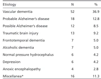
Table 3. Etiology of presenile dementia (n = 141).
Etiology N %
Vascular dementia 52 36.9
Probable Alzheimer’s disease 18 12.8 Possible Alzheimer’s disease 12 8.5
Traumatic brain injury 13 9.2
Frontotemporal dementia 7 5.0
Alcoholic dementia 7 5.0
Normal pressure hydrocephalus 6 4.2
Depression 6 4.2
Anoxic encephalopathy 4 2.8
Miscellanea* 16 11.3
594 Arq Neuropsiquiatr 2004;62(3-A)
There was a male predominance in the sample (61%) with similar distribution between gender: in age (p= 0.3302) and educational level (p= 0.7469). Mean age of men was 54.2 years and median val-ue of 58 years. Women had mean age of 52.1 years and median value of 54 years. The mean schooling was 3.72 and 3.53 years, for men and women, res-pectively. There was no difference at age of onset between gender, a mean of 49.1 years in males and 52.4 years in females. In Table 2 we show the fre-quency by five years interval. An increased pre-valence after 45 years old was observed. After 50 years there was more than an half of the sample in both sexes (61.8% of men and 56.3% of women).
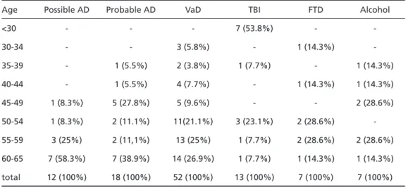
VaD dementia was the most frequent etiology (36.9%) followed by AD (probable AD: 12.8% and possible AD: 8,5%). Traumatic brain injury (TBI) was the prime cause among nondegenerative de-mentias (9.2%), see (Table) 3. In Table 4 we report frequencies by age groups by dementia.
Analysis of variance (ANOVA) was performed to compare dementia groups by demographic vari-ables and initial scores on the MMSE. There were differences related to: age at onset in the first evaluation, F (22,118) =3.8917, p <0.0001); age of onset by history, F (21,111) =3.2482, p < 0.0001; years of schooling, F (20,95) = 2.6638, p= 0.0007; and length of dementia, F (21,111) = 1.7125, p= 0.0388. In the post-hocanalysis by Tukey’s test, we observed differences between TBI and VaD patients by educa-tional level (p= 0.0471), with greater level for the first and more educated group. TBI subjects were different by age at the first evaluation when com-pared to AD and VaD, these results were replicat-ed in the analysis by age of onset, comparing with: VaD (p= 0.0032), possible AD (p= 0.0002), and
pro-bable AD (p= 0.0013). In both situations age of TBI subjects was minor.
Familiar history was reported by 22 patients (15.6% of sample): probable AD, 9 subjects (40.9%); possible AD, 3 cases (13.6%); FTD, 3 cases (13.6%); and VaD, 3 cases (13.6%). These findings of famil-iar history correspond to 50% in probable AD, 25% of cases of possible AD, 42.8% of FTD, and only 5.8% of VaD. The most frequent risk factors for cerebrovascular disease were: systemic hyper-tension (41.8%), smoking (25.5%), dyslipidemia (24.1%), alcoholism (20.6%), and diabetes (17%). In VaD patients 78.8% reported previous stroke, and presented more risks for cerebrovascular dis-ease, as expected: systemic hypertension (57.7%), dyslipidemia (32.7%), smoking (30.8%), diabetes (21.1%), and alcoholism (17.3%). Seizures were associated with dementia in 16 patients, five with VaD and one AD subject.
DISCUSSION
About half of evaluated patients (46.6%) in our clinic met criteria for presenile dementia, in con-trast to other preliminary study that an early onset of dementia has occurred in 28.6%14; these
find-ings confirm the obligation to pay attention for this diagnosis by clinicians and neurologists in gen-eral, in front of a young people with cognitive com-plaints. Our patients exhibited similar age on diag-nosis time compared to another studies, Panegyres et al.23observed a mean age of 54.2 years among
their 150 patients and Harvey et al.24in their
preva-lence study showed a little more old subjects (mean 58.7 years). Also there was a predominance of ma-les in studies of early onset dementia23,24.
Table 4. Prevalence of dementia types by age group.
Age Possible AD Probable AD VaD TBI FTD Alcohol
<30 - - - 7 (53.8%) -
-30-34 - - 3 (5.8%) - 1 (14.3%)
-35-39 - 1 (5.5%) 2 (3.8%) 1 (7.7%) - 1 (14.3%)
40-44 - 1 (5.5%) 4 (7.7%) - 1 (14.3%) 1 (14.3%)
45-49 1 (8.3%) 5 (27.8%) 5 (9.6%) - - 2 (28.6%)
50-54 1 (8.3%) 2 (11.1%) 11(21.1%) 3 (23.1%) 2 (28.6%) -55-59 3 (25%) 2 (11,1%) 13 (25%) 1 (7.7%) 2 (28.6%) 2 (28.6%)
Arq Neuropsiquiatr 2004;62(3-A) 595
AD is the most frequent etiology in studies with early onset dementia2,4,23-27, although in one report
VaD is the most prevalent (Southampton area)26as
well as in the Japanese study28. In our clinic VaD
was the most important cause of dementia, with a frequency of 36.9% and increasing prevalence after 50 years. These differences with other occiden-tal studies may be dueto particular sample characte-ristics: lower socioeconomic level causing under risk factor control for cerebrovascular diseases, facilita-ted reference to our clinic, heterogeneous racial population (although with no Japanese descen-dents). The second more frequent type was AD, with increasing prevalence in older ages. Among young adults we had TBI and FTD as common eti-ologies, but the second was less frequent than other studies4, 23. This fact does not seem to us a
diagnostic failure, and we could not explain these findings. Maybe our lower prevalence of FTD fol-lows the general prevalence of this type of demen-tia in Brazil. We do not have data about this preva-lence by epidemiological surveys. Alcohol demen-tia was more frequent in epidemiological study in England24 than in our clinic perhaps due to
evalu-ation by psychiatrists in our environment.
One point must be highlighted: patients spend a long time before to look for medical helping, as we could see observing age between onset of cog-nitive impairment and age of first evaluation, a mean of three years to go to medical assistance.
We can conclude that some differences exist bet-ween presenile and senile dementia in Brazil among studies. It is necessary more studies in different re-gions of our country about early onset dementia, and the ones with senile dementia clearly has showed AD as principal cause of dementia9-13.
Collaborative researches can clarify this picture.
REFERENCES
1. McGonigal G, Thomas B, Mcquade C, et al. Epidemiology of Alzheimer’s presenile dementia in Scotland, 1974-88. Br Med J 1993;306:680-683. 2. Newens AJ, Forster DP, Kay DW, et al. Clinically diagnosed presenile
dementia of the Alzheimer type in the Northern Health Region: ascertain-ment, prevalence, incidence and survival. Psychol Med 1993;23:631-644. 3. Treves T, Korczyn ADK, Zilber N, et al. Presenile dementia in Israel.
Arch Neurol 1986;43:26-29.
4. Harvey RJ, Skelton-Robinson M, Rossor MN. The prevalence and caus-es of dementia in people under the age of 65 years. J Neurol Neurosurg Psychiatry 2003;74:1206-1209.
5. Hoffman A, Rocca WA, Brayne C, et al. The prevalence of dementia in Europe: a collaborative study of 1980-1990 findings. Int J Epidemiol 1991;20:736-748.
6. Jorm AF, Korten AE, Henderson AS. The prevalence of dementia: a quan-titative integration of the literature. Acta Psychiatr Scand 1987;76:465-479. 7. Kokmen E, Beard CM, Offord KP, et al. Prevalence of medically diag-nosed dementia in a defined United States population: Rochester, Minnesota, January 1 1975. Neurology 1989;39:773-776.
8. Varma AR, Adams W, Lloyd JJ, et al. Diagnostic patterns of regional atrophy on MRI and regional cerebral blood flow change on SPECT in young onset patients with Alzheimer’s disease, frontotemporal demen-tia and vascular demendemen-tia. Acta Neurol Scand 2002;105:261-269. 9. Vale FAC, Miranda SJC. Clinical and demographic features of patients
with dementia attended in a tertiary outpatient clinic. Arq Neuropsi-quiatr 2002;60:548-552.
10. Silva DW, Damasceno BP. Demência na população de pacientes do Hospital das Clínicas da UNICAMP. Arq Neuropsiquiatr 2002;60:996-999. 11. Caixeta L. Epidemiologia das diferentes formas de demência em Goiás.
Arq Neuropsiquiatr 2003;61(Suppl 2):S19.
12. Takada LT, Caramelli P, Radanovic M, et al. Prevalence of potentially reversible dementias in a dementia outpatient clinic of a tertiary uni-versity-affiliated hospital in Brazil. Arq Neuropsiquiatr 2003;61:925-929. 13. Canineu PR, Damasceno BP, Silva MC. Prevalência de demências na população de pacientes idosos (60 anos) internados no serviço de saúde Dr Cândido Ferreira da Prefeitura Municipal de Campinas. Arq Neuro-psiquiatr 2003;61(Suppl 2):S21.
14. Engelhardt E, Laks J, Leibing A, et al. Demência pré-senil: aspectos pre-liminares. Arq Neuropsiquiatr 2001;59(suppl 3):S3-S4.
15. American Psychiatric Association. Diagnostic and statistical manual of mental disorders, 4.Ed. (DSM-IV). Washington DC: APA, 1994. 16. McKhann G, Drachman D, Folstein M, Katzman R, Price D, Stadlan
EM. Clinical diagnosis of Alzheimer’s disease: report of the NINCDS/ ADRDA Work Group under the auspices of Department of Health and Human Services Task Force on Alzheimer’s disease. Neurology 1984; 34:939-944.
17. Roman GC, Tatemichi TK, Erkinjuntti T, et al. Vascular dementia: diag-nostic criteria for research studies. Report of the NINDS/ AIREN International Workshop. Neurology 1993;43:250-260.
18. McKeith IG, Galasko D, Kosaka K, et al. Consensus guidelines for the clinical and pathological diagnosis of dementia with Lewy bodies (DLB): report of the consortium on DLB international workshop. Neuro-logy 1996;47:1113-1124.
19. McKeith IG, Perry EK, Perry RH. Report of the second dementia with Lewy body international workshop: diagnosis and treatment. Consortium on dementia with Lewy bodies. Neurology 1999;53:902-905. 20. The Lund and Manchester Groups. Clinical and neuropathological
cri-teria for frontotemporal dementia. J Neurol Neurosurg Psychiatry. 1994;57:416-418.
21. Petersen, RC, Stevens, JC, Ganguli MC, et al. Early detection of demen-tia: mild cognitive impairment (an evidence-based review): report of the Quality Standards Subcommittee of the American Academy of Neurology. Neurology 2001;56:1133-1142.
22. Brucki SMD, Nitrini R, Caramelli P, Bertolucci PHF, Okamoto IH. Su-gestões para o uso do Mini-Exame do Estado Mental no Brasil. Arq Neu-ropsiquiatr 2003;61:777-781.
23. Panegyres PK, Dvaies SR, Connor CF. Early-onset dementia. Med J Aust 2000:173:279-280.
24. Harvey RJ, Rossor MN, Skelton-Robinson M, Garralda E. Young onset dementia: epidemiology, clinical symptoms, family burden, support and outcome. Dementia Research Group. London.1998.
25. Elberling TV, Stokholm J, Hogh P, Waldemar, G. Diagnostic profile of young and middle-aged memory clinic patients. Neurology 2002;59:1259-1262. 26. Woodburn KJ, Johnstone EC. Early-onset dementia in Lothian, Scotland: an analysis of clinical features and patterns of decline. Health Bull (Edinb) 1999;57:384-392.
27. Delaney N, Rosenvinge H. Presenile dementia: suffers, careers and services. Int J Geriatr Psychiatry 1995;10:597-601.