To date, despite modern advances in understan-ding, monitoring, and treating increased intracra-nial pressure (ICP), patients suffering from uncon-trollable intracranial hypertension due to
trauma-tic brain swelling most often either die or survive in an extremely disabled state1-3. It is well known that intracranial hypertension can lead to a reduction in cerebral perfusion pressure (CPP) and a potential
1Staff Physician, Division of Neurosurgery, Hospital das Clínicas, São Paulo University Medical School, São Paulo SP, Brazil (FMUSP); 2Staff Physician, Division of Neurology, Hospital das Clínicas; 3Chairman, Division of Neurosurgery, Hospital das Clínicas FMUSP.
Received 14 July 2003, received in final form 25 February 2004. Accepted 30 March 2004.
Dr. Edson Bor-Seng-Shu - Rua Loefgreen 1272 - 04040-001 São Paulo SP - Brasil. E-mail: [email protected]
TRANSCRANIAL DOPPLER SONOGRAPHY IN TWO
PATIENTS WHO UNDERWENT DECOMPRESSIVE
CRANIECTOMY FOR TRAUMATIC BRAIN SWELLING
Report of two cases
Edson Bor-Seng-Shu
1, Manoel Jacobsen Teixeira
1, Roberto Hirsch
2,
Almir Ferreira de Andrade
1, Raul Marino Jr
3ABSTRACT - The role of decompressive craniectomy in the treatment of severe posttraumatic cerebral swelling remains quite a controversial issue. To the best of our knowledge, there is no study demonstrating the ef-fect of decompressive craniectomy on cerebral blood flow (CBF) velocity by means of transcranial Doppler sonography (TCD). We present two patients who developed traumatic brain swelling and uncontrollable intracranial hypertension with coma and signs of transtentorial herniation. One patient underwent bifrontal, while the second, unilateral, frontotemporoparietal decompressive craniectomy with dural expansion. In both patients, TCD examinations were performed immediately before and after surgery to study the cere-bral hemodynamic changes related to the operations. Pre and postoperative TCD examinations demonstra-ted a significant increase in blood flow velocity in the intracranial arteries in both subjects. In conclusion, our cases suggest that decompressive craniectomy with dural expansion may result in elevation of CBF veloci-ty in patients with massive brain swelling. The increase in CBF velociveloci-ty appears to occur not only in the decom-pressed hemisphere, but also on the opposite side.
KEY WORDS: brain swelling, cerebral hemodynamics, decompressive craniectomy, head injury, intracranial hypertension, transcranial Doppler sonography.
Doppler transcraniano em doentes com tumefação encefálica pós-traumática submetidos à craniectomia descompressiva: relato de dois casos
RESUMO - Atualmente, as controvérsias sobre os benefícios da craniectomia descompressiva no tratamen-to de doentes com tumefação encefálica pós-traumática ainda existem. Não há estudos disponíveis na lite-ratura médica sobre os efeitos da craniectomia descompressiva na hemodinâmica encefálica avaliados pelo Doppler transcraniano. Estudamos dois doentes com tumefação encefálica pós-traumática associada à hiperten-são intracraniana e evidências de herniação transtentorial. Um deles foi submetido à craniectomia bifrontal e outro, à craniectomia frontoparietotemporal unilateral. O primeiro doente apresentou seqüela neurológi-ca moderada e reintegração social, enquanto o segundo manteve-se em estado vegetativo. Exames de Doppler transcraniano realizados imediatamente antes e após a cirurgia revelaram aumento significativo da veloci-dade do fluxo sangüíneo nas artérias intracranianas de ambos os doentes. Concluiu-se que a craniectomia descompressiva com plástica de ampliação da dura-máter pode resultar em elevação da velocidade do fluxo sangüíneo encefálico nestes doentes. O aumento da velocidade do fluxo sangüíneo pode ocorrer nos hemisfé-rios cerebrais do lado operado e, também, do lado oposto.
decrease in cerebral blood flow (CBF). As a conse-quence, secondary ischemic insults and increases in cerebral swelling may occur4-10.
If conventional medical therapy fails in patients with refractory intracranial hypertension due to ce-rebral swelling, decompressive craniectomy with dural augmentation may be indicated as a last re-sort11. However, the actual benefit of this surgical procedure on patient outcome remains a controver-sial issue in neurosurgical literature. There is no con-clusion as to how effective this surgical treatment
pressive craniectomy21,22. To the best of our knowl-edge, there is no work demonstrating the effect of decompressive craniectomy on cerebral blood flow velocity by means of transcranial Doppler sonogra-phy (TCD) in head-injured patients.
In this study we present two patients with trau-matic brain swelling and uncontrollable intracra-nial hypertension treated by decompressive craniec-tomy with dural opening. TCD, performed pre- and postoperatively, demonstrated a significant increase in blood flow velocity (BFV) in the intracranial ar-teries after surgical decompression. The significan-ce of these findings from a significan-cerebral hemodynamic point of view have been discussed.
CASES
Patient 1.A 28-year-old man was admitted to the hos-pital on October 25th, 1998, about 30 minutes after fal-ling from a height (> 5 meters). On admission, his Glas-gow coma scale (GCS) score was 12 while CT scan showed a diffuse traumatic subarachnoid hemorrhage, a brain swelling, and a small left-frontal contusion without sig-nificant mass effect. This patient underwent the place-ment of an ICP monitor and was transferred to neurolo-gical intensive care unit. Three days later, the patient de-teriorated neurologically to a GCS score of 6, and the ICP reached values between 35-40 mmHg, which was con-trolled by elevation of the head, mannitol solution and sedative drugs for a further 2 days, after which the ICP became uncontrollable, reaching 60 mmHg. At this time, a slight right hemiparesis was also noted. CT scan was
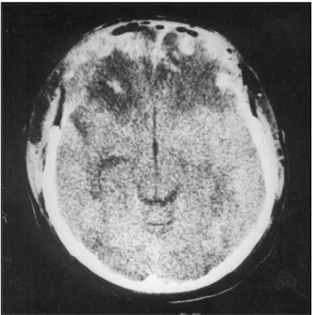
Fig 1. CT scan of patient 1 after bifrontal decompressive craniec-tomy.
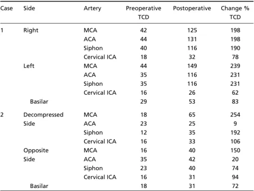
repeated and demonstrated worsening of the brain swelling. Both frontal and left temporal lobes were hy-poatenuated. A large bifrontal decompressive craniecto-my with dural expansion graft was indicated (Fig 1). TCD was performed immediately before and after surgery ac-cording to the technique proposed by Aaslid et al.23. The
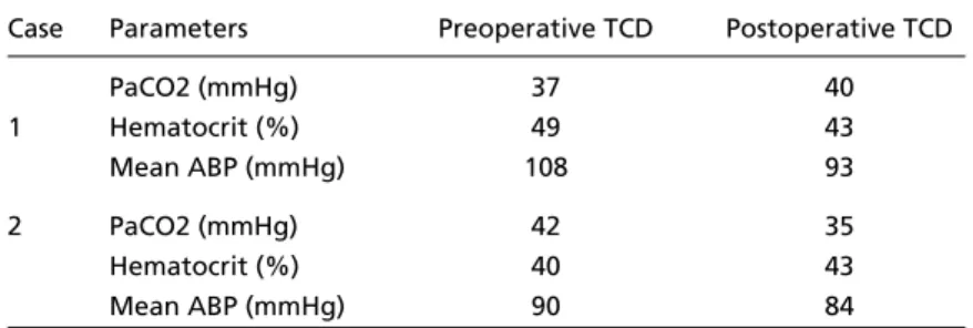
results are shown in Tables 1 and 2. The physiological parameters such as mean arterial blood pressure, blood gases, and hematocrit were recorded during TCD
stud-ies (Table 3). Subsequently, the patient recovered slow-ly. Four months after this trauma, the patient was awake, oriented, and walking with a mild hemiparesis. In the 2-year-follow-up, the value of the Glasgow Outcome Scale score was 4 (moderate disability and social reintegration).
Patient 2. A 22-year-old man was admitted to the hos-pital on March 24th, 1999, with a 30 minutes history of falling from a bus in motion. The initial GCS score was
Fig 3. Transcranial Doppler spectral waveforms obtained from the right middle cerebral artery of patient 2. A, before decompressive craniectomy, the cerebral circulation was characterized by reduced blood flow velocity and high pulsatility index (PI) (18 cm/s and 7.09, respectively). Note that during the diastolic phase, the blood flow velocity decreases continuously reaching zero value, and straight away, there is a rever-sion of flow direction (see arrow). This finding can indicate the presence of critical intracranial hyperten-sion with a severe impairment of cerebral blood flow. B, immediately after surgery, the blood flow was restored to a unidirectional pattern, with more acceptable flow dynamics in terms of flow velocity and PI (65 cm/s and 0.92, respectively).
8, but this patient rapidly deteriorated to a GCS score of 6 and developed anisocoria with a greater right pupil. CT scan revealed a thin subdural hematoma on the right side (greatest thickness: 0.4 mm) and brain swelling,
main-ly in the right hemisphere. The midline shift from the right to the left was of 14 mm at the anterior septum, and the mesencephalic cisterns were compressed, as well as the right lateral ventricle (Fig 2A). TCD performed soon
TCD TCD
1 Right MCA 42 125 198
ACA 44 131 198
Siphon 40 116 190
Cervical ICA 18 32 78
Left MCA 44 149 239
ACA 35 116 231
Siphon 35 116 231
Cervical ICA 16 26 62
Basilar 29 53 83
2 Decompressed MCA 18 65 254
Side ACA 23 25 9
Siphon 12 35 192
Cervical ICA 16 33 106
Opposite MCA 16 40 150
Side ACA 35 42 20
Siphon 23 40 74
Cervical ICA 16 31 94
Basilar 18 31 72
aTCD, transcranial Doppler sonography; MCA, middle cerebral artery; ACA, anterior cerebral artery; ICA,
internal carotid artery.
Table 2. Changes in Pulsatility Index before and after Decompressive Craniectomya.
Case Side Artery Preoperative Postoperative
TCD TCD
1 Right MCA 1.38 1.15
ACA 1.11 1.00
Siphon 1.19 1.15
Cervical ICA 1.16 0.97
Left MCA 1.42 1.16
ACA 1.21 0.88
Siphon 1.42 1.19
Cervical ICA 1.16 1.32
Basilar 1.21 1.34
2 Decompressed MCA 7.09 0.92
Side ACA 3.90 0.83
Siphon 5.27 1.33
Cervical ICA 3.18 1.09
Opposite MCA 5.87 1.30
Side ACA 1.35 0.89
Siphon 3.20 1.38
Cervical ICA 3.29 1.05
Basilar 3.00 1.10
aTCD, transcranial Doppler sonography; MCA, middle cerebral artery; ACA, anterior
after CT disclosed a significantly decreased BFV and an important increase in the pulsatility index (PI) (Tables 1,2). There was an oscillating flow pattern characterized by a discrete reversed-flow direction during the diastolic phase in the middle and anterior cerebral arteries (Fig 3A). In the basilar artery, the end diastolic BFV reached zero value (Fig 4A). This patient underwent a large right frontotemporoparietal decompressive craniectomy with dural augmentation. Postoperatively, TCD demonstrat-ed an important increase in BFV along with a decrease in PI, in relation to the preoperative values (Tables 1, 2). There was no longer an oscillating flow pattern in the intracranial arteries (Fig 3B) and the diastolic blood flow velocity was no longer zero (Fig 4B). The physiological parameters such as mean arterial blood pressure, blood gases, and hematocrit were recorded at the moment of each TCD study (Table 3). Approximately 24 hours lat-er, this patient developed anisocoria with a greater left pupil and an increase in tension at the craniectomy site. CT scan disclosed a large left fronto-temporo-parietal subdural hematoma of delayed development (Fig 2B). This patient was brought to the operating room and the hematoma was evacuated by craniotomy (Fig 2C). On discharge from hospital, 5 months on, this patient remai-ned in a vegetative state. In a two-year follow-up, the patient had not improved neurologically.
DISCUSSION
Transcranial Doppler sonography was first intro-duced by Aaslid et al.23in 1982, and quickly allowed new perspectives for assessing cerebrovascular he-modynamics. TCD has been validated scientifical-ly and its use has become routine practice in recent years, in the diagnostic study of patients with seve-re head injury and ceseve-rebrovascular disease24. TCD allows a rapid, noninvasive, reproducible, and dy-namic examination of intracranial circulation. The hemodynamic parameter, measured in real-time in the major intracranial arteries, is blood flow veloc-ity. The direction of the blood flow is also record-ed5,24,25. Since the reliability of findings is operator
dependent5,24,25, for this study, all examinations were performed by an experienced TCD operator (senior author) so that the depth and angle of ves-sel insonation could be maintained as similar as pos-sible, pre and postoperatively. TCD monitoring cannot give quantitative blood flow data, such as flow rate (in ml/min) or tissue perfusion (in ml /100g/min)25. However, it is possible to use TCD to follow relative changes in flow in a specific artery, over a period. Considering the principle that giv-en the diameter of the evaluated arterial segmgiv-ent remains constant, any change in flow velocity cor-relates well with a change in cerebral blood flow in the territory of that vessel5,25,26. This principle ena-bles TCD to be used to evaluate the effect of var-ious therapies, such as decompressive craniectomy, on relative changes in cerebral blood flow. Both of our patients presented a significant increase in CBF velocity after surgical decompression.
Currently, it is well known that the increase in ICP provokes changes in cerebral circulation, which may be evaluated with TCD hemodynamic param-eters5,6,9,24,25,27,28. Researchers in TCD are now trying to estimate the cerebral perfusion pressure8and pre-dict the intracranial pressure curves29in a noninvasi-ve manner. An elevation in ICP causes an increase in cerebrovascular resistance (CVR), probably as a result of cerebral microcirculatory or venous com-pression. This elevation in CVR results in an increase in the pulsatility index (PI), which is defined by the formula (systolic velocity - diastolic velocity)/mean velocity. In patients with intact cerebral autoregu-lation, increases in ICP will cause an increase in the PI and no change in the mean flow velocity, if the CPP remains in the autoregulatory range. Further increases in ICP that bring the CPP below the range of autoregulation, will cause a further increase in PI, as well as a decrease in mean flow velocity. When ICP reaches the level of the diastolic systemic blood
Table 3. Physiological Parameters During the Pre- and Postoperative Transcranial Doppler Sonography (TCD) Examinationsa.
Case Parameters Preoperative TCD Postoperative TCD
PaCO2 (mmHg) 37 40
1 Hematocrit (%) 49 43
Mean ABP (mmHg) 108 93
2 PaCO2 (mmHg) 42 35
Hematocrit (%) 40 43
Mean ABP (mmHg) 90 84
aTCD, transcranial Doppler sonography; PaCO2, arterial blood CO2 partial pressure; ABP,
phase of circulation. With a further increase in ICP, a to-and-fro, also called oscillating flow, pattern may appear, when the flow progress in the systole and, during diastole, a critically high ICP, CVR and a dis-tended intracranial arteries eject the blood in a ret-rograde direction. When net forward flow is seri-ously reduced, severe ischemic brain damage or bra-in death may occur. With a persistent level of crit-ically raised ICP, the intracranial waveform degrades to become a small systolic spike and then disappe-ars altogether5,6,9. Case 2 presented a cessation of diastolic flow in the basilar artery (Fig 4A) and an oscillating progressive flow in the middle cerebral arteries (Fig 3A), translating to a critical compromi-se of intracranial circulation resulting from compromi-severe intracranial hypertension. Comparing the PI in bo-th of our patients before and after decompression, it is evident that, preoperatively, the value of the PI was higher, indicating raised ICP. After surgical decompression, there was a significant decrease in PI (Table 2). The CBF of case 2 was restored to a uni-directional pattern and an acceptable value for dias-tolic velocity (Figs 3B and 4B). These cerebral hemo-dynamic findings could only be explained by a re-duction of ICP due to decompressive craniectomy. The diagnosis of posttraumatic vasospasm is important since it is a potential contributor to sec-ondary ischemic injury, besides being a factor for worsening brain swelling30,31. Various studies have been published showing that vasospasm may occur in between 18.6 to 68 % of severe head-injured pa-tients during the posttraumatic course30,31. Once diagnosed, rational therapy strategies to increase CBF may be planned and instituted. The TCD crite-ria for diagnosing cerebral vasospasm have been improved over the years. The criteria adopted by Martin et al.31, which require both a threshold middle cerebral artery velocity of 120 cm/s and a “Lindegaard” hemispheric ratio (mean MCA blood velocity divided by the ipsilateral extracranial inter-nal carotid artery blood velocity) greater than 3, per-mit differentiation between elevated velocities se-condary to increased CBF, and those which are ele-vated chiefly as a result of arterial narrowing31,32. For the diagnosis of vasospasm in the anterior cere-bral arteries, a velocity of 130 cm/s or 140 cm/s is required so that the specificity of the TCD would be 96% to 100%33. Taking all this into account on when analyzing postoperative TCD data, case 1
pre-CT scan and developed hemiparesis during the post-traumatic course. This fact could increase the pos-itive predictive value of these TCD findings. We be-lieve that before surgical decompression, case 1 had already presented vasospasm, which could not be demonstrated by TCD due to concomitant raised ICP, since the sensitivity of TCD in the diagnosis of vasospasm is reduced in the presence of elevated ICP34. In theory, this is a dangerous cerebral hemody-namic condition, in which the vasospasm in the lar-ge arteries (M1 and A1) leads to increased macro-vasculature resistance while at the same time, in-tracranial hypertension increases the microvascula-ture resistance such that the effect on CBF reduc-tion is more significant. In case 1, probably the asso-ciation of vasospasm and intracranial hypertension resulted in CBF reduction to an ischemic threshold level where the vicious circle of low CBF, with ische-mia, cerebral swelling and intracranial hyperten-sion could be established and maintained. We be-lieve that the indication of decompressive craniecto-my in this patient played an important role in bre-aking this vicious circle, providing the possibility of reducing ICP and increasing CBF velocity.
using SPECT, which may support our data in terms of the CBF increase after this operation.
In conclusion, our cases suggest that decompres-sive craniectomy with dural expansion may result in an elevation of CBF velocity in patients with mas-sive cerebral swelling. The increase in CBF velocity appears to occur not only in the decompressed he-misphere, but also on the opposite side. Prospective studies concerning this issue are warranted.
REFERENCES
1. Marshall LF, Gautille T, Klauber MR. The outcome of severe closed head injury. J Neurosurg 1991;75:28-36.
2. Polin RS, Shaffrey ME, Bogaev CA, et al. Decompressive bifrontal craniectomy in the treatment of severe refractory posttraumatic cere-bral edema. Neurosurgery 1997;41:84-94.
3. Marmarou A, Fatouros PP, Barzó P, et al. Contribution of edema and cerebral blood volume to traumatic brain swelling in head-injured patients. J Neurosurg 2000;93:183-193.
4. Muizelaar JP, Becker DP, Lutz HA, Newlon PG. Cerebral ischemia after severe head injury: its role in determining clinical status and its possi-ble treatment. In Villani R, (ed). Advances in neurotraumatology. Ams-terdam: Excerpta Medica, 1984:92-98.
5. Newell DW. Transcranial Doppler ultrasonography. Neurosurg Clin N Am 1994;5:619-631.
6. Hassler W, Steinmetz H, Gawlowski J. Transcranial Doppler ultraso-nography in raised intracranial pressure and in intracranial circulato-ry arrest. J Neurosurg 1988;68:745-751.
7. Morgalla MH, Krasznai L, Buchholz R, et al. Repeated decompressive craniectomy after head injury in children: two successful cases as a result of improved neuromonitoring. Surg Neurol 1995;43:583-590. 8. Czosnyka M, Matta BF, Smielewski P, Kirkpatrick PJ, Pickard JD.
Cerebral perfusion pressure in head-injured patients: a noninvasive asses-sment using transcranial Doppler ultrasonography. J Neurosurg 1998; 88:802-808.
9. Ducrocq X, Hassler W, Moritake K, et al. Consensus opinion on diagno-sis of cerebral circulatory arrest using Doppler-sonography. Task Force Group on cerebral death of the Neurosonology Research Group of the World Federation of Neurology. J Neurol Sci 1998;159:145-150. 10. Yoo DS, Kim DS, Cho KS, Huh PW, Park CK, Kang JK. Ventricular
pressu-re monitoring during bilateral decomppressu-ression with dural expansion. J Neurosurg 1999;91:953-959.
11. Bullock R, Chesnut RM, Clifton G, et al. Guidelines for the manage-ment of severe head injury: Brain Trauma Foundation. J Neurotrauma 1996;13:639-734.
12. Kunze E, Meixensberger J, Janka M, Sörensen N, Roosen K: Decom-pressive craniectomy in patients with uncontrollable intracranial hyper-tension. Acta Neurochir (Wien) 1998;(Suppl)71:S16-S18.
13. Kleist-Welch Guerra W, Gaab MR, Dietz H, Mueller JU, Piek J, Fritsch MJ. Surgical decompression for traumatic brain swelling: indications and results. J Neurosurg 1999;90:187-196.
14. De Luca GP, Volpin L, Fornezza U, et al. The role of decompressive cra-niectomy in the treatment of uncontrollable post-traumatic intracranial hypertension. Acta Neurochir 2000;(Suppl)76:S401-S404.
15. Meier U, Zeilinger FS, Henzka O. The use of decompressive craniecto-my for the management of severe head injuries. Acta Neurochir Suppl 2000;(Suppl)76:S475-S478.
16. Münch E, Horn P, Schürer L, Piepgras A, Paul T, Schmiedek P. Mana-gement of severe traumatic brain injury by decompressive craniecto-my. Neurosurgery 2000;47:315-323.
17. Taylor A, Butt W, Rosenfeld J, et al. A randomized trial of very early de-compressive craniectomy in children with traumatic brain injury and sustained intracranial hypertension. Childs Nerv Syst 2001;17:154-162. 18. Whitfield PC, Kirkpatrick PJ, Czosnyka M, Pickard JH. Management of severe traumatic brain injury by decompressive craniectomy. Neuro-surgery 2001;49:225-226.
19. Coplin WM, Cullen NK, Policherla PN, et al. Safety and feasibility of craniectomy with duraplasty as the initial surgical intervention for se-vere traumatic brain injury. J Trauma 2001;50:1050-1059.
20. Csókay A, Együd L, Nagy L, Pataki G. Vascular tunnel creation to im-prove the efficacy of decompressive craniotomy in posttraumatic cere-bral edema and ischemic stroke. Surg Neurol 2002;57:126-129. 21. Rinaldi A, Mangiola A, Anile C, Maira G, Amante P, Ferraresi A.
Hemo-dynamic effects of decompressive craniectomy in cold induced brain oedema. Acta Neurochir Suppl (Wien) 1990;(Suppl)51:S394-S396. 22. Yamakami I, Yamaura A. Effects of decompressive craniectomy on
regional cerebral blood flow in severe head trauma patients. Neurol Med Chir (Tokyo) 1993;33:616-620.
23. Aaslid R, Markwalder TM, Nornes H. Noninvasive transcranial Doppler ultrasound recordings of flow velocity in basal cerebral arteries. J Neurosurg 1982;57:769-774.
24. Babikian VL, Feldmann E, Wechsler LR, et al. Transcranial Doppler ultra-sonography: year 2000 update. J Neuroimaging 2000;10:101-115. 25. Martin NA, Doberstein C. Cerebral blood flow measurement in
neurosur-gical intensive care. Neurosurg Clin N Am 1994;5:607-618.
26. Kirkham FJ, Padayachee TS, Parsons S. Transcranial measurement of blo-od velocities in the basal cerebral arteries using pulsed Doppler ultra-sound: velocity as an index of flow. Ultrasound Med Biol 1986;12:15-21. 27. Chan KH, Miller D, Mark Dearden N, Andrews PJD, Midgley S. The effect of changes in cerebral perfusion pressure upon middle cerebral artery blood flow velocity and jugular bulb venous oxygen saturation after severe brain injury. J Neurosurg 1992;77:55-61.
28. Homburg AM, Jakobsen M, Enevoldsen E. Transcranial Doppler record-ings in raised intracranial pressure. Acta Neurol Scand 1993;87:488-493. 29. Schmidt B, Klingelhöfer J, Schwarze JJ, Sander D, Wittich I. Noninvasive prediction of intracranial pressure curves using transcranial Doppler ultrasonography and blood pressure curves. Stroke 1997;28:2465-2472. 30. Lee JH, Martin NA, Alsina G, et al. Hemodynamically significant cere-bral vasospasm and outcome after head injury: a prospective study. J Neurosurg 1997;87:221-223.
31. Martin NA, Patwardhan RV, Alexander MJ, et al. Characterization of cerebral hemodynamic phases following severe head trauma: hypoperfu-sion, hyperemia and vasospasm. J Neurosurg 1997;87:9-19. 32. Lindegaard KF, Nornes H, Bakke SJ, Sorteberg W, Nakstad P. Cerebral
vasospasm diagnosis by means of angiography and blood flow veloc-ity measurements. Acta Neurochir 1989;100:12-24.
33. Sloan MA. Transcranial Doppler monitoring of vasospasm after sub-arachnoid hemorrhage. In Tegeler CH, Babikian VL, Gomez CR, (eds). Neurosonology. St. Louis: Mosby, 1996:156-171.