Article
Printed in Brazil - ©2016 Sociedade Brasileira de Química0103 - 5053 $6.00+0.00
*e-mail: [email protected]; [email protected]
Agarose Based Magnetic Solid-Phase Extraction-Magnetic Field Agitation for
Determination of Trace Amounts of Molybdenum in Beans
Narges Poursheikhi, Payman Hashemi,* Mehdi Safdarian, Fariba Nazari Serenjeh and Faezeh Hesami
Department of Chemistry, Faculty of Science, Lorestan University, 6713817133 Khoramabad, Iran
A magnetic solid-phase extraction method based on agarose coated magnetic nanoparticles (ACMNPs) coupled to a magnetic field agitation (MFA) device was developed and used for the preconcentration and graphite furnace atomic absorption spectrometric determination of trace amounts of MoVI in beans. The formation of the nanoparticles and their encapsulation in agarose
micro-flakes was conducted in a single step. The nanomagnetic agarose particles were activated by an epichlorohydrin method, functionalized by phenylephrine and used for selective preconcentration of MoVI. The influence of different analytical parameters such as pH, extraction time, type and
volume of eluent and amount of adsorbent on the preconcentration of MoVI were investigated. No
important interferences were observed for the determination of the analyte in presence of several other metal ions. The capacity of the adsorbent for MoVI at pH 4.0, was 13.2 µg per mL of its packed
volume. Eight replicated analyses at the optimized conditions resulted in a recovery of 95.3% with a relative standard deviation of 4.9%. The detection limit of the method (3σ) for MoVI was
49 ng L−1. The method was successfully applied to the determination of MoVI in beans samples.
Keywords: agarose coated magnetic nanoparticles, magnetic solid-phase extraction, magnetic field agitation, molybdenum(VI), beans
Introduction
Molybdenum is an essential trace element for both animals and plants. In plants, this element is necessary for the fixation of atmospheric nitrogen by bacteria to begin the protein synthesis. Deficiency or excess of molybdenum can cause damage to plants, and hence its routine control
is highly recommended for healthy plant growth.1
Thus, monitoring of molybdenum in environmental and biological samples is necessary in order to know the exposure level of this element.2 There are many analytical
techniques available for the direct determination of molybdenum such as colorimetry,3 inductively coupled
plasma-mass spectrometry (ICP-MS),4 inductively
coupled plasma-atomic emission spectrometry (ICP-AES) and graphite furnace atomic absorption spectrometry (GFAAS).5 The two crucial parameters in all analytical
chemical procedures are sensitivity and selectivity. Atomic absorption spectrometry is now probably the most used technique for determination of metals in different samples.3
A number of methods such as solvent extraction,6 cloud
point extraction,7 dispersive liquid-liquid microextraction,8
co-precipitation9 and solid phase extraction (SPE)10,11
have been used for the separation and preconcentration of molybdenum. Among different separation and preconcentration techniques, SPE is advantageous in terms of simpler and faster operation, higher enrichment factors with better recoveries, quicker phase separation, lower cost and reduced consumption of organic solvents.12
The use of magnetic materials in solid phase extraction has received considerable attention in recent years taking into account many advantages arising from the inherent characteristics of magnetic particles. Magnetic solid phase extraction (MSPE) methodology overcomes problems such as column packing and phase separation, which can be easily performed by applying an external magnetic field.13,14 The
final size of the magnetic adsorbent particles may be in the micrometer or nanometer range. Generally, a magnetic solid phase is made from magnetic iron oxides to create magnetic properties for the adsorbent.15 The particles are coated by a
Agarose-based adsorbents, due to their hydrophilicity and chemical resistance in a wide pH range of 0-14, have shown to be excellent support materials for the preconcentration of metal ions.17 In a recent work in our
laboratory,18 a single step method was developed for the
formation of nanomagnetic agarose micro-flakes to be used for the separation, preconcentration and determination of PdII in aqueous solutions after being functionalized by
iminodiacetic acid.
In the present study, the agarose coated magnetic nanoparticles (ACMNPs) are functionalized by phenylephrine (PhP), a known medicine, to be coupled to a magnetic field agitation (MFA) system and used for preconcentration of molybdenum in some beans, before determination by GFAAS.
Experimental
Reagents and materials
Agarose, ethanol, hydrochloric acid, nitric acid and other chemicals used in the experiments were purchased from Merck (Darmstadt, Germany) and used without additional purification. Ammonium molybdate tetrahydrate, (NH4)6Mo7O24.4H2O, and phenylephrine hydrochloride
were purchased from Sigma-Aldrich. MoVI stock solution
(100 mg L−1) was prepared by dissolving appropriate
amount of the salt in 5 mL of 2% nitric acid and diluted to 100 mL volume by double-distilled water.
Apparatus
A GFAAS instrument with pyrolytic coated graphite tubes and deuterium background correction (Shimadzu, AA 6650, Japan) equipped with an automatic sample injection system was used for determination of MoVI under optimal
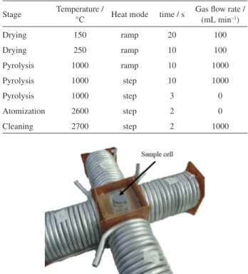
conditions. A hollow-cathode lamp operated at 7 mA was used and absorbances were measured at 313.3 nm with a slit width of 0.5 nm. The analytical conditions used for the determination of MoVI is shown in Table 1.
A totally glass Hamilton (model WSC/4D) double distiller was used for preparation of doubly distilled water. A Shimadzu (Japan) model 1650PC double-beam spectrophotometer was used for running absorption spectra. Fourier transform infrared (FTIR) spectra were recorded by a Shimadzu, model 8400 (Japan), spectrometer in the transmittance mode. A Philips transmission electron microscope (TEM) (model EM208, Philips) was used in order to investigate the topology and size of nanoparticles. A Memmert type (SV 1422) shaker water bath (Germany) was used for shaking the solutions
and mixtures. ACMNPs were stirred with a home-made magnetic field agitation (MFA) device, designed by Safdarian et al.18 in the Department of Chemistry, Lorestan
University (Figure 1).
Preparation of ACMNPs
Preparation of ACMNPs was performed by a one-pot process reported elsewhere.18 In this procedure,
preparation of iron oxide nanoparticles and their coating by agarose is performed in one step. Briefly, 0.2 g FeCl3
and 0.073 g FeCl2 were dissolved in 1 mol L−1 HCl
(12 mL). Then, 0.05 g agarose was dissolved in 2 mol L−1
NaOH (25 mL) under heating to 80 °C and 2 mL of the Fe3+/Fe2+ solution was added to it, dropwise. The
addition was made under nitrogen atmosphere at reflux conditions (90 °C) and magnetic stirring (800 rpm). The color of the bulk solution changed to black immediately, indicating the production of Fe3O4 nanoparticles. By
this method, magnetic micro-flake agarose particles were produced. The final mixture was cooled to ambient temperature and the prepared ACMNPs were separated by a permanent magnet and washed with water and 50% (v/v) water/ethanol. To obtain more uniform particles and remove coarse particles, the particles were passed through a sieve of 120 µm before being stored under 20% (v/v) ethanol at 4 °C.
Table 1. GFAAS analytical conditions for Mo determination
Stage Temperature /
°C Heat mode time / s
Gas flow rate / (mL min−1)
Drying 150 ramp 20 100
Drying 250 ramp 10 100
Pyrolysis 1000 ramp 10 1000
Pyrolysis 1000 step 10 1000
Pyrolysis 1000 step 3 0
Atomization 2600 step 2 0
Cleaning 2700 step 2 1000
Activation and functionalization of ACMNPs
For partial crosslinking of the agarose particles and their epoxy activation, an epichlorohydrin activation method was used as reported elsewhere.19 After activation,
ACMNPs were functionalized by the PhP ligand. For this purpose, 1 mL of a 20% v/v suspension of ACMNPs was added to 10 mL of 0.1 mol L−1 ligand solution (adjusted
on pH 10 by 2 mol L−1 NaOH solution). The mixture was
then shaken for 24 h, washed with water and stored under 20% (v/v) ethanol at 4 °C (as a 20% suspension) for later uses. Figure 2 shows a schematic representation of the preparation procedure for ACMNPs and their activation and functionalization by phenylephrine.
Preparation of beans samples
Pea, corn, bean and lentil samples were purchased from local markets. The beans samples were ground to a fine powder using a porcelain mortar. To 1.00 g of each powdered sample, 6 mL of concentrated HNO3 (65%) and
2 mL hydrogen peroxide (30%) were added and the mixture was gently heated until the sample was completely digested. After digestion, the volume of the sample was made up to 100 mL with distilled water.20
The MSPE-MFA procedure
In the MSPE-MFA procedure, 5 mL of the sample or MoVI test solution were transferred into the sample cell
of the MFA device, surrounded by four electric magnets, and 200 µL of the 20% suspension of the ACMNPs was added to this solution. The MFA device was programmed to change the magnetic field in a circular manner around the sample cell, in 500 and 1000 ms periods, so that the adsorbent particles were rotated in the solution.18 After
20 min of agitation, the magnetic particles were collected on a wall of the vessel, by keeping one of the electrical magnets on, and the solution was withdrawn by a syringe.
Then, the particles were washed with water and finally desorbed by 500 µL of a 1 mol L−1 HCl solution for the
determination by GFAAS method. The steps have been summarized in Figure 3.
Results and Discussion
Spectrophotometric studies
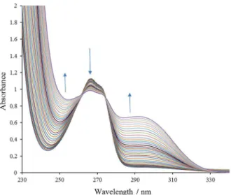
To study complex formation between MoVI and PhP
ligand, UV-Vis absorption spectra at constant concentration of the ligand and increasing concentrations of the metal ion were recorded. As shown in Figure 4, PhP shows a minimum absorbance at 244 nm and a maximum at 266 nm. By addition of MoVI, the minimum is increased
and the maximum is decreased and a shoulder appears at about 290 nm. Appearance of two clear isobestic points indicates the formation of a complex between the metal ion and the ligand. Study of the absorbance changes at 266 nm as a function of metal ion to ligand concentration ratios shows that MoVI-PhP complex is formed in a 1:2 ratio in
low metal concentrations (Figure 5). However, this ratio is
Figure 2. Schematic representation of ACMNPs and their activation and functionalization by phenylephrine.
Figure 3. The steps of the MSPE-MFA method.
Figure 4. Absorption spectra of MoVI-PhP solutions. PhP concentration:
5 × 10⁻⁴ mol L−1; and MoVI concentration: 2.53 × 10⁻⁵-2.18 × 10⁻³ mol L−1.
eventually changed in the higher metal concentrations and a 2:1 complex is formed as shown in the mole ratio curve.
Characterization of the adsorbent
Characterization of the ACMNPs was performed by optical microscopy, TEM and FTIR spectrometry. The optical microscopy images showed that the ACMNPs are in the shape of micro-flakes with a relatively narrow size distribution in the range of 90-130 µm. Iron oxide nanoparticles usually have a high affinity to aggregate together. However, polysaccharide matrices can avoid this and act as stabilizers for their synthesis.21 Agarose is also
a polysaccharide and as the TEM image (Figure 6) shows, the nanoparticles have been fairly dispersed in the agarose matrix in the ACMNPs. By the study of the TEM images, the average sizes of the Fe3O4 nanoparticles were estimated to be
in the range of 10-14 nm. Figure 7 compares the FTIR spectra of the Fe3O4 MNPs, agarose micro-flakes and ACMNPs. An
OH stretching band has shifted from 3445 cm−1 (in agarose)
to 3418 cm−1 (in ACMNPs) suggesting the formation of
hydrogen bonds between agarose and hydroxyl functional groups onto the surface of the magnetic nanoparticle.15
Optimization of the MSPE-MFA procedure
Preliminary experiments showed that the phenylephrine-ACMNPs adsorbent can efficiently adsorb MoVI and can
probably be used for the MSPE of this metal ion in combination with GFAAS. In order to avoid grinding of the agarose micro-flakes during stirring by magnetic bars, the home-made MFA system was used for efficient agitation of the magnetic agarose particles during the adsorption/ desorption procedures.18 Effects of several experimental
parameters on the preconcentration of MoVI by the
MSPE-MFA method were carefully studied and optimized.
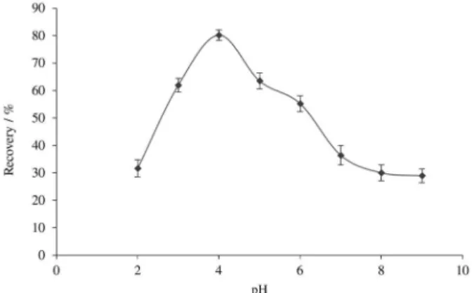
Effect of pH
The solubility of metal ions in water is pH-sensitive, because many of these ions will precipitate in high pH values. In the low pH region, due to competition of protons with metal ions, stability of metal complexes will be greatly decreased. Therefore, optimization of pH is a priority in SPE experiments. The samples were buffered by 0.02 mol L−1 phosphate buffer for stabilization of pH during
this study. As shown in Figure 8, the maximum recovery for MoVI was obtained at pH 4.
Effect of the amount of the sorbent
The amount of sorbent is a significant parameter in MSPE. Increasing the amount of sorbent requires higher volumes of the eluent to wash out metal ions that in turn reduces the preconcentration factor. On the other hand, reducing the amount of adsorbent may reduce the extraction efficiency. Given these issues to achieve the best results, the effect of the PhP-ACMNPs adsorbent amount was
Figure 5. Relative absorbance as a function of metal to PhP ligand
concentrations ratios for MoVI at 266 nm. Figure 6. TheTEM image of ACMNPs.
studied in a constant eluent volume of 500 µL. As shown in Figure 9, an increase of the sorbent amount up to 200 µL increased the recovery. Then, the recovery gradually decreased in higher sorbent volumes probably due to the inefficient desorption by the aforementioned volume of the eluent. Since increasing the eluent volume was not suitable, 200 µL was selected as the optimum volume of the sorbent in subsequent experiments.
Effects of volume and concentration of elution solvent
Hydrochloric acid was found to be an efficient eluent
for desorption of MoVI from the PhP-ACMNPs. Using
various volumes of a 1 mol L−1 solution of HCl, indicated
that a minimum volume of 500 µL was necessary for the complete desorption of the analyte.
Elution solvent concentration was another important factor to be studied. Acid concentration should be high enough to break down the metal-ligand bound but should not be so high to destroy the agarose based adsorbent. Due to the destructive effects of acid concentrations above 1 mol L−1 on agarose, the highest possible concentrations of
hydrochloric acid, i.e., 1 mol L−1, was chosen as the optimal
acid concentration. Using lower acid concentrations reduced the recovery of the analyte.
Effect of extraction time
The required time for an efficient extraction depends on the kinetics of adsorption and the mixing efficiency of the solution. In a previous work in our laboratory,18 it was
shown that the MFA device, due to its efficient and direct agitation of the ACMNPs, is more efficient than magnetic stirring and shaking methods for mixing of the solution. Using the same MFA device in this work, after 20 min the interaction between the adsorbent and the MoVI sample
solution reached to a plateau as shown in Figure 10. This indicates that the extraction time in this work is even shorter than the previous work (30 min). This should be due to a faster sorption kinetics of MoVI on the PhP
functionalized sorbent compared to the sorption of PdII on
the iminodiacetic acid functionalized sorbent.18
The analytical performances of the MSPE-MFA method
Eight replicated analysis at the optimized conditions for a sample containing 10 µg L−1 of MoVI (eluent volume,
500 µL; eluent concentration, 1 mol L−1, adsorbent amount,
200 µL; extraction time, 20 min; pH, 4.0) resulted in a recovery of 95.3% with an relative standard deviation (RSD) of 4.9%. The calibration curve of the method (6 points) was linear over a range from 1 to 150 µg L−1
with a slope of 0.0051 (± 0.0001) L µg−1, an intercept of
0.1375 (± 0.007) and a correlation coefficient (r) of 0.9990. The limit of detection (LOD) of the MSPE-MFA method was calculated from three times of the standard deviation of blank (n = 20) to be 49 ng L−1 for the analyte.
Figure 8. Effect of pH of test solutions on the recovery of MoVI by the
MSPE-MFA method. MoVI concentration, 10 µg L−1; amount of adsorbent,
100 µL; eluent, 500 µL of 1 mol L−1 HCl.
Figure 9. Effect of the amount of the sorbent on the recovery of MoVI by
the MSPE-MFA method. The sample pH was 4.0 and other conditions were as in Figure 8.
Figure 10. Effect of the extraction time on the recovery of MoVI by the
MSPE-MFA method. MoVI concentration, 10 µg L−1; amount of adsorbent,
An important characteristic of a sorbent is its adsorption capacity. The capacity of a sorbent indicates the number of active sites on it or the ability of a certain amount of the sorbent to form complex with a metal ion. The capacity of the PhP-ACMNPs sorbent was calculated with a method described elsewhere.18 The calculated capacity
of the sorbent for MoVI at pH 4.0 was calculated to be
13.2 µg per mL of its packed volume. The capacity and magnetic properties of the sorbent was not significantly changed within at least 30 times of uses during a period of three months.
Table 2 compares the performance of the proposed method for the determination of molybdenum with some other reported methods. As shown in the table, the performances of the MSPE-MFA technique is comparable or better than many of the other reported methods.
Interference effect of metal ions
Preconcentration procedures for small quantities of metal ions can be influenced by other ions present in the sample. One of the important characteristics of an SPE method is its selectivity, which is the ability of the method to preconcentrate and measure the analyte without interference from other species present in the sample solution. The effects of potentially interfering ions such as Cu2+, Ni2+, Pb2+, Fe2+, Cr3+, Co2+, Zn2+, Mg2+, Sr2+ on the
preconcentration/separation and determination of Mo6+
were examined under the optimum conditions described above. For this purpose, 5 mL of binary mixtures of Mo6+
(10 µg L−1) and various metal ions with a concentration
equivalent to 100 times the concentration of Mo6+
(1000 µg L−1) were studied by the proposed method.
Relative errors caused by the interfering ions on the extraction of Mo6+, were calculated using known relations.22
As shown in Figure 11, only Fe3+ caused an error of about
7% and the error produced by the other metal ions was less than 3%. Therefore, the method showed a good selectivity
towards MoVI, with little or no interference from common
metal ions.
Application of the method
To test the applicability of the proposed method to real samples, MoVI concentration in some beans samples were
determined after preconcentration on the PhP-ACMNPs by the proposed method. The analytical results have been summarized in Table 3. As the results show, the recoveries for the spiked beans samples are well comparable with the
Table 2. Comparison of the proposed method with some other reported procedures for molybdenum determination
Determination method LOD / (ng L−1) RSD / % Linear range /
(µg L−1) Enrichment factor Reference
Reflectance spectroscopy 200000 3.3 500-12000 − 3
GFAAS and ICP-AES 50 1-3 − 100 5
Fiber optic spectrophotometry 1430 2.8 < 100 72.6 8
GFAAS 100 2.8-3.1 0.2-20 − 9
GFAAS 80 2-3 0.2-4 10 10
GFAAS 49 4.9 1-150 10 this work
LOD: limit of detection; RSD: relative standard deviation; GFAAS: graphite furnace atomic absorption spectrometry; ICP-AES: inductively coupled plasma-atomic emission spectrometry.
Table. 3. Analytical results for the determination of MoVI in real beans
samples by the proposed method, under the optimized conditions (mean ± s, n = 3)
Sample Added / (µg g−1) Found / (µg g−1) Recovery / %
Pea − 1.91 ± 0.01 −
1 3.04 ± 0.06 104.4
Corn − 1.62 ± 0.06 −
1 2.81 ± 0.02 107.6
Bean − 1.11 ± 0.04 −
1 2.33 ± 0.01 110.4
Lentil − 1.03 ± 0.04 −
1 2.14 ± 0.02 105.4
Figure 11. Effect of interfering ions on the relative error of MoVI
mean recovery of the method for standard MoVI solutions.
Therefore, the method can be used for MoVI determinations
in beans samples.
Conclusions
One-step method for the synthesis of nanomagnetic agarose micro-flake particles is simple, fast and without the need to any organic solvent. MFA device is efficient for increasing the rate of adsorption in MSPE and attainment of equilibrium in a reasonable time. The functionalized ACMNPs with PhP is relatively selective to MoVI and its application in the MSPE-MFA system for
the preconcentration and GFAAS determination of MoVI
in beans samples was successful. The proposed method is simple, sensitive and selective and can be used for the preconcentration of MoVI in different samples.
Acknowledgements
The authors would like to thank the Chemistry Graduate Program of the Chemistry Department of Lorestan University, Khoramabad, Iran.
References
1. Diaz-Guemes, M.; Bhatti, A.; Dollimore, D.; Thermochim. Acta 1986, 106, 125.
2. Shrivas, K.; Agrawal, K.; Harmukh, N.; J. Hazard. Mater. 2009, 161, 325.
3. Filik, H.; Aksu, D.; Apak, R.; Boz, İ.; Sens. Actuators, B 2009, 141, 491.
4. Vanhoe, H.; Vandecasteele, C.; Versieck, J.; Dams, R.; Anal. Chem. 1989, 61, 1851.
5. Burba, P.; Willmer, P.; Fresenius’ J. Anal. Chem. 1986, 324, 298.
6. Sastre, A. M.; Alguacil, F. J.; Chem. Eng. J. 2001, 81, 109. 7. Paleologos, E. K.; Giokas, D. L.; Karayannis, M. I.; TrAC,
Trends Anal. Chem. 2005, 24, 426.
8. Gharehbaghi, M.; Shemirani, F.; Food Chem. Toxicol. 2011, 49, 423.
9. Burguera, J.; Burguera, M.; Rondon, C.; Talanta 2002, 58, 1167. 10. Ferreira, S. L.; dos Santos, H. C.; Campos, R. C.; Talanta 2003,
61, 789.
11. Vassileva, E.; Furuta, N.; Spectrochim. Acta, Part B 2003, 58, 1541.
12. Das, D.; Gupta, U.; Das, A. K.; TrAC, Trends Anal. Chem. 2012, 38, 163.
13. Giakisikli, G.; Anthemidis, A. N.; Anal. Chim. Acta 2013, 789, 1.
14. Aguilar-Arteaga, K.; Rodriguez, J.; Barrado, E.; Anal. Chim. Acta 2010, 674, 157.
15 Mashhadizadeh, M. H.; Karami, Z.; J. Hazard. Mater. 2011, 190, 1023.
16. Zhang, M.; Cheng, D.; He, X.; Chen, L.; Zhang, Y.; Chem. - Asian J. 2010, 5, 1332.
17. Hashemi, P.; Rahmani, Z.; Talanta 2006, 68, 1677.
18. Safdarian, M.; Hashemi, P.; Adeli, M.; Anal. Chim. Acta 2013, 774, 44.
19. Matsumutu, I.; Mizuno, Y.; Nobuko, S.; J. Biochem. 1979, 85, 1091.
20. Soylak, M.; Kaya, B.; Tuzen, M.; J. Hazard. Mater. 2007, 147, 832.
21. Chang, P. R.; Yu, J.; Ma, X.; Anderson, D. P.; Carbohydr. Polym. 2011, 83, 640.
22. Parham, H.; Zargar, B.; Heidari, Z.; Hatamie, A.; J. Iran. Chem. Soc. 2011, 8, S9.
Submitted: July 29, 2015