DOI: 10.1590/0004-282X20150099
VIEW AND REVIEW
An update in the management of spinal
metastases
Atualização no manejo das metástases na coluna vertebral
Andrei F. Joaquim1, Ann Powers2, Ilya Laufer2, Mark H. Bilsky2
Advances in cancer treatment over the past decade, such biologics and immunotherapy are improving patient
out-comes and consequently life expectancy. he most common
sites for metastases in the general population with cancer are the liver and lungs, followed by bone. Considering bone
me-tastases, the majority will afect the spine1. Some studies
es-timate that up to 40% of all patients with cancer will have spinal metastases (SM) during the course of their disease2,3.
he most frequent histologic types of cancer that give
rise to bone metastases are breast, prostate and lung can-cer4. Most of the spinal metastases are diagnosed following
the diagnosis of the primary cancer. However, in about 10%
of the patients, SM is the irst manifestation of an unknown
primary tumor1.
Spinal cord compression is expected in up to 20% of the patients with SM. Approximately 95% of the patients with
SM will demonstrate epidural metastases, mainly afecting
the vertebral body and the pedicle regions, whereas 5% will present with intradural and less than 1% with intramedullary metastases5,6. Symptomatic spinal cord compression occurs
more frequently in the thoracic spine, followed by cervical
and then lumbar. he greater incidence of SM in the thorac -ic spine is attributed to the higher number of vertebrae and small canal diameter7.
1Universidade Estadual de Campinas, Departamento de Neurologia, Divisão de Neurocirurgia, Campinas SP, Brazil; 2Memorial Sloan Kettering Cancer Center, Department of Neurosurgery, New York NY, USA.
Correspondence: Andrei F. Joaquim; UNICAMP, Disciplina de Neurocirurgia; Rua Antonio Lapa, 280/sala 506; 13025-240 Campinas SP, Brasil; E-mail: [email protected]
Conflict of interes: There is no conlict of interest to declare.
Received 05 January 2015; Received in inal form 15 April 2015; Accepted 04 May 2015.
ABSTRACT
The best clinical treatment for spinal metastases requires an integrated approach with input from an interdisciplinary cancer team. The principle goals of treatment are maintenance or improvement in neurologic function and ambulation, spinal stability, durable tumor control, and pain relief. The past decade has witnessed an explosion of new technologies that have impacted our ability to reach these goals, such as separation surgery and minimally invasive spinal procedures. The biggest advance, however, has been the evolution of stereotactic radiosurgery that has demonstrated durable tumor control both when delivered as deinitive therapy and as a postoperative adjuvant even for tumors considered markedly resistant to conventional external beam radiation. In this paper, we perform an update on the management of spinal metastases demonstrating the integration of these new technologies into a decision framework NOMS that assesses four basic aspects of a patient’s spine disease: Neurologic, Oncologic, Mechanical Instability and Systemic disease.
Keywords: spinal metastases, bone metastases, diagnosis, management, treatment, NOMS.
RESUMO
O tratamento dos pacientes com metástases na coluna requer uma abordagem multidisciplinar por equipe especializada em oncologia. Os objetivos básicos do tratamento são a manutenção/ melhora da função neurológica com preservação da deambulação, manutenção da estabilidade da coluna, controle tumoral e alívio da dor. A última década testemunhou uma explosão de novas tecnologias que auxiliaram a atingir os objetivos terapêuticos, como a cirurgia de separação e procedimentos minimamente cirúrgicos minimamente invasivos. Contudo, o maior avanço terapêutico constitui-se do uso da radiocirurgia no tratamento das metástases de coluna, que possibilita bom controle local tanto como terapia deinitiva ou no pós-operatório de tumores, mesmo os considerados radioresistentes à radioterapia convencional. No presente artigo, realizamos atualização do manejo das metástases de coluna, apresentando a integração das novas tecnologias em um algoritmo de decisão “NOMS” que inclui os quatros aspectos básicos dos pacientes com metástases na coluna: Neurologic, Oncologic,
Mechanical Instability e Systemic disease.
he optimal clinical management of these patients re -quires integrated decisions by an interdisciplinary cancer team comprised of medical and radiation oncologists and spine surgeons, as well as all other health care professionals (e.g. rehabilitation team, nurses, and other involved medical specialties)8,9. Survival is based on the primary tumor
histol-ogy, systemic disease status, and also patient comorbidities, resulting in a very heterogeneous group8. Of note, only 10 to
20% of the patients with SM will be alive after two years
af-ter the diagnosis, which must be taken into account when
considering a patient’s treatment8,9. With the introduction of
newer biologics and immunotherapy for tumors such as re-nal cell carcinoma and melanoma, overall survival is improv-ing leadimprov-ing to need to provide a more durable solution for spine metastases. Considering the life expectancy, treatment
of patients with SM is palliative with the aim of achieving ive
principle goals: 1) pain control, 2) improvement in quality of life, 3) spine stabilization, 4) maintenance or improvement of neurological status, and 5) local disease control8.
New treatment modalities have improved the treatment of spine metastases over the past decade. Surgical advances in-clude less invasive approaches, such as separation surgery,
pedi-cle screw ixation which can be placed via either an open or per -cutaneous approach and cement augmentation techniques9,10.
However, by far, the greatest impact has been the evolution and integration of spine stereotactic radiosurgery (SRS), typically de-livered as 16 to 24 Gy single fraction or 24 to 30 Gy in 3 fractions9.
he ability to deliver high-dose conformal radiation in ablative
doses has improved local control rates from 30% at 3 months us-ing conventional external beam radiotherapy (cEBRT) (e.g. 30 Gy
in 10 fractions) to over 90% when used as deinitive therapy or as
postoperative adjuvant11,12,13,14,15.
In this review, we perform an update based on the NOMS
decision framework of the concepts involved in the manage -ment of SM, considering the basic assess-ments necessary to choose the best treatment modality for an individual patient.
PATIENT’S EVALUATION – TREATMENT DECISION PROCESS
Many scoring system have been proposed to estimate the prognosis of patients with SM, an important factor in
the decision of treatments to be performed. he modiied Tokuhashi Scoring System is a prognostic scale that evalu
-ates patients Karnofsky score, neurological status, number of
bone metastases outside spine, metastasis to major organs and the primary tumor site16. he system is used to estimate
survival, which is very important in the decision-making.
Another popular scoring system developed to aid in pre-dicting survival is the Tomita score that considers the pri-mary tumor site, visceral metastases and bone metastases17.
Both systems were proposed for helping in the choice of pal-liative versus aggressive surgery based on estimated patient
survival. However, these algorithms have been criticized be-cause they do not consider the presence of instability, or the
signiicant impact of advances in radiation therapy, such as
radiosurgery. Further, these algorithms have not reassessed the extended survivals demonstrated in tumors such as renal cell carcinoma and melanoma with the integration of newer biologics and immunotherapy18.
Rather than using algorithms that are ixed in time and
constrained by technology available at the time they were
proposed, a new decision framework NOMS has been devel -oped that considers four sentinel decision points in decision
making8. NOMS consists of four fundamental assessments:
Neurologic, Oncologic, Mechanical Instabilty, and Sytemic
Disease8. Unlike the Tokuhashi and Tomita algorithms, the
advantage of the NOMS framework is that it incorporates
both new technologies and evidenced based medicine as they become available. Considering these four assessments, the interdisciplinary team can determine the optimal treatment consisting of radiation therapy (Table 1), surgery, systemic therapy, or a combination of these. In the NOMS decision
framework, the neurologic assessment principally relects
the degree of epidural spinal cord compression (ESCC) as well as the presence or absence of myelopathy and/or func-tional radiculopathy8. Spinal cord compression is based on
a validated scoring system using magnetic resonance (MR) axial T2-weighed images8.his scoring system is used to dif
-ferentiate no or minimal ESCC (0-1c) from high-grade spinal ESCC 2-3 (Table 2)8,19. he oncologic consideration is pred
-icated on the known cytotoxicity and the durability of the
response to current treatment modalities including cEBRT, SRS, chemotherapy, hormones, immunotherapy, or biolog-ics. In terms of malignant tumors, surgery plays a very lim-ited role in tumor control. Mechanical instability has
recent-ly been deined for neoplastic disease and a scoring system,
spinal instability neoplastic score (SINS), has been developed to aid in this assessment20.he recognition of spinal instabil
-ity resulting from tumor is imperative because an unstable spine will not respond to radiation and/or chemotherapy, but requires an intervention, such as brace application, percuta-neous cement augmentation and/or pedicle screws, or open
surgery. he inal assessment in NOMS relects the extent of
systemic disease, medical co-morbidities and expected
sur-vival, which all impact the decision to ofer not only surgical
treatment, but also radiation or systemic therapy.
he neurologic and oncologic assessments are consid -ered in combination8. From the oncologic perspective,
radia-tion is the mainstay of therapy for tumor control. With few exceptions, such as myeloma and lymphoma, systemic
ther-apy has little impact in this regard. It is unknown whether the
newer biologics, such as sorafenib for renal cell carcinoma, have the same responses in bone metastases as visceral dis-ease even if the driver mutation is present21. Our experience
setting of high-grade spinal cord compression or
symptomat-ic lesions. he two modalities of radiation currently available
for the treatment of spine metastases are conventional exter-nal beam radiation (cEBRT - e.g. 30 Gy in 10 fractions) and stereotactic radiosurgery (SRS - e.g. 16 to 24 Gy single frac-tion or 24 to 30 Gy in 3 to 5 fracfrac-tions)10,11,12,13,14,15,22. hese hypo
-fractionated radiation doses are ablative to the tumor while sparing normal tissue tolerance. Mounting evidence suggests
that the radiobiology of SRS is diferent from conventionally
fractionated radiation23. In combination with the oncologic
considerations, the neurologic assessment is one of the criti-cal determinants in deciding between these modalities as cEBRT can be used in the setting of high-grade ESCC (ESCC 0 to 3), but the safe delivery of SRS requires a 2 to 3 mm mar-gin on the spinal cord in order to avoid radiation myelitis24.
Although groups are studying the use of SRS in the setting of high-grade spinal cord compression, most commonly SRS
use is restricted to tumors conined to the bone or with mini -mal epidural impingement (ESCC 0-1c)8,24.
Radiosensitive tumors: Conventional External Beam Radiation
Patients with radiosensitive tumors can be treated efec -tively with cEBRT regardless of the degree of ESCC8. A review of
the literature shows that tumor histology is perhaps the most important factor in determining response to cEBRT9. Among
patients who underwent cEBRT in the setting of spinal metas-tases, the mean ambulation rate was 81% (range from 58% to 100%)9. However, only 6% to 67% percent of non-ambulatory
patients’ recovered ambulation, with reports in the higher range thought to be attributable to the large number of favor-able histologies in those series11,13. Literature analysis reveals
that all authors classify lymphoma, seminoma, and myeloma as radiosensitive histologies and supports the use of cEBRT to treat these tumors, regardless of the degree of ESCC or
neu-rologic deicit11,13,25,26,27. On the other hand, solid tumors
ex-hibit a wide range of radiosensitivity25-29. Radiosensitive
sol-id tumor histologies include breast, prostate, ovarian, and
neuroendocrine carcinomas. Renal, thyroid, hepatocellular, colon, and non-small cell lung carcinomas, sarcoma, and mel-anoma represent radioresistant tumors25,26,27,28,29. Solid tumors
with radioresistant histologies generally require SRS to achieve durable local control, whereas radiosensitive solid tumors may be treated with cEBRT or SRS15,,25,26,27,28,29.
Radioresistant Tumor without spinal cord compression (ESCC 0 to 1c): SRS
Unlike the poor responses to cEBRT, responses to SRS now
demonstrate greater than 90% durable response rates when
used as deinitive treatment in patients with minimal or no
spinal cord compression (ESCC 0 to 1c). In a review of 413 pa-tient undergoing single fraction SRS with doses ranging from 18 to 24 Gy, Yamada, et al., reported 3–year recurrence rates of 4% when censored for death that were histology indepen-dent with a dose response demonstrating that those who re-ceived greater 24 Gy had a better local tumor control than those who received less than 24 Gy12. Guckenberger et al. re
-ported a multi-institutional retrospective review of 301 pa-tients with 387 vertebral body metastases29. he median
treatment dose was 24 Gy in 3 fractions with 2-year local con-trol rates reported at 83.9%. In this study, tumor histology (i.e. non-small lung carcinoma, melanoma, and renal cell carci-noma) were associated with worse outcomes for local tumor
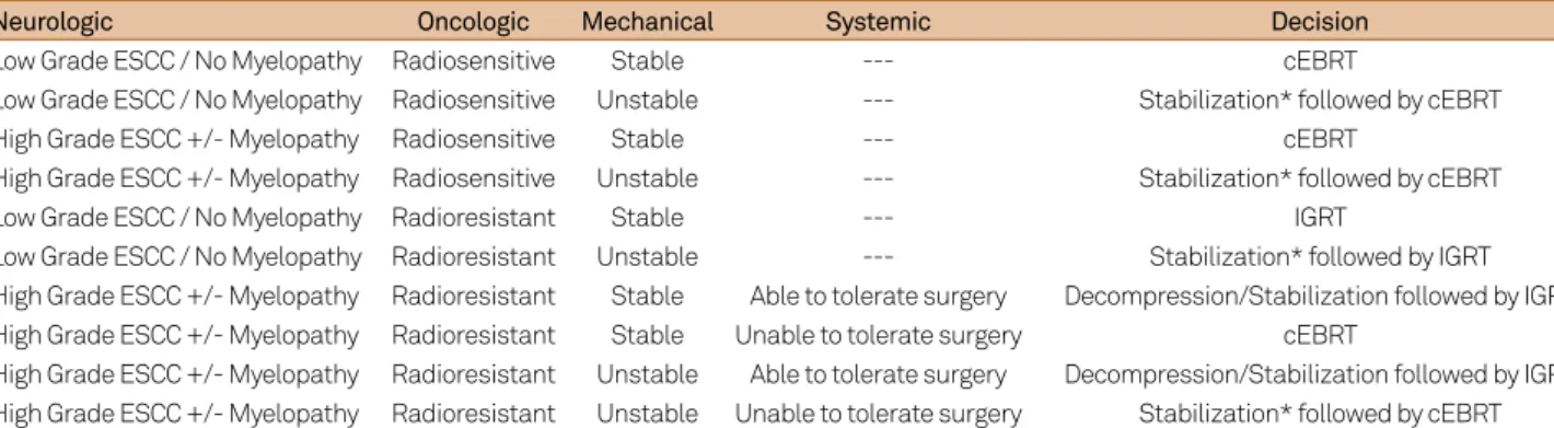
Table 1.Current NOMS decision framework.
Neurologic Oncologic Mechanical Systemic Decision
Low Grade ESCC / No Myelopathy Radiosensitive Stable --- cEBRT
Low Grade ESCC / No Myelopathy Radiosensitive Unstable --- Stabilization* followed by cEBRT
High Grade ESCC +/- Myelopathy Radiosensitive Stable --- cEBRT
High Grade ESCC +/- Myelopathy Radiosensitive Unstable --- Stabilization* followed by cEBRT
Low Grade ESCC / No Myelopathy Radioresistant Stable --- IGRT
Low Grade ESCC / No Myelopathy Radioresistant Unstable --- Stabilization* followed by IGRT High Grade ESCC +/- Myelopathy Radioresistant Stable Able to tolerate surgery Decompression/Stabilization followed by IGRT High Grade ESCC +/- Myelopathy Radioresistant Stable Unable to tolerate surgery cEBRT
High Grade ESCC +/- Myelopathy Radioresistant Unstable Able to tolerate surgery Decompression/Stabilization followed by IGRT High Grade ESCC +/- Myelopathy Radioresistant Unstable Unable to tolerate surgery Stabilization* followed by cEBRT
ESCC Scale 0 and 1 = Low Grade ESCC; ESCC Scale 2 and 3 = High Grade ESCC; * Stabilization options include percutaneous cement augmentation, percutaneous pedicle screw instrumentation and open instrumentation. In patients with signiicant systemic comorbidities limiting their ability to tolerate open surgery, stabilization may be limited to cement augmentation and / or percutaneous screw augmentation.
NOMS: neurologic, oncologic, mechanical instability and systemic disease; ESCC: epidural spinal cord compression; cEBRT: conventional External Beam Radiotherapy; IGRT image-guided radiation therapy.
Table 2.Epidural spinal cord compression scoring system based on MR axial T2-weighted images.
0: tumor conined to bone,
1: tumor extension into the epidural space without deformation of the spinal cord,
1a: epidural impingement but no deformation of the thecal sac; 1b: deformation of the thecal sac but without spinal cord abutment; 1c: deformation of the thecal sac with spinal cord abutment but without compression
2: spinal cord compression but CSF is visible and 3: spinal cord compression without visible CSF
control29. he application of SRS has transformed tumors his
-torically considered resistant to cEBRT into very responsive
tumors. he responses to SRS are histology independent un
-like those seen with cEBRT.
Radioresistant Tumors with High-grade spinal cord compression (ESCC 2 to 3): Surgery + SRS
From the neurologic and oncologic perspective, surgery is reserved for patients with radioresistant tumors who have high-grade spinal cord compression. Surgery is used to pre-serve or restore neurologic function and stabilize the spine.
he justiication for surgery in patients with high-grade spinal
cord compression is primarily based on a prospective
random-ized trial published by Patchell et al. comparing surgery fol -lowed by cEBRT to cEBRT alone30.Statistically signiicant im
-provements were found in the surgical group with regard to maintenance and recovery of ambulation, bowel and bladder continence, narcotic requirements, and survival. Despite limi-tations, this study was sentinel in establishing that
neurologi-cal outcomes were signiicantly better in treating myelopathic
patients with radioresistant tumors. Radiosensitive tumors, such as lymphoma and myeloma, were excluded as this would have heavily biased the study toward the radiation arm and would not have answered the principal question regarding the best treatment for solid tumor malignancies. At the time the study was designed, instability was not recognized as a contra-indication to radiation as a primary treatment. Without
recog-nizing this concept, 30% in each arm were classiied as unsta
-ble by the Cybulski criteria, which biased the outcome toward
the surgical arm. Additionally, the duration of compression was longer in the irradiated arm potentially biasing the
im-proved outcomes toward surgery. he deinition of ambulation was also fairly limited, i.e., the ability to walk two steps: how
-ever, using this deinition, the diferences in both maintaining and recovering ambulation were signiicant.
Finally, despite surgery providing neurologic salvage, du-rable tumor control remained a problem when using cEBRT as a postoperative adjuvant with recurrence rates of approxi-mately 70% at one-year follow-up.A major risk factor for re -currence is radioresistant-tumor histology8. he transition
to using of SRS as a postoperative adjuvant has substantially improved local control rates compared to cEBRT while re-ducing the need for aggressive surgical approaches, such as
en bloc spondylectomy.
M - Mechanical instability
Mechanical instability is a separate assessment from the neurologic and oncologic evaluation as no amount of radia-tion will stabilize an unstable spine. Mechanical instability in the setting of neoplastic spinal disease can be evaluated used the SINS(Table 3), a new comprehensive classiication
system recently proposed that is based on patient symptoms and radiographic criteria in an attempt to predict eventual spinal instability20.
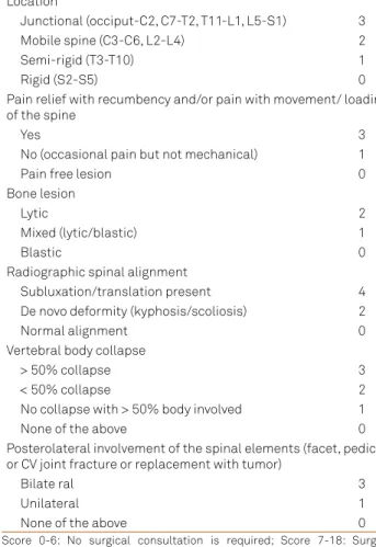
he SINS includes the following assessments: 1) tumor loca -tion, 2) type and presence of pain, 3) bone lesion quality, 4) spinal alignment, 5) extent of vertebral body collapse and 6) postero-lateral elements involvement. A qualitative score is them
ob-tained, and instability is suggested according to the inal score. he minimum score is 0 and maximum is 18. Stability is denoted
when the score is 0 to 6; indeterminate instability is proposed
when the score of 7 to 12 and frank instability is determined
when the injury has a score of 13 to 18. Surgical consultation is recommended when patients had a score of 7 to 18. Mechanical instability is an indication for surgical stabilization, regardless of tumor radioresistance to cEBRT or histology20.
S - Systemic disease
Systemic disease and medical comorbidities evaluate the ability of the patient to tolerate a proposed treatment and can
inluence patient’s survival as well as the oncological status. he oncology team can help in stratify surgical risk accord -ing to tumor histology. Some tumors have median survival of less than 6 months, such as non-small cell lung carcinoma10.
In such situations, surgical treatment can be not advisable.
he same is true, for instance, in the setting of a severe heart
ischemic disease or chronic pulmonary obstructive disease,
Table 3.The SINS - Spinal Instability Neoplastic Score Element of SINS Score.
Location
Junctional (occiput-C2, C7-T2, T11-L1, L5-S1) 3
Mobile spine (C3-C6, L2-L4) 2
Semi-rigid (T3-T10) 1
Rigid (S2-S5) 0
Pain relief with recumbency and/or pain with movement/ loading of the spine
Yes 3
No (occasional pain but not mechanical) 1
Pain free lesion 0
Bone lesion
Lytic 2
Mixed (lytic/blastic) 1
Blastic 0
Radiographic spinal alignment
Subluxation/translation present 4
De novo deformity (kyphosis/scoliosis) 2
Normal alignment 0
Vertebral body collapse
> 50% collapse 3
< 50% collapse 2
No collapse with > 50% body involved 1
None of the above 0
Posterolateral involvement of the spinal elements (facet, pedicle or CV joint fracture or replacement with tumor)
Bilate ral 3
Unilateral 1
None of the above 0
where a surgical procedure can be prohibitive. In summary, the extent of systemic disease and functional status as as-sessed by patient comorbidities can preclude surgical treat-ment in selected cases.
DIAGNOSTIC WORK-UP
Basic routine tests of patients with SM include: complete
blood count with diferential, coagulation test, renal function assessment, electrolytes, among others. Speciic evaluation is
guided according to clinical presentation and tumor charac-teristics and primary site. Radiological assessment of system-ic metastases is critsystem-ical except in cases where urgent
treat-ment is required, such as a progressive acute deicit.
Although plain radiographies and CT scanning can be use-ful for surgical planning and to evaluate spinal deformities, MR is the imaging modality of choice for SM evaluation30. Typically
sagittal screening of the entire spinal axis is undertaken to as -sess for occult lesions outside the symptomatic area that may
bear on decision making. he most important sequences for
sagittal screening are T1-weighted and STIR images in which
tumor is hypointense and hyperintense, respectively. Speciic MRI sequences can help radiologists to diferentiate degener -ative changes and osteoporotic fractures from metastases8,31.
As previously noted, the degree of spinal cord compression is predicated on axial T2-weighted sequences. Most commonly images are obtained with and without gadolinium contrast to further evaluate the degree of spinal cord compression and to rule out leptomeningeal and intramedullary tumors. More re-cently perfusion images are being explored to assess the viabil-ity of tumors. Dynamic contrast-enhanced MR perfusion im-ages particularly evaluating plasma volume are very sensitive
and speciic for assessing treatment responses following SRS
even before changes are seen on standard MR imaging32.
Positron Emission Tomography (PET-CT) is also useful imaging modalities for screening systemic disease and other
metastases that can inluence the treatment planned PET has often been used to diferentiate pathologic from osteoporotic or traumatic spine fractures. Laufer et al. reviewed 82 patients who had needle biopsy of a suspicious lesion within 6 week of a luorodeoxyglucose (FDG) PET scan33. he mean standard
-ized uptake value (SUV) for tumor was 7.1 versus 2.1 for benign
lesions. In patients with lytic or mixed lytic/blastic from sol-id tumor metastases, there was a 100% concordance between
FDG-PET and needle biopsy when using an SUV cutof of 233.
MEDICAL MANAGEMENT
Considering medication, treatment of pain is of para-mount importance. Pain is the most common clinical mani-festation of SM31. Metastatic spine patients typically present
with either biologic or instability pain. Biologic pain is night
or morning pain that resolves over the course of the day. he
suspected pathophysiology is the diurnal variation in
endog-enous steroid secretion which decreases during sleep. he lare pain results from inlammatory mediators secreted by
the tumor. Identifying the cause of pain is important to guide proper treatment: biologic pain often responds to steroids and radiation, but pain secondary to spinal instability may require a surgical procedure for stabilization. Medication
for SM included non-steroidal anti-inlammatories and ste -roids. Steroids can be also used in the setting of neurologi-cal impairment due to tumor compression: generally, a load-ing 10 mg dose of dexamethasone followed by 4 mg every 6 hours can decrease neurological symptoms secondary to compression as well as local pain7. However, doses should
be decreased as soon as possible to avoid side efects such
as cognitive dysfunction, gastrointestinal bleeding and glu-cose intolerance31. Patients on long-term steroids are placed
on trimethoprim/sulfamethoxazole three times per week as
pneumocystic prophylaxis. Opioids are also important
medi-cation to control pain, especially because the risk of addic -tion is not a major concern in cancer patients. Some patients also may require antiepileptic drugs for treating neuropathic pain, such as tricyclics and gabapentin. Considering patient’s systemic status and general condition is important prior to prescribe any pain medication34.
SURGICAL PROCEDURES
Biopsy
Biopsy is advisable in all patients without a diagnosis of a
primary tumor and for spine tumors that represent the irst
sign of metastasis35. It can be performed via a percutaneous
needle biopsy guided by luoroscopy or CT scan or an open
biopsy. Especially in the setting of spinal cord compression, it is important to have a fast tissue analysis to evaluate tumor sensitivity to radiation therapy35. In the setting of acute and
rapid progressive neurological deicits, waiting for histopato -logical analysis can result in severe neuro-logical impairment and a surgical procedure for decompression and obtain tis-sue samples can be preferred than a needle biopsy. Biopsy
should also be considered when radiological indings are in
-consistent with tumor, requiring diferential diagnosis, such
as an infection process or in primary bone tumors.
Open surgery
One obvious but basic principle for surgical treatment is that the patient can tolerate the procedure. Most authors considered that life expectancy should be of more than 3 months to justify a surgical approach. However, patients’
with severe pain due to instability or in speciic selected cas
Instability, evaluated by the SINS, is an indication for sur-gery that is independent of other variables, such as tumor histology or radiosensitivity8,20. Modern spinal
instrumenta-tion with anterior and/ or posterior ixainstrumenta-tion can decrease the
pain and improve neurological function secondary to spinal instability10,35. Minimally invasive techniques can be used to
decrease surgical morbidity. Of note, bone graft is typically used in spine procedures, but the expectation of arthrodesis is low due to the short life and the use of radiation and sys-temic therapy.
Simple laminectomy without instrumentation is not routinely used because it will potentially create iatrogenic instability of the spine involved by tumor30. However, in
se-lected cases of tumor involving only the posterior elements or epidural tumor without bone involvement, laminectomy is a reasonable surgical option. Of note, these patients can require further procedures for spine stabilization,
consider-ing the potential risk of development of a deformity resultconsider-ing
from radiation10.
Surgery can provide spinal stabilization and spinal cord de-compression, but local tumor control is made dependent on
efective radiation therapy. cEBRT can achieve local control
in more than 80% of the cases in radiosensitive tumors after 2 years22. However, control rates with cEBRT are of less than 50%
after 2 years in radioresistant histologies36. Before
image-guid-ed techniques for radiation therapy, more aggressive excisions were required for treating radioresistant tumors, which result-ed in increasresult-ed morbidity and extendresult-ed procresult-edures in these severely ill patients. SRS has demonstrated local control of up to 90% even in histologies considered poor responsible for cE-BRT, such as melanoma and renal cell carcinoma14,15. he need
for cytoreductive surgery has decreased over the past 10 years for treating SM with the high tumor control rates obtained us-ing SRS has changed the paradigm of maximal tumor resection to the era of “separation” surgery – a procedure that separates
the tumor from the dural sac to allow good conditions for de-livery radiation therapy after the procedure10.
Separation surgery is used to decompress the spinal cord
in order to reconstitute the spinal luid space in conjunction
with a long screw-rod instrumented fusion (Figure)10.Tumor
in the vertebral body and large paraspinal masses are not re-sected which reduces the time and morbidity from more ex-tensive surgical approaches and aggressive tumor resection.
his decompression provides a safe margin to deliver a cyto -toxic radiation dose to the tumor within the constraints of spinal cord tolerance. Stabilization depends on the degree of
instability, but usually ixation is performed with pedicle or
lateral mass screws two levels above and two levels below the
afected level, with cement augmentation in cases where poor
bone quality is observed. In this context, anterior reconstruc-tion is rarely required, decreasing blood loss and surgical time
signiicantly10. Moulding et al. reported the outcomes using
high-dose (18-24 Gy) single fraction SRS after separation sur-gery resulted in local disease control rates with an estimated
1-year local failure risk of only 6.3%24. In a review of 186
pa-tients undergoing separation surgery with postoperative SRS,
Laufer et al. reported a 1-year estimated cumulative incidence
of recurrence of 16.4%10. Patients receiving low-dose
hypofrac-tionated radiation (eg.30 Gy in 5 fractions) had local recur-rence rate of 22.6% compared to high-dose hypofractionated (24 to 30 Gy in 3 fractions) or high-dose single fraction (24 Gy) with local recurrence rates of 4.1% and 9.0%, respectively. Of the tumors treated, 144 (77%) were considered radioresistant to cEBRT and 91 (49%) had previously failed cEBRT10.
Some authors proposed en bloc resection for metastatic tumors of the spine to avoid local recurrence and to improve long term survival37. Potential indications may include a
soli-tary metastatic tumor in the setting of a well controlled pri-mary cancer. Most of the evidence for this aggressive treat-ment is based on small case series and the morbidity of the
Figure.A and B) A 59 years-old man with renal cell carcinoma metastatic to T9 causing high-grade spinal cord compression. Patient was ambulatory without neurologic deicit. C) Patient underwent separation surgery with T8-10 laminectomy, circumferential spinal cord decompression and D) T7-T12 posterolateral instrumentation and fusion.
A
B
C
procedure should strongly taken into account the context of
an oncologic patient with spinal metastases and a limited life expectancy. Further studies are necessary to demonstrate the value of aggressive surgery compared with the advantag-es of new treatment modalitiadvantag-es in local disease control and long term survival of spinal metastases.
MINIMALLY INVASIVE AND PERCUTANEOUS PROCEDURES
Advancements in surgical instrumentation resulted in the development of the armamentarium for minimally in-vasive surgical (MIS) techniques. In an attempt to decrease the morbidity of surgical procedures, allowing faster wound healing and shorter hospital stays, percutaneous
augmenta-tion techniques such as vertebroplasty and kyphoplasty as well as percutaneous pedicle screws ixation and decom
-pression may play a role in the treatment of SM. he prin -ciple indication is mechanical instability without spinal cord compression. While percutaneous vertebral body cement augmentation is somewhat controversial in the osteoporotic
population, Berenson et al. reported the results of a prospec -tive, randomized trial in patients with painful vertebral
com-pression fractures resulting from cancer showing signiicant
improvement in the Roland Morris Disability Questionnaire
in patients undergoing kyphoplasty compared to controls38.
he integration of percutaneous pedicle screws as an adjunct
to vertebral body cement augmentation is principally at the thoracolumbar junction in tumors creating a burst fracture
with posterior element involvement. hese patients typically
need a posterior tension band in addition to anterior support.
he indications for MIS are similar to open surgical, but it
is important to recognize surgeons and techniques limitation. For instance, circumferential decompression using MIS can be challenge even in experience hands, as well as
approach-ing vascularized tumors due to diicult in obtain hemostasis.
FINAL REMARKS
Spinal metastases management require an integrate
de-cision of an interdisciplinary cancer team. he NOMS de
-cision framework has facilitated the choice of the best way to manage these patients. he evaluation of instability due
to SM can be performed using the SINS. cEBRT and SRS re-sulted in high rates of local tumor control in radiosensitive and radioresistant histologies. Separation surgery provides optimal conditions for postoperative radiation therapy and avoids the morbidity of larger excisional surgeries. Minimally invasive surgical techniques, such as percutaneous pedicle screws, endoscopic decompressions and percutaneous ver-tebral augmentation can decrease surgical morbidity and po-tentially improves patient’s outcomes.
References
1. Delank KS, Wendtner C, Eich HT, Eysel P. The treatment of spinal metastases. Dtsch Arztebl Int. 2011;108(5):71-9. doi:10.3238/arztebl.2011.007
2. Walsh GL, Gokaslan ZL, McCutcheon IE, Mineo MT, Yasko AW, Swisher SG et al. Anterior approaches to the thoracic spine in patients with cancer: indications and results. Ann Thorac Surg. 1997;64(6):1611-8. doi:10.1016/S0003-4975(97)01034-5
3. Ortiz Gómez JA. The incidence of vertebral body metastases. Int Orthop. 1995;19(5):309-11. doi:10.1007/BF00181116
4. Greenlee RT, Murray T, Bolden S, Wingo PA. Cancer statistics, 2000. CA Cancer J Clin. 2000;50(1):7-33. doi:10.3322/canjclin.50.1.7
5. Perrin RG, Livingston KE, Aarabi B. Intradural extramedullary spinal metastasis: a report of 10 cases. J Neurosurg. 1982;56(6):835-7. doi:10.3171/jns.1982.56.6.0835
6. Schick U, Marquardt G, Lorenz R. Intradural and extradural metastases. Neurosurg Rev. 2001;24(1):1-5. doi:10.1007/PL00011959
7. Bucholtz JD. Metastatic epidural spinal cord compression. Semin Oncol Nurs. 1999;15(3):150-9. doi:10.1016/S0749-2081(99)80002-3
8. Laufer I, Rubin DG, Lis E, Cox BW, Stubbleield MD, Yamada Y et al. The NOMS framework: approach to the treatment of spinal metastatic tumors. Oncologist. 2013;18(6):744-51. doi:10.1634/theoncologist.2012-0293
9. Bilsky MH, Laufer I, Burch S. Shifting paradigms in the treatment of metastatic spine disease. Spine (Phila Pa 1976). 2009;34(22 suppl):S101-7. doi:10.1097/BRS.0b013e3181bac4b2
10. Laufer I, Iorgulescu JB, Chapman T, Lis E, Shi W, Zhang Z et al. Local disease control for spinal metastases following “separation surgery”
and adjuvant hypofractionated or high-dose single-fraction stereotactic radiosurgery: outcome analysis in 186 patients. J Neurosurg Spine. 2013;18(3):207-14. doi:10.3171/2012.11.SPINE12111
11. Maranzano E, Latini P. Effectiveness of radiation therapy without surgery in metastatic spinal cord compression: inal results from a prospective trial. Int J Radiat Oncol Biol Phys. 1995;32(4):959-67. doi:10.1016/0360-3016(95)00572-G
12. Yamada Y, Lovelock DM, Yenice KM, Bilsky MH, Hunt MA, Zatcky J et al. Multifractionated image-guided and stereotactic intensity-modulated radiotherapy of paraspinal tumors: a preliminary report. Int J Radiat Oncol Biol Phys. 2005;62(1):53-61. doi:10.1016/j.ijrobp.2004.09.006
13. Gerszten PC, Mendel E, Yamada Y. Radiotherapy and radiosurgery for metastatic spine disease: what are the options, indications, and outcomes? Spine (Phila Pa 1976). 2009;34(22 Suppl):S78 -92. doi:10.1097/BRS.0b013e3181b8b6f5
14. Garg AK, Shiu AS, Yang J, Wang XS, Allen P, Brown BW et al. Phase 1/2 trial of single-session stereotactic body radiotherapy for previously unirradiated spinal metastases.
Cancer. 2012;118(20):5069-77. doi:10.1002/cncr.27530
15. Joaquim AF, Ghizoni E, Tedeschi H, Pereira EB, Giacomini LA. Stereotactic radiosurgery for spinal metastases: a literature review. Einstein (São Paulo). 2013;11(2):247-55. doi:10.1590/S1679-45082013000200020
17. Tomita K, Kawahara N, Kobayashi T, Yoshida A, Murakami H, Akamaru T. et al. Surgical strategy for spinal metastases. Spine (Phila Pa 1976). 2001;26(3):298-306. doi:10.1097/00007632-200102010-00016
18. Yamashita T, Siemionow KB, Mroz TE, Podichetty V, Lieberman IH. A prospective analysis of prognostic factors in patients with spinal metastases: use of the revised Tokuhashi score.Spine (Phila Pa 1976). 2011;36(11):910-7. doi:10.1097/BRS.0b013e3181e56ec1
19. Bilsky MH, Laufer I, Fourney DR, Groff M, Schmidt MH, Varga PP et al. Reliability analysis of the epidural spinal cord compression scale. J Neurosurg Spine. 2010;13(3):324-8. doi:10.3171/2010.3.SPINE09459
20. Fisher CG, DiPaola CP, Ryken TC, Bilsky MH, Shaffrey CI, Berven SH et al. A novel classiication system for spinal instability in neoplastic disease: an evidence-based approach and expert consensus from the Spine Oncology Study Group. Spine (Phila Pa 1976). 2010;35(22):E1221-9. doi:10.1097/BRS.0b013e3181e16ae2
21. Heng DY, Signorovitch J, Swallow E, Li N, Zhong Y, Qin P et al. Comparative effectiveness of second-line targeted therapies for metastatic renal cell carcinoma: a systematic review and meta-analysis of real-world observational studies. PLoS One. 2014;9(12):e114264. doi:10.1371/journal.pone.0114264
22. Rades D, Karstens JH, Hoskin PJ, Rudat V, Veninga T, Schild SE et al. Escalation of radiation dose beyond 30 Gy in 10 fractions for metastatic spinal cord compression. Int J Radiat Oncol Biol Phys. 2007;67(2):525-31. doi:10.1016/j.ijrobp.2006.09.025
23. Garcia-Barros M, Paris F, Cordon-Cardo C, Lyden D, Raii S,
Haimovitz-Friedman A et al. Tumor response to radiotherapy regulated by endothelial cell apoptosis. Science. 2003;300(5622):1155-9. doi:10.1126/science.1082504
24. Moulding HD, Elder JB, Lis E, Lovelock DM, Zhang Z, Yamada Y et al. Local disease control after decompressive surgery and adjuvant high-dose single-fraction radiosurgery for spine metastases. J Neurosurg Spine. 2010;13(1):87-93. doi:10.3171/2010.3.SPINE09639
25. Katagiri H, Takahashi M, Inagaki J, Kobayashi H, Sugiura H, Yamamura S et al. Clinical results of nonsurgical treatment for spinal metastases. Int J Radiat Oncol Biol Phys. 1998;42(5):1127-32. doi:10.1016/S0360-3016(98)00288-0
26. Gerszten PC, Burton SA, Ozhasoglu C, Welch WC. Radiosurgery for spinal metastases: clinical experience in 500 cases from a single institution. Spine (Phila Pa 1976). 2007;32(2):193-9. doi:10.1097/01.brs.0000251863.76595.a2
27. Gilbert RW, Kim JH, Posner JB. Epidural spinal cord compression from metastatic tumor: diagnosis and treatment. Ann Neurol. 1978;3(1):40-51. doi:10.1002/ana.410030107
28. Rades D, Fehlauer F, Schulte R, Veninga T, Stalpers LJ, Basic H et al. Prognostic factors for local control and survival after radiotherapy of metastatic spinal cord compression. J Clin Oncol. 2006;24(21):3388-93. doi:10.1200/JCO.2005.05.0542
29. Rades D, Fehlauer F, Stalpers LJ, Wildfang I, Zschenker O, Schild SE et al. A prospective evaluation of two radiotherapy schedules
with 10 versus 20 fractions for the treatment of metastatic spinal
cord compression: inal results of a multicenter study. Cancer.
2004;101(11):2687-92. doi:10.1002/cncr.20633
30. Guckenberger M, Mantel F, Gerszten PC, Flickinger JC, Sahgal
A, Létourneau D et al. Safety and eficacy of stereotactic body
radiotherapy as primary treatment for vertebral metastases:
a multi-institutional analysis. Radiat Oncol. 2014;9(1):226. doi:10.1186/s13014-014-0226-2
31. Patchell RA, Tibbs PA, Regine WF, Payne R, Saris S, Kryscio RJ et al.
Direct decompressive surgical resection in the treatment of spinal cord compression caused by metastatic cancer: a randomised trial.
Lancet. 2005;366(9486):643-8. doi:10.1016/S0140-6736(05)66954-1
32. Mazura JC, Karimi S, Pauliah M, Banihashemi MA, Gobin YP, Bilsky MH et al. Dynamic contrast-enhanced magnetic resonance
perfusion compared with digital subtraction angiography for the
evaluation of extradural spinal metastases: a pilot study. Spine (Phila
Pa 1976). 2014;39(16):E950-4. doi:10.1097/BRS.0000000000000409
33. Laufer I, Lis E, Pisinski L, Akhurst T, Bilsky MH. The accuracy
of [(18)F]luorodeoxyglucose positron emission tomography
as conirmed by biopsy in the diagnosis of spine metastases
in a cancer population. Neurosurgery. 2009;64(1):107-13.
doi:10.1227/01.NEU.0000335176.98788.A1
34. Quinn JA, DeAngelis LM. Neurologic emergencies in the cancer
patient. Semin Oncol. 2000;27(3):311-21.
35. Mendel E, Bourekas E, Gerszten P, Golan JD. Percutaneous
techniques in the treatment of spine tumors: what are the diagnostic
and therapeutic indications and outcomes? Spine (Phila Pa 1976).
2009;34(22 Suppl):S93-100. doi:10.1097/BRS.0b013e3181b77895
36. Brown PD, Stafford SL, Schild SE, Martenson JA, Schiff D. Metastatic
spinal cord compression in patients with colorectal cancer. J
Neurooncol. 1999;44(2):175-80. doi:10.1023/A:1006312306713
37. Cloyd JM, Acosta FL Jr, Polley MY, Ames CP. En bloc resection
for primary and metastatic tumors of the spine: a systematic
review of the literature. Neurosurgery. 2010; 67(2):435-44.
doi:10.1227/01.NEU.0000371987.85090.FF
38. Berenson J, Plugmacher R, Jarzem P, Zonder J, Schechtman
K, Tillman JB et al. Balloon kyphoplasty versus non-surgical
fracture management for treatment of painful vertebral body
compression fractures in patients with cancer: a multicentre,
randomised controlled trial. Lancet Oncol. 2011;12(3):225-35.