Images in Clinical Hematology
396 Rev Bras Hematol Hemoter. 2012;34(5):396
The utility of multiparametric low cytometry for the detection of minimal residual
disease in acute lymphoblastic leukemia
The quantiication of the amount of minimal residual disease (MRD) in the bone marrow (BM) of patients with acute lymphoblastic leukemia (ALL) at several points of the treatment of the disease has proven to be an important independent predictor of treatment outcome(1-5). More recently, treatment protocols have been designed to adjust chemotherapy according to the presence or absence of MRD on day 8 or 15 of the induction therapy and at the end of induction (day 29) in order to deliver suficient chemotherapy to cure the patient and minimize acute and long-term side effects. However, this approach is only justiied in controlled clinical studies.
Flow cytometry, an essential component of the diagnostic work-up in ALL, is used to determine the cell lineage of leukemic blasts, to quantify antigen expression and to detect aberrant antigen expressions. These data are essential for the detection of abnormal cell populations in the BM in the study of MRD. It has been shown that leukemic blasts may be distinguished from normal lymphoid precursors in up to 87% of ALL cases. Moreover, if the technique is well standardized, it is able to detect one abnormal cell among 10,000 to 100,000 normal BM cells(3). Several different panels of antibody combinations have been described with some also considering the cost-effectiveness and simplicity(1,5). However, combinations of at least four colors are essential to give reliable results.
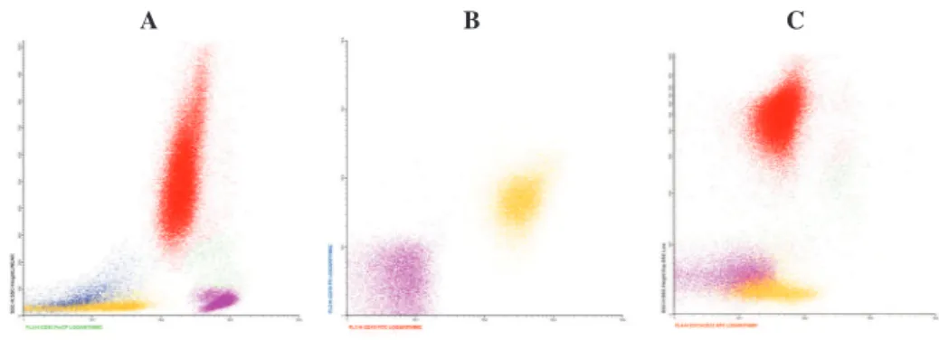
The following igure presents the analysis of a case of B-cell ALL. The abnormal blasts may be distinguished from the normal ones by their deicient and variable expression of CD45 (Figure 1). Fernanda Gonçalves Pereira Cunha
Felipe Franco da Rocha
Irene Gyongyver H. Lorand-Metze
Universidade Estadual de Campinas – UNICAMP, Campinas, SP, Brazil
Conlict-of-interest disclosure:
The authors declare no competing inancial
interest
Submitted: 9/26/2012 Accepted: 10/10/2012
Corresponding author: Irene Lorand-Metze Centro de Hematologia e Hemoterapia de Campinas Universidade Estadual de Campinas- Unicamp
Rua Carlos Chagas, 450 – Cidade Universitária “Prof. Zeferino Vaz” 13083-878 Distrito de Barão Geraldo, Campinas, SP, Brazil
Phone: 55 19-3521 8740 [email protected]
www.rbhh.org or www.scielo.br/rbhh
DOI: 10.5581/1516-8484.20120098
Figure 1 – Minimal residual diseasestudy of a B-cell acute lymphoblastic leukemia case. Phenotype at diagnosis: CD45dim variable, CD19+, CD10+ and aberrant expression of CD13/
CD33. Residual blasts in yellow (dim and variable expression of CD45, positive for CD19, CD10 and CD13+CD33). A: CD45/SSC plot B: CD10/CD19 plot C: CD13+CD33/SSC plot
A B C
References
1. Patkar N, Abu Alex AA, Bargavi B, Ahmed R, Abraham A, George B, et al. Standardizing minimal residual disease by low cytometry for precursor b lineage acute lymphoblastic leukemia in a developing country. Cytometry B Clin Cytom. 2012;82(4):252-8.
2. Coustan-Smith E, Song G, Clark C, Key L, Liu P, Mehrpooya M, et al. New markers for minimal residual disease detection in acute lymphoblastic leukemia. Blood. 2011; 117(23):6267-76.
3. Campana D. Minimal residual disease in acute lymphoblastic leukemia. Hematology Am Soc Hematol Educ Program. 2010;2010:7-12.
4. Borowitz MJ, Devidas M, Hunger SP, Bowman WP, Carroll AJ, Carroll WL, Linda S, Martin PL, Pullen DJ, Viswanatha D, Willman CL, Winick N, Camitta BM; Children’s Oncology Group. Clinical signiicance of minimal residual disease in childhood acute lymphoblastic leukemia and its relationship to other prognostic factors: a Children’s Oncology Group study. Blood. 2008;111(12):5477-85
5. Delbuono E, Maekawa YH, Latorre MR, Seber A, Petrilli AS, Braga JAP, Lee ML. Simpliied low cytometric assay to detect minimal residual disease in childhood with acute lymphoblastic leukemia. Rev Bras Hematol Hemoter. 2008;30(4):281-6.