167
Key words: blabber neoplasm, cystectomy, laparoscopy, urinary diversion
Int Braz J Urol. 2013; 39: 167-72
__________________
Submitted for publication: August 21, 2012
__________________
Accepted after revision: January 18, 2013 Purposes: We retrospectively assessed our experience with the W-shaped orthotopic ileal
pouch, which was constructed with non –absorbable titanium staples. For these purpose, we discuss the results of bladder capacity, urinary continence and early and long-term postoperative complications.
Materials and Methods: We included in the study 17 patients who underwent radical cystoprostatectomy followed by construction of an orthotopic W-shaped ileal pouch be-tween October 2000 and November 2009. A 65-70 cm segment of ileum was isolated and prearranged into a W- configuration, leaving two 10 cm intact segments on both sides of the ileal fragment. In our technique we entirely anatomized all adjacent limbs in order to create a sphere-shaped pouch. The ureters were directly anastomized to both intact segments of the ileal division. All our patients underwent pouchscopy 6 months after operation and annually.
Results: Mean operative time for neobladder reconstruction and ureteral anastomoses was 87 ± 7.67 minutes. In one patient a leak from the ileo-ileal anastomosis was confirmed on the 3rd day after operation. In 2 cases unilateral stricture of the ureteral-neobladder anastomosis was documented. Staple lines were mostly covered with ileal mucosa after 6 months. The mean functional bladder capacity was 340 ± 27.6 mL and 375 ± 43.4 mL at 6 and 12 months, respectively. First-year daytime and nighttime continence was good and acceptable in 90% and 78% of patients, while it increased to 95% during the 2nd year.
Conclusions: The long term follow-up shows that non-absorbable titanium staples can be safely used for creation of an orthotopic ileal neobladder. However, these data should be further validated in a larger series of patients.
INTRODUCTION
Since the first report of successful labo-ratory and clinical data of stapled bladder closu-re with titanium staples (1), several studies have adopted this technique with encouraging results (2-6). This technique possesses several advanta-ges, such as decreased operative time, excellent
tissue adaptation and presumably watertight clo-sure. However, the possibility of urinary stone for-mation on the staples requires long term follow--up (1). In this study we describe our technique for W-shaped orthotopic ileal pouch construction with non-absorbable titanium staples and report the results of long term follow-up.
Radical cystectomy with W-shaped orthotopic ileal
neobladder constructed with non-absorbable titanium
staples-long term follow-up
_______________________________________________
Sergey Kravchick, Leonid Lobik, Adrian Paz, Eugeny Stepnov, David Ben-Dor, Shmuel Cytron
Department of Urology (SK, LL, AP, ES, SC) and Department of Pathology (DBD), Barzilai Med. Center, Ashkelon, Israel
ABSTRACT
ARTICLE
INFO
MATERIALS AND METHODS
Between October 2000 and November 2009 seventeen males with invasive high grade carcinoma of urinary bladder underwent radical cystoprostatectomy with orthotopic W-shaped ileal pouch, which was constructed with non--absorbable staples. Six patients underwent this operation with a laparoscopic approach, in which case the pouch was created after surgical speci-men removal through an additional 7 cm incision. The criteria for inclusion in the study were: non--metastatic disease, negative biopsies from prosta-tic urethra, adequate renal function (serum creati-nine < 1.5mg/dL), normal liver function, no active inflammatory bowel disease or previous extensi-ve bowel resection (for laparoscopic procedure), physical and mental ability to live with a bladder substitute and the ability to perform self cathete-rization if needed, and compliance with routine follow-up.
Technique
Open (n-11) radical cystoprostatectomy was performed in the usual manner. In all lapa-roscopic cases (n-6) we used a 5-port transperi-toneal approach (7-9) and a 2-arm spring-loaded
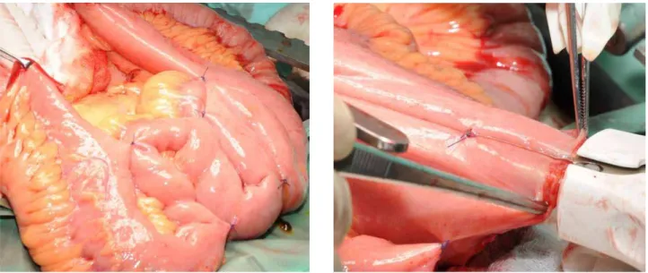
articulating instrument holder (Endoholder). In laparoscopic cases the surgical specimen was re-moved intact through an additional 7 cm inci-sion. Afterwards we isolated a 65-70 cm segment of ileum 20 cm proximally from the ileocecal valve. The most dependent part of the segment, that could easily reach the top of the symphy-sis pubis, was pointed and marked with a su-ture. The ileum was then divided between bo-wel clamps and a standard bobo-wel anastomosis was performed with staples. The mesenteric trap was closed. A 40-45 cm segment of the isolated ileum was prearranged into W configuration, le-aving two intact segments on both sides of the ileal fragment in order to decrease the tension on the ureteral anastomoses. In our version of the W-shaped technique all adjacent limbs were entirely anastomosed “side by side”. For this purpose, an opening was created on the poin-ted distal loop of the segment and a 80X3.5 mm mechanical stapler (Multifire GIA - US Surgical) was inserted through the opening and fired (Fi-gures 1 A and B). Later, additional openings were made on the proximal part of the segment and 60/80X3.5 mm mechanical staplers were inser-ted and fired in order to create a sphere-shaped pouch (Figures 1 C and D).
Figure 1A - A 40-45 cm segment of the isolated ileum is pre-arranged into W configuration, leaving two intact segments on both sides of the ileal fragment. An opening was created on the pointed distal loop.
169
Afterwards, 2 single J stents were inserted up to the renal pelvis and the ureters were directly anastomosed to the intact segments of the ileal division, according to the Wallace technique. The distal ends of the stents were taken out through the separate small openings on the lateral walls of the pouch. Finally, the proximal openings in the pouch, which were made for the staplers insertion, were closed with running absorbable sutures, le-aving the distal outlet open. The neobladder was placed inside the abdominal cavity and a 22 Fr silicone Foley catheter was inserted in the urethra. The pouch was anastomosed to the urethra with six interrupted 3-0 monocryl sutures over a 22F Foley. All knots were tied in the outside manner.
Postoperative and follow-up protocols
The urethral Foley catheter was irriga-ted with 50 mL of saline every 4 to 6 hours for the first 2 to 3 postoperative days. Starting from the 4th day it was irrigated with 100 mL every 8 hours. The Jackson-Pratt drains were removed as drainage decreased to less than 100 mL. If the cystogram on postoperative day 14 excluded any urinary extravasation, the left ureteral stent was removed first, followed 24 hours later by the right stent. The urethral catheter was removed between days 18-21.
During the first months all patients were instructed to void in a sitting position by
rela-xation of the pelvic floor, followed by slight ab-dominal straining. Effectiveness of emptying was maintained by hand pressure on the lower abdo-men and bending forward. We asked our patients to perform daily self catheterization and pouch irrigation during the first month in order to de-crease the chance of mucous blockage. Through the first 2 months patients were instructed to void every 3-4 hours during the day and every 4 hours at night. All our patients underwent pouchscopy 6 months after operation and annually.
We carried out cystometry 6/12 months postoperatively in all patients. Maximal neoblad-der capacity was identified based on the maximal discomfort in the lower abdomen or urethral le-akage. We asked patients to fill out voiding charts to assess functional neobladder capacity and pos-tvoiding residual volume (measured by catheter insertion). We also inquired that the patients gra-de their day and night time continence as good (no need for pad), acceptable (1-2 pads) or poor (> 2 pads). To exclude the detrimental effect (obs-truction) of the anti- peristaltic segment of ileum which was used for uretero-ileal anastomosis, all patients underwent renal scan.
RESULTS
Patients’ ages ranged from 47 to 72 ye-ars with a mean age of 68 ± 6.23. Mean time for
Figure 1C - The additional opening is made on the proximal part of the segment and 60/80X3.5 mm mechanical staplers is inserted and fired.
orthotopic ileal-neobladder reconstruction and ureteral anastomoses was 87 ± 7.67 minutes. No tumor was found in 2 postoperative specimens. In the other patients pathology reported T1 low grade (n-2), T1 high grade (n-4), T2 high grade (n-5), T2 high grade with CIS (n-1) and T3 a-b high grade TCC (n-3). Positive lymph nodes were found in 4 specimens. No sign of leakage was found on posto-perative cystograms. Three patients suffered from upper tract UTI in the early postoperative period (Clavien Grade 2). In one patient a leak was con-firmed from the intestinal anastomosis (ileo-ileal) on the 3rd day after operation (Clavien Grade 3b).
Five patients died from metastatic disease in the first 3 years after operation and two patients died from concomitant diseases (2 and 6 years, respectively). Mean follow-up was 64.76 ± 23.6 months. Seven patients remained in follow-up > 5 years (6-8 years) and six patients ≥ 3 years (3-5 ye-ars), while 4 patients were followed for ≤ 2 years.
During the first six months after operation daytime continence was good and acceptable in 90% (no or 1-2 pads), while nighttime continence was good and acceptable in 78% of patients. Howe-ver, starting from the second half of the year dayti-me continence increased to 95%. One half year after surgery the mean maximal neobladder capacity was 395 ± 65.3 mL, while one year after operation it was 463 ± 59.4 mL. The mean functional bladder capacity was 340 ± 27.6 mL and 375 ± 43.4 mL at 6 and 12 months, respectively. Only three patients still used self catheterization 3 years after operation.
In 2 patients UTI developed 6 to 37 months after operation. Creatinine rose > 0.5 mg/dL from the “baseline” in 5 patients, while renal scan confir-med deterioration of the renal function in three of them (Clavien Grade 4a): two in the right and one in the left unit. In 8 patients treatment with vitamin B12 was started due to anemia. In two cases mild unilateral stricture of the right ureteral-neobladder anastomosis was suspected on control CT urogra-phy. In both cases renal scan (99mTc-MAG3) was followed by furosemide administration with a sepa-rate 20-min assessment and was interpreted as an equivocal result.
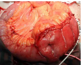
Cystoscopic control of the pouch revealed that the staple lines were mostly covered with ile-al mucosa 6 months after operation (Figures 2 A and B). Small calculi 0.5 - 1 cm were found in 4 patients 2-6 years after operation and in all the-se cathe-ses the calculi pasthe-sed spontaneously or were removed with a basket during follow-up flexible cystoscopy. We failed to find staple material in the nucleus of the stones.
DISCUSSION
The experience of general surgeons, who pioneered staplers for anastomosis construction during bowel resection and reconstruction, has shown that this device may decrease operative time, simplify tissue adaptation and guarantee leak proof closure (10-12). Consequently, the re-sults of operation can become less dependent on
171
the operators’ experience. Since 1993 urologists also began using stapling devices for laparoscopic bladder closure (1,2). Kerbl et al. performed labo-ratory and clinical studies and reported no intra- and postoperative complications, with no episodes of stone formation 3-5 months after operation (1). However, at that time, these authors warned that previous experience with stainless steel staples in ileal loop diversion and Kock pouch had shown stone occurrence in 1.8-10% and in 16.7% of the patients, respectively. Later on, Shalhav et al. dis-cussed the results of long-term follow-up (greater than 7 years) after bladder closure with titanium staples and reported no stone formation on the closure line or within the bladder (3). In addition, Grubb et al. reported that Endo-GIA stapler used for renal pelvis closure possessed no risk for stone formation in the short term period (4).
Endoscopic control of neobladder in our study showed that urothelium had covered the staple line six months after operation. These re-sults are in accordance with the data reported by Abreu et al. (5,6). However, small stones were found in 4(26%) of 17 patients. In all of these ca-ses, the stones passed spontaneously or were re-moved with a basket. Subsequent analysis failed to find staple material in the nucleus of the cal-culi. Therefore, we agree that calculi formation in neobladder might be explained by another cause, including metabolic changes and other processes that take place in the pouch (13,14). Although the rates of stone formation in our study were a little bit higher than those previously reported (5,6,13,15,16), we believe that long-term follow--up and relative frequent endoscopic control mi-ght explain this discrepancy.
Ureteral stricture is one of the most con-troversial issues in ileal pouch construction, be-cause it can be-cause renal damage. In an attempt to decrease their incidence, one should avoid exces-sive ureteral mobilization, devascularization and anastomotic tension. Based on the previous study of Montie et al., we used chimneys on the both si-des of ileal loop in order to advance them towards the end of ureters (17). Anastomotic strictures oc-curred in two cases and were successfully treated with minimal endoscopic procedures. Although the stricture rate in our study was higher than
that reported by Fontana et al., it is still compa-rable with the results of other studies (18). We also failed to find any sign of obstruction of the uretero-ileal anastomosis on the antiperistaltic site of neobladder.
Our version of the W-neobladder cons-truction provided comparable maximal and func-tional bladder capacity due to the long detubu-larized segment and entirely anastomosed limbs. The storage capacity of our reservoir was much better than that reported by Montie et al. (17). As a result, good/acceptable daytime and nighttime continence was reported in 90% and 78% of the patients six months after operation. By the end of the first year daytime continence improved and reached 95%. These results are very similar to those previously reported by other authors, who constructed different types of neobladder (19,20). However, it must be emphasized that other studies had reported superior functional bladder capacity at the end of the first year (21). In these studies absorbable sutures were used, and this fact makes us to suggest that staplers line may restrain blad-der functional capacity during the first years.
We presume that our study comes from a relatively “low volume” hospital and this fact can inversely affect the results of long-term follow-up (22). However, the success rates and the percenta-ge of complications are very comparable with the data reported by “high volume hospitals/surgeons”. In this context we would like to agree that mecha-nical staplers simplify the operation and makes its results less dependent on operator experience.
CONCLUSIONS
CONFLICT OF INTEREST
None declared.
REFERENCES
1. Kerbl K, Chandhoke P, McDougall E, Figenshau RS, Stone AM, Clayman RV: Laparoscopic stapled bladder closure: laboratory and clinical experience. J Urol. 1993; 149: 1437-9; discussion 1439-40.
2. Chandhoke PS, Clayman RV, Kerbl K, Figenshau RS, McDougall EM, Kavoussi LR, et al.: Laparoscopic ureterectomy: initial clini-cal experience. J Urol. 1993; 149: 992-7.
3. Shalhav AL, Dunn MD, Portis AJ, Elbahnasy AM, McDougall EM, Clayman RV: Laparoscopic nephroureterectomy for upper tract transitional cell cancer: the Washington University experi-ence. J Urol. 2000; 163: 1100-4.
4. Grubb RL 3rd, Sundaram CP, Yan Y, Chen C, McDougall EM, Clayman RV: Use of titanium staples during upper tract laparo-scopic reconstructive surgery: initial experience. J Urol. 2002; 168: 1366-9.
5. Abreu SC, Messias FI, Argollo RS, Guedes GA, Araujo MB, Fonseca GN: Laparoscopic assisted radical cystoprostatectomy with Y-shaped orthotopic ileal neobladder constructed with non-absorbable titanium staples through a 5 cm Pfannensteil incision. Int Braz J Urol. 2005; 31: 362-7; discussion 368-9. 6. Abreu SC, Araújo MB, Silveira RA, Regadas RP, Pinheiro DG,
Messias FI, et al.: Laparoscopic-assisted radical cystectomy with U-shaped orthotopic ileal neobladder constructed using nonabsorbable titanium staples. Urology. 2006; 68: 193-7. 7. Gill IS, Fergany A, Klein EA, Kaouk JH, Sung GT, Meraney AM, et
al.: Laparoscopic radical cystoprostatectomy with ileal conduit performed completely intracorporeally: the initial 2 cases. Urol-ogy. 2000; 56: 26-9; discussion 29-30.
8. Moinzadeh A, Gill IS: Laparoscopic radical cystectomy with uri-nary diversion. Curr Opin Urol. 2004; 14: 83-7.
9. Basillote JB, Abdelshehid C, Ahlering TE, Shanberg AM: Lapa-roscopic assisted radical cystectomy with ileal neobladder: a comparison with the open approach. J Urol. 2004; 172: 489-93. 10. Kracht M, Hay JM, Fagniez PL, Fingerhut A: Ileocolonic anas-tomosis after right hemicolectomy for carcinoma: stapled or hand-sewn? A prospective, multicenter, randomized trial. Int J Colorectal Dis. 1993; 8: 29-33.
11. Gustavsson K, Gunnarsson U, Jestin P: Postoperative complica-tions after closure of a diverting ileostoma--differences accord-ing to closure technique. Int J Colorectal Dis. 2012; 27: 55-8.
12. Leung TT, MacLean AR, Buie WD, Dixon E: Comparison of sta-pled versus handsewn loop ileostomy closure: a meta-analysis. J Gastrointest Surg. 2008; 12: 939-44.
13. Terai A, Ueda T, Kakehi Y, Terachi T, Arai Y, Okada Y, et al.: Uri-nary calculi as a late complication of the Indiana continent uri-nary diversion: comparison with the Kock pouch procedure. J Urol. 1996; 155: 66-8.
14. Osther PJ, Poulsen AL, Steven K: Stone risk after bladder sub-stitution with the ileal-urethral Kock reservoir. Scand J Urol Nephrol. 2000; 34: 257-61.
15. Fontana D, Bellina M, Fasolis G, Frea B, Scarpa RM, Mari M, et al.: Y-neobladder: an easy, fast, and reliable procedure. Urology. 2004; 63: 699-703.
16. Turk TM, Koleski FC, Albala DM: Incidence of urolithiasis in cystectomy patients after intestinal conduit or continent urinary diversion. World J Urol. 1999; 17: 305-7.
17. Montie JE, Pontes JE, Parulkar BG, Selby T: W-stapled ileal neo-bladder formed entirely with absorbable staples. J Urol. 1994; 151: 1188-92.
18. Shaaban AA, Abdel-Latif M, Mosbah A, Gad H, Eraky I, Ali-El-Dein B, et al.: A randomized study comparing an antireflux sys-tem with a direct ureteric anastomosis in patients with ortho-topic ileal neobladders. BJU Int. 2006; 97: 1057-62.
19. Madersbacher S, Möhrle K, Burkhard F, Studer EU: Long-term voiding pattern of patients with ileal orthotopic bladder substi-tutes. J Urol. 2002; 167: 2052-7.
20. Rogers E, Scardino PT: A simple ileal substitute bladder after radical cystectomy: experience with a modification of the Studer pouch. J Urol. 1995; 153: 1432-8.
21. Marim G, Bal K, Balci U, Girgin C, Dinçel C: Long-term urody-namic and functional analysis of orthotopic “W” ileal neoblad-der following radical cystectomy. Int Urol Nephrol. 2008; 40: 629-3.
22. Goossens-Laan CA, Gooiker GA, van Gijn W, Post PN, Bosch JL, Kil PJ, et al.: A systematic review and meta-analysis of the relationship between hospital/surgeon volume and outcome for radical cystectomy: an update for the ongoing debate. Eur Urol. 2011; 59: 775-83.