ORIGINAL ARTICLE
228
Key words:
Carcinoma, Renal Cell; Biological Markers;
Proteomics;Proteins; MASP2 protein, human
[Supplementary Concept]
Int Braz J Urol. 2013; 39: 228-39
__________________
Submitted for publication: May 10, 2012
__________________
Accepted after revision: November 11, 2012
Purpose: To screen proteins/peptides in urine of Renal Cell Carcinoma (RCC) patients by SELDI-TOF (Surface Enhanced Laser Desorption Ionization - Time of Flight) in search of possible biomarkers.
Material and Methods: Sixty-one urines samples from Clear Cell RCC and Papillary RCC were compared to 29 samples of control urine on CM10 chip. Mass analysis was perfor-med in a ProteinChip Reader PCS 4,000 (Ciphergen Biosystems, Fremont, CA) with the software Ciphergen Express 3.0. All chips were read at low and at high laser energy. For statistical analysis the urine samples were clustered according to the histological clas-sification (Clear Cell and Papillary Carcinoma). For identification urine was loaded on a SDS PAGE gel and bands of most interest were excised, trypsinized and identified by MS/MS. Databank searches were performed in Swiss-Prot database using the MASCOT search algorithm and in Profound.
Results: Proteins that were identified from urine of controls included immunoglobulin light chains, albumin, secreted and transmembrane 1 precursor (protein K12), mannan--binding lectin-associated serine protease-2 (MASP-2) and vitelline membrane outer layer 1 isoform 1. Identification of immunoglobulins and isoforms of albumin are quite common by proteomics and therefore cannot be considered as possible molecular ma-rkers. K12 and MASP-2 play important physiological roles, while vitellite membrane outer layer 1 role is unknown since it was never purified in humans.
Conclusions: The down expression of Protein K-12 and MASP-2 make them good can-didates for RCC urine marker and should be validated in a bigger cohort including the other less common histological RCC subtypes.
INTRODUCTION
Urine analysis is a non-invasive method of clinical analysis and has been used primarily to monitor diseases of the urogenital tract. In a
pe-riod of 24 hours, normal urine shows 150 milligra-ms of proteins and peptides that are derived from a variety of sources. Most of them come from the glomerular filtrate of plasma, while others from
Urine screening by Seldi-Tof, followed by biomarker
identification, in a Brazilian cohort of patients with
Renal Cell Carcinoma (RCC)
_______________________________________________
Gilda Alves, Denise A. Pereira, Vanessa Sandim, Antonio A. Ornellas, Niko Escher, Christian Melle,
Ferdinand von Eggeling
Applied Genetic Laboratory, Hematology Division, National Institute of Cancer, (GA, DAP, VS); Division of Urology, National Institute of Cancer, (AAO); Division of Urology, Hospital Mário Kroeff, Rio de Janeiro, RJ, Brazil (AAO); Core Unit Chip Application, Institute of Human Genetics, Jena University Hospital, Jena, (NE, FE); Alere Technologies GmbH, Jena, (NE) and Biomolecular Photonics Group, Jena University Hospital, Germany (CM)
ABSTRACT
ARTICLE
INFO
_________________________________________________________ ___________________
IBJU |URINE SCREENING BY SELDI-TOF IN PATIENTS WITH RENAL CELL CARCINOMA
229
the process of apoptosis and the cleavage of mem-brane proteins that are secreted. High molecular weight proteins (albumin, for example) are able to pass through the glomerular filtrate. Small pro-teins, or peptides (< 10 kDa) are easily filtered by the glomerulus and constitute an important source of information, even if the peptide is the result of proteolysis of larger proteins circulating in plasma (1-3). Urine is especially attractive for biomarker discovery in urological diseases since any change in concentration of proteins in plasma will reflect in the urine.Mass Spectroscopy (MS) is an analytical technique that measures the mass-to-charge ra-tio of charged particles. It is used for determining the composition of a sample or molecule, and for elucidating the chemical structures of molecules, such as peptides and other chemical compounds. MS works by ionizing chemical compounds; the ions are then accelerated through a potential di-fference and focused into a beam. The ion beam passes through a magnetic field which bends the charged stream. Lighter components or compo-nents with more ionic charge will deflect in the field more than heavier or less charged compo-nents. The final element of a MS is the detector. The detector records either the charge induced or the current when an ion passes by or hits a surfa-ce. In a scanning instrument, the signals will ge-nerate a mass spectrum, a record of ions expressed as m/z (mass/charge).
MS/MS is the tandem mass spectrometry consisting of two mass spectrometers in series connected by a chamber known as a collision cell. The sample to be examined is essentially sorted and weighed in the first mass spectrometer, then broken into pieces in the collision cell, and a piece or pieces sorted and weighed in the second mass spectrometer. Identification by MS/MS is conside-red more accurate.
The technology of SELDI-TOF (Surface Enhanced Laser Desorption Ionization - Time of Flight) is a kind of mass spectrometer (MS). The instrument enables the separation of proteins and peptides by their physical properties (hydropho-bic, hydrophilic, acidic, basic, with affinity for metal etc.) on a solid surface called Chip. The time-of-flight (TOF) analyzer measures the time
they take to reach the detector. Ions velocities will depend on their masses and lighter ions will reach the detector first. The SELDI-TOF is very sensitive instrument and is able to detect proteins in fluids like urine without previous processing.
In spite of some new drugs for treatment (4), RCC is a disease that would benefit from im-provement of early detection. More than 30% of patients show locally advanced or metastatic disease at diagnosis. Furthermore, approximate-ly 40% of patients undergoing curative surgery for localized disease relapse. In search of possible biomarkers, we have screened proteins/peptides in individual samples of urine from 61 RCC Brazi-lian patients of the two most common histological subtypes (Clear Cell (CC) and Papillary).
MATERIAL AND METHODS
Patients and Controls
Patients and control individuals inclu-ded in this study were adults of both genders, smokers and nonsmokers. This study was appro-ved by the Ethics Committee of Instituto Nacio-nal de Câncer (INCA), Rio de Janeiro, Brazil, re-gistration # 38/05.
IBJU
|
URINE SCREENING B
Y SELDI-T
OF IN P
A
TIENTS WITH RENAL CELL CARCINOMA
230
Table 1 - Patient data, histopathological findings, tumor stage and survival analysis.
Pts. Gender Histological Type Furhman Grade Stage Follow-up (months) Deaths by Disease
1 Female ccRCC I T1aNxM0 55
2 Female ccRCC I T2aNxMx 62
3 Male ccRCC I T1aNxMx 1
4 Female ccRCC I T1aNxM0 66
5 Male ccRCC I T2aN0M0 37
6 Female ccRCC II T1bNxM0 61
7 Male ccRCC II T1bNxM0 67
8 Male ccRCC II T3aNxMx 66
9 Female ccRCC II T1bNxMx 65
10 Male ccRCC II T1bNxMx 75
11 Male ccRCC II T1aN0M0 64
12 Male ccRCC II T1aNxMx 9
13 Male ccRCC II T3aNxMx 61
14 Male ccRCC II T3aNxMx 61
15 Female ccRCC II T1bNxMx 60
16 Female ccRCC II T1aNxMx 59
17 Male ccRCC II T1bN0Mx 47
18 Female ccRCC II T1bNxMx 54
19 Male ccRCC II T1bNxMx 36
20 Male ccRCC II T2aNxMx 36
IBJU
|
URINE SCREENING B
Y SELDI-T
OF IN P
A
TIENTS WITH RENAL CELL CARCINOMA
2
31
22 Female ccRCC II T2aNxM0 29
23 Male ccRCC II T1bNxMx 32
24 Female ccRCC II T3aNxMx 9
25 Male ccRCC II T3aNxMx 30
26 Male ccRCC II T1bNxM0 27
27 Female ccRCC II T3aNxMx 25
28 Male ccRCC II T3aNxMx 11
29 Male ccRCC II T3aN2Mx 40
30 Female ccRCC III T3aNxMx 3
31 Male ccRCC III T3aNxMx 64
32 Female ccRCC III T1N0M0 63
33 Male ccRCC III T3aNxM0 56
34 Male ccRCC III T3aNxMx 1
35 Male ccRCC III T3aNxMx 15
36 Female ccRCC III T2aNxMx 54
37 Male ccRCC III T3bN0Mx 22
38 Male ccRCC III T3aNxMx 58
39 Male ccRCC III T3aN1Mx 2
40 Male ccRCC III T3bNx Mx 53
41 Male ccRCC III T3aNxMx 33
42 Male ccRCC III T2aN0M0 32
43 Male ccRCC III T1bN1M0 28
44 Female ccRCC III T2aN0Mx 26
IBJU
|
URINE SCREENING B
Y SELDI-T
OF IN P
A
TIENTS WITH RENAL CELL CARCINOMA
232
46 Female ccRCC IV T2bNxMx 64
47 Male ccRCC IV T1bN0M0 7
48 Female ccRCC IV T3aNxM0 30
49 Female ccRCC IV T2bNxMx 6
50 Male ccRCC IV T3aN2Mx 22
51 Female ccRCC IV T2bN2Mx 6
52 Male ccRCC IV T2N0M0 1
53 Female ccRCC IV T1bN0M0 27
54 Male Papillary RCC type I -- T1aNxM0 5
55 Female Papillary RCC type I II T1bN0M0 30
56 Female Papillary RCC type I II T2aNxM0 50
57 Female Papillary RCC type I II T1NxM0 39
58 Female Papillary RCC type II II T3aN0M0 48
59 Male Papillary RCC type II II T2bN1M0 55
60 Male Papillary RCC type II III T2bNxMx 14
IBJU |URINE SCREENING BY SELDI-TOF IN PATIENTS WITH RENAL CELL CARCINOMA
233
glomerulopathies, chronic kidney disease, urinary tract infections, lithiasis and prostatic diseases.Twenty-nine control urine samples were obtained from over 40 years old healthy individu-als from Laboratory of Molecular Biology, Depart-ment of Urology, Jena University Hospital, Ger-many. Control individuals had no indications of kidney abnormalities.
Urine Collection
The 10-20 mL first-void urine was discar-ded; the following 50-70 mL urine was collected, centrifuged at 1,000g for 10 minutes and the su-pernatant saved (5). Phenylmethanesulfonyl flu-oride (PMSF) was added to the urine samples at final concentration of 1mM, aliquoted and frozen at -70oC.
Binding conditions of urine proteins to the chips
Aliquots of urine were thawed in ice and were then vortexed at full power (6). Thereafter, all procedures were carried out at room tempera-ture. All experiments were performed in duplica-te. Three kinds of chips, IMAC30, Q-10 and CM10 (BIORAD), were tested.
The operating mechanism of the Protein-Chip IMAC30 is the reversible binding of proteins to the surface through a coordinated metal inte-raction. The ProteinChip IMAC30 array incorpora-tes nitrilotriacetic acid (NTA) groups and is capa-ble of forming stacapa-ble octahedral complexes with polyvalent metal ions, including Cu2+,Ni2+, Fe3+, and Ga3+. After loading the array surface with the desired metal ion, two free sites are available from the formed octahedral complex for interac-tion with specific amino acid residues (such as His) or posttranslational modifications such as phos-phate groups. To generate selectivity, binding and washing buffers may contain increasing concen-trations of competitors, such as imidazole, which compete with the coordinated metal on the NTA group for binding to the protein or peptide.
Q10 incorporates quaternized ammonium groups (positively charged) and thus acts as a strong anion exchanger. The Q10 surface binds peptides and proteins that are negatively charged at a given pH. By maintaining the pH of the bin-ding or washing buffer at alkaline conditions (e.g.,
pH 8), an overall net negative charge is imparted on a greater number of proteins within the sample (therefore more binding). By decreasing the pH of the binding or washing buffer, an overall net po-sitive charge is imparted on the proteins, resulting in less binding (i.e., more specificity).
CM10 incorporates a carboxylate chemis-try (negatively charged) and thus acts as a weak cationic exchanger. CM10 surface binds proteins that are positively charged at a given pH. To ge-nerate selectivity, the pH of the binding buffer is increased or decreased, depending on the need. By decreasing the pH of the binding and washing bu-ffers, an overall net positive charge is imparted on a greater number of proteins within the sample (therefore more binding). By increasing the pH of the binding and washing buffers, an overall net negative charge is imparted on the proteins, resul-ting in less binding (i.e., more specificity).
The effect of prefractioning of the urine samples was tested applying the Protein Chip Se-rum Fraction kit (BioRad, cat. K10-0007) accor-ding to manufacture’s protocol. The increase of the peak intensity was also tested with the addi-tion of a denaturing soluaddi-tion (9M urea, 2% CHAPS (3-[(3-Cholamidopropyl) dimethylammonio]-1--propanesulfonate hydrate), 50mM TRIS (Tris (hydroxymethyl)aminomethane)-HCl pH 9.0) to the urine before mixing to the binding buffer.
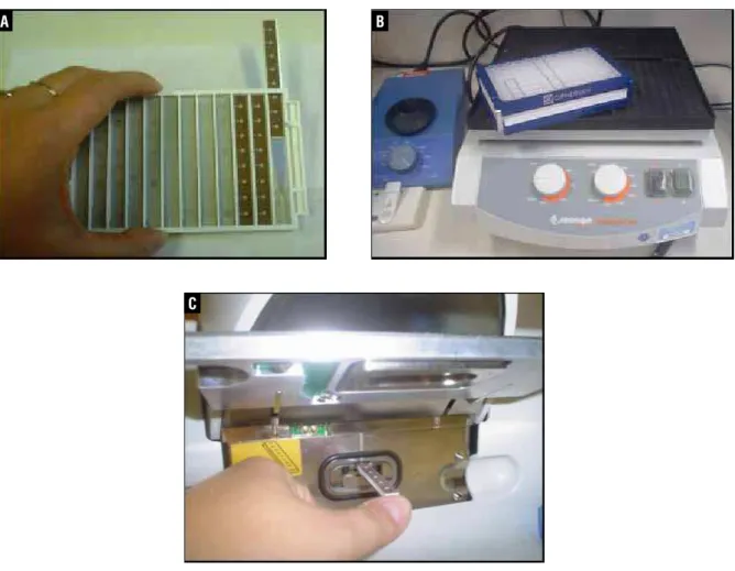
Alternatively, 50µL of pure urine were lo-aded with appropriate buffers in a bioprocessor (Ciphergen) for 90 minutes on a shaker. The chips were then washed, on a shaker, three times with the same buffers and two times with deionized wa-ter and air dried. A saturated solution of sinapinic acid (SPA) was prepared and 2x 0.5µL was applied in each spot. Figure-1a shows the assembling of chips into a frame that will go into a bioprocessor (Figure-1b); Figure-1c shows a chip being inserted into the SELDI-TOF).
Spectral data processing and statistical analysis
Biosyste-IBJU |URINE SCREENING BY SELDI-TOF IN PATIENTS WITH RENAL CELL CARCINOMA
234
Figure 1 A-C - Assembling of chips into the bioprocessor and insertion of a chip into the SELDI-TOF.
A B
C
ms, Fremont, CA) with the software Ciphergen Express Client 3.0. All chips were read at low laser energy (2,200 KJ) and at high laser energy (3,500KJ). Low laser energy and high laser ener-gy focused mass range (m/z) of 1,000 to 20,000 Da and 20,000 to 200,000 Da, respectively. Peaks corresponding to albumin and hemoglobin were excluded from the analysis.
Urine pool fractionation with RP beads followed by SDS page gel
RP resins are a family of polymeric me-dia useful for sample fractionation. RP beads are porous spherical beads, containing a surface area that is chemically stable to maximize extraction efficiency. The beads resin shows special
characte-ristics such as: non-polar, non-ionic, ultra-clean, highly crossed-linked polymers that differ in par-ticle size, pore size and surface area. The pore size large is selected according elements to enter into the pore and to adsorb onto the pore surface. Among the applications are exchange solvents, separate hydrophobic molecules by size and de-salt samples.
IBJU |URINE SCREENING BY SELDI-TOF IN PATIENTS WITH RENAL CELL CARCINOMA
235
The sample was fractionated into six fractions by sequential elution with 100µL aliquots of elution buffer with increasing concentrations of acetoni-trile in 0.1% trichloroacetic acid (10-100% aceto-nitrile). Two µL of each fraction was directly spot-ted on the CM10 and analyzed by SELDI-TOF for selection. The fraction containing the protein of interest was concentrated in a vacuum centrifuge and loaded on a precast 12% SDS-PAGE gel (In-vitrogen). Protein fractions co-migrated with the molecular maker Page Ruler Plus (Fermentas, cat. SM 1811). The gel was stained with Simply Blue Safe Stain (Enhanced Coomassie, Invitrogen).Gel band trypsinization
The band of interest was excised from the stained gel and destained overnight with a mix of methanol 50% and acetic acid 10%. The gel piece was dehydrated in 100% acetonitrile for 30 mi-nutes, dried in a vacuum centrifuge. Ten ng/mL of sequencing grade trypsin (Promega, UK) in 50 mM ammonium bicarbonate was added to dried gel piece and incubated at 37oC overnight.
MS/MS Analysis
The resulting peptides were analyzed in a MALDI TOF/TOF with a Bruker ultraflex devi-ce (Bruker Daltonics, Bremen, Germany). Brie-fly, samples were prepared in dried-droplet-pre-paration with α-hydroxy-cyano-cinnamic acid (CHCA). Peptide mass fingerprint and peptide fragmentation spectra of each sample were
sub-mitted for identification using MASCOT interface (MASCOT inhouse server 2.1.03, Matrix Scien-ce, London, UK) for search in the NCBI database. Hits were considered significant according to the MASCOT score (P = 0.05).
RESULTS
Binding conditions to the chips
Tests were performed with 3 different chips (IMAC30, Q-10 and CM10, BIORAD). The quanti-ty of peaks that were produced in each condition was considered. The protocol applying fractions produced by the Protein Chip Serum Fraction kit did not improve the number of peaks and their intensity, no matter the chip. The protocol of p-denaturing of the urine has not improved the re-sults as well. The highest number of peaks were generated when pure urine samples were applied on CM10 chip therefore it was selected to perform the screening of RCC urines.
Peaks selection and identification
For analysis, the urine samples were clus-tered according to the histological classification and compared to 29 control samples. We observed 22 peaks (m/z) that were statistically overexpres-sed (p < 0.05) in the control group. The most sig-nificant peaks are listed at Table-2. An example of differently expressed peak is shown in Figure-2.
The selected RP fractions of the control uri-ne pool were loaded in a SDS PAGE gel (Figure-3)
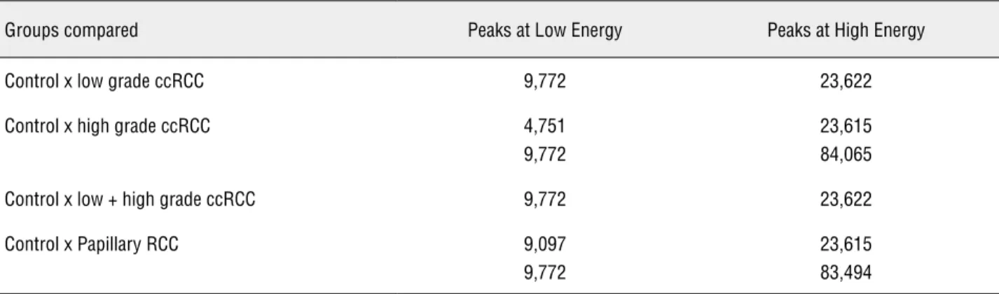
Table 2 – Peaks (m/z) found overexpressed in the Control urine group in comparison to different histological/grade types of Renal Cell Carcinoma (RCC) urines.
Groups compared Peaks at Low Energy Peaks at High Energy
Control x low grade ccRCC 9,772 23,622
Control x high grade ccRCC 4,751
9,772
23,615 84,065
Control x low + high grade ccRCC 9,772 23,622
Control x Papillary RCC 9,097
9,772
23,615 83,494
IBJU |URINE SCREENING BY SELDI-TOF IN PATIENTS WITH RENAL CELL CARCINOMA
236
Figure 2 - Spectra showing the peak of approximately 23,600 that is underexpressed in patients.
Figure 3 - Isolation of bands of interest from a Coomassie Blue stained 12% SDS PAGE gel from fraction eluted with 60% acetonitrile. Arrows indicate the bands of interest of approximately 9,770 (A) and 23,600 (B).
and bands of most interest (A and B) were excised for identification. Table 3 shows the results of the MS/MS analysis of bands A and B (Figure-3).
COMMENTS
IBJU |URINE SCREENING BY SELDI-TOF IN PATIENTS WITH RENAL CELL CARCINOMA
237
Proteins that were identified from urine of controls by MS/MS analysis were immunoglobu-lin light chain, albumin, secreted and transmem-brane 1 precursor (protein K12), mannan-binding lectin-associated serine protease-2 (MASP-2) and vitelline membrane outer layer 1 isoform 1 (Ta-ble-3). Identification of immunoglobulins and isoforms of albumin are quite common by prote-omics and therefore cannot be considered as pos-sible molecular markers. On the other hand, the other 3 identified proteins should be taken into consideration. The peptides sequences (Table-3) from each one of these proteins were checked on uniprot data bank (www.uniprot.org) and they all corresponded 100% to the proteins assigned.Expression of mRNA of K12 gene (SECTM1) was detected at the highest levels in peripheral blood leukocytes and breast cancer cell
lines. Western blots showed that the K12 protein exists as a cluster of bands around 27 kDa, and extractions using nonionic detergents or high pH conditions demonstrate that it behaves as an in-tegral membrane protein. Immunofluorescence localization studies reveal that K12 is not detec-table on the cell surface, but instead is found in a perinuclear Golgi-like pattern and colocalizes with a well-known Golgi marker. In addition, an approximately 20-kDa soluble form of the K12 protein derived from the N-terminal domain is specifically secreted by cells into the culture me-dium. Immunohistochemical analysis of periphe-ral blood cells shows that K12 is found in leu-kocytes of the myeloid lineage, with the strongest staining observed in granulocytes and no detecta-ble expression in lymphocytes. Based on its range of expression, it is possible that K12 is a protein Table 3 - Protein identified by MS/MS.
Band NCBI1 Human Protein Score2 Average
mass3 (KDa)
Peptide4
A gi|306999 Immunoglobulin
light chain
81 R.LLIYGASTR.A
-.EIVLTQSPGTLSLSPGER.A
A gi|23241675 Albumin 195 46,442 K.FQNALLVR.Y
K.LVNEVTEFAK.T K.KVPQVSTPTLVEVSR.N
B gi|4506869 Secreted and
transmembrane 1 precursor
113 27,307 R.DSHAGLYMWHLVGHQR.N
R.AHGQESAIFNEVAPGYFSR.D
B gi|3297879 MASP-2; mannan-binding
lectin-associated serine protease-2
62 77,176 R.APGKDTFYSLGSSLDITFR.S
B gi|32698964 Vitelline membrane outer
layer 1 isoform 1
146 22,034 R.GLGDDTALNDAR.L
R.VEAPTTLGDNTAANNVR.F K.VEPPQGIPGDDTALNGIR.L
1NCBI: National Center for Biotechnology Information: numbering refers to the protein in database
2Score: ideal value is above 35
3Theoretical molecular weight;
IBJU |URINE SCREENING BY SELDI-TOF IN PATIENTS WITH RENAL CELL CARCINOMA
238
with potential importance in hematopoietic and/ or immune system processes (8).Mannan-binding lectin (MBL) is an oligo-meric serum lectin that plays a role in innate im-munity by activating the complement system. In human, two types of MBL-associated serine prote-ase (MASP-1 and MASP-2) and a truncated pro-tein of MASP-2 (small MBL-associated propro-tein; sMAP or MAp19) are complexed with MBL (9). Af-ter activation by auto-catalytic cleavage, MASP-2 cleaves C2 and C4, leading to their activation and to the formation of C3 convertase.
Vitelline membrane outer layer 1 isoform 1 belongs to the family VMO1 that is conserved in the chicken, in the mouse and in human. In chi-ckens, this protein participates in the construction of the vitelline membrane portion of the egg shell, a rigid structure required to maintain the shape of the egg (10). The role of this protein in humans is unknown, actually it was never purified. It was inferred from electronic annotation and probably is secreted.
Amazingly, the down expression of pro-tein K-12 and MASP-2 were observed in two his-tological subtypes (ccRCC and Papillary) which show differentiated origin in the kidney and dis-tinguished genetics alterations. Papillary RCCs are characterized by trisomy of chromosomes 3q, 7, 8, 12, 16, 17, and 20 and loss of the Y. On the other hand, chromosome 3p deletion is the most typi-cal genetic alteration in ccRCC, present in 75.8% of cases which coincides with von Hippel-Lindau disease (11). This fact creates the possibility that a subtype independent RCC urine marker could be in the way. K-12 and MASP-2 should be tested in the urine of other RCC histological subtypes, although they are much less frequent than ccRCC and Papillary.
CONCLUSIONS
The down expression of Protein K-12 and MASP-2 make them good candidates for RCC uri-ne makers and should be validated in a bigger co-hort including the other less common histological RCC subtypes. Probably, these proteins were found in the control urine as a consequence of blood filtration role developed by the kidney. The lower
abundance of these proteins in the patients’ urine could be a consequence of their degradation or of kidney failure.
ACKNOWLEGMENTS
We thank Prof. Dr. Kestin Junker of Labo-ratory of Molecular Biology, Department of Uro-logy, Jena University Hospital, 07740 Jena, Ger-many, for providing some of urine control samples and Annett Urbanek for technical assistance at SELDI-TOF.
This work was supported by PROBRAL CA-PES/DAAD 293/08 and Programa de Oncobiologia (Rio de Janeiro, Brazil).
CONFLICT OF INTEREST
None declared.
REFERENCES
1. Rindler MJ, Naik SS, Li N, Hoops TC, Peraldi MN: Uromod-ulin (Tamm-Horsfall glycoprotein/uromucoid) is a phos-phatidylinositol-linked membrane protein. J Biol Chem. 1990; 265: 20784-9.
2. Pisitkun T, Shen RF, Knepper MA: Identification and pro-teomic profiling of exosomes in human urine. Proc Natl Acad Sci U S A. 2004; 101: 13368-73
3. Pisitkun T, Johnstone R, Knepper MA: Discovery of urinary biomarkers. Mol Cell Proteomics. 2006; 5: 1760-71. 4. Ather MH, Masood N, Siddiqui T: Current management
of advanced and metastatic renal cell carcinoma. Urol J. 2010; 7: 1-9.
5. Schaub S, Rush D, Wilkins J, Gibson IW, Weiler T, Sang-ster K, et al.: Proteomic-based detection of urine proteins associated with acute renal allograft rejection. J Am Soc Nephrol. 2004; 15: 219-27.
6. Mataija-Botelho D, Murphy P, Pinto DM, Maclellan DL, Lan-glois C, Doucette AA: A qualitative proteome investigation of the sediment portion of human urine: Implications in the biomarker discovery process. Proteomics Clin Appl. 2009; 3: 95-105.
IBJU |URINE SCREENING BY SELDI-TOF IN PATIENTS WITH RENAL CELL CARCINOMA
239
8. Slentz-Kesler KA, Hale LP, Kaufman RE: Identification andcharacterization of K12 (SECTM1), a novel human gene that encodes a Golgi-associated protein with transmem-brane and secreted isoforms. Genomics. 1998; 47: 327-40. 9. Matsushita M, Thiel S, Jensenius JC, Terai I, Fujita T: Proteo-lytic activities of two types of mannose-binding lectin-associ-ated serine protease. J Immunol. 2000; 165: 2637-42.
10. Guérin-Dubiard C, Pasco M, Mollé D, Désert C, Croguennec T, Nau F: Proteomic analysis of hen egg white. J Agric Food Chem. 2006; 54: 3901-10.
11. Algaba F, Akaza H, López-Beltrán A, Martignoni G, Moch H, Montironi R, et al.: Current pathology keys of renal cell carcinoma. Eur Urol. 2011; 60: 634-43.