Key words:
Ureteroscopy; Calculi; Stents; Emergencies
Int Braz J Urol. 2013; 39: 203-8
__________________
Submitted for publication: June 06, 2012
__________________
Accepted after revision: January 15, 2013 Purpose: Emergency double-J (DJ) stenting following “uncomplicated” ureteroscopic
(UURS) stone treatment is both morbid and costly. Our study aims at identifying those patients who are more likely to require such an extra procedure. Handling of this com-plication will also be highlighted.
Materials and Methods: 319 cases of UURS cases were selected out of 903 patients, who were admitted for URS stone treatment at King Abdullah University Hospital during the period from May, 2003 to December, 2010. Thirty-eight of them (11.9%) had emergency post-URS DJ stenting within 24 hours of discharge. The medical records of all UURS cases were retrospectively reviewed. Comparison in demographic and stone-related variables was made using 2-paired t-test with P < 0.05. Operative findings of 38 stented patients were outlined.
Results: Significant risk factors for emergency stenting were noted in males with larger (> 1.5 cm) and proximal stones (38 stented vs. 281 unstented). Operative risk factors among the 38 patients were: initial procedure time > 45 minutes (42.1%), ureteral wall edema (21.1%), repeated access for stones > 1.5 cm (21.1%), impacted stone (10.5%) and ignored or missed stones/fragments (4.6%).
Conclusions: The need for emergency DJ stenting following UURS stone treatment is not uncommon. The routine insertion is impractical and weakly-supported. With risk-factor stratification, selective and individualized DJ stenting policy is recommended.
INTRODUCTION
Fragmentation and clearance of ureteric stones can either be achieved by extracorporeal shock wave lithotripsy (ESWL) or ureteroscopy stone treatment (URS). URS stone removal has been found to carry a better overall stone-free rate compared to ESWL. The Current European guide-lines recommend primary use of URS in treatment of most ureteric stones (1).
The insertion of double-J (DJ) stent du-ring URS stone extraction is controversial. Since the pioneering report by Hosking et al. (2) and the radical characterization by Moon (3), urologists started to adopt a more selective policy.
Complications of DJ stent insertion inclu-de disturbing storage lower urinary tract symp-toms, pain, hematuria, infection and poorer
qua-Emergency double-J stent insertion following
uncomplicated Ureteroscopy: risk-factor analysis and
recommendations
_______________________________________________
Yousef S. Matani, Mohammed A. Al-ghazo, Rami S. Al-azab, Osamah Bani-hani, Daher K. Rabadi
Department of General Surgery and Urology (YSM, MAA, RSA, OB) and Department of Anaesthesia (DKR) Jordan University of Science and Technology, Faculty of Medicine, King Abdullah University Hospital, Irbid-Jordan
ABSTRACT
ARTICLE
INFO
lity of life (4,5). It is, however, thought to reduce post URS obstruction, facilitate clearance of stone fragments and decrease stricture rate (6,7).
The definition of uncomplicated URS (UURS) is both lacking and weakly standardized. Denstedt et al. defined UURS as a procedure with “no evidence of perforation or lack of clinically important edema”. Free flow of contrast into the bladder on retrograde pyelography is exclusive of edema (8). Other studies used an endoscopic, non--validated grading of ureteric edema on a scale of 0 (mild) to 2 (severe) (6,9).
Our study will look at risk-group stratifi-cation of patients who might require stenting du-ring their initial “UURS” and address the concept of “prophylactic” DJ stent use.
MATERIALS AND METHODS
From May 2003 to December 2010, 903 pa-tients had undergone semirigid URS with Holmium laser (365 micron; 0.5-1.4J/5-10 Hz) lithotripsy. All patients were admitted to the urology department at King Abdullah University Hospital and their medical records retrospectively analyzed. All patients had a preoperative consent. Imaging studies included kid-ney-ureter-bladder (KUB) X-ray and non-enhanced computed tomography (NECT).
Among a total of 903 patients, 319 un-derwent primary “straightforward” UURS which was defined based on the following selection criteria:
1. All had single and unilateral ureteric stone;
2. Intra-operative perforation was not documented;
3. DJ stent and/or ureteric catheter were not inserted;
4. Ureteric dilatation and/or usage of access catheter were not used;
5. Children and pregnant ladies were excluded;
6. Stone free after the procedure was documented (defined as complete removal and/or residual stone frag-ments < 3 mm in diameter).
URS was performed, using 8/9.8 semirigid ureteroscope (Richard Wolf, Germany), under ge-neral anesthesia in all patients. Urine cultures were
negative. Prophylactic antibiotic was given at induc-tion as a single 1 g IV ceftriaxone. Subsequently, 500mg oral ciprofloxacin tablets were given twice daily for 24 hours.
The stone size and location were determined by KUB and NECT films. They were divided into pro-ximal, middle and distal third ureteral stones. URS stone extraction was achieved by Dormia basket and/or forceps.
Thirty-eight out of 319 UURS had emer-gency stent insertion within 24 hours of initial URS due to intolerable colic and significant discomfort. Diagnostic URS was performed, prior to stenting, for defining a possible etiology or injury. A height-ma-tched length 6F DJ stent was used. Discharge was made within 24 hours and the stent was removed after 1-2 weeks.
The demographic features, stone-related factors and operative URS findings were analyzed and tabulated. Comparison between those stented and un-stented (38 vs. 281) groups was made using 2-tailed t-test statistics. A P < 0.05 was taken as the level of significance. The analysis was performed with computer software (Statistical Package for the Social Sciences, version 16.0).
RESULTS
Thirty eight of the 319 UURS (11.9 %) pa-tients had emergency stent insertion. The proce-dure was complication-free. The mean operative time was 25 minutes. The demographic and sto-ne-related variables of the study group are listed in Table-1. Twenty seven patients were men and 11 women (2.5:1), with a mean age of 38.2 ye-ars (range 28-62). The stones included 9 proxi-mal (23.7%), 11 mid-ureter (28.9%), and 18 distal stones (47.4%). Average stone diameter was 10.2 cm (range 7-23 mm).
Significant preoperative risk variables in-cluded male sex (P = 0.037) and proximal stones (P = 0.018). Average ages were comparable (38.2 vs. 39.1 years, P = 0.30). Average stone diameter was 1.2 cm and 0.94 cm in the stented and un--stented groups, respectively (P = 0.06).
repe-ated access through the ureteral orifice for larger stones (> 1.5 cm) in 8 (21.1%), localized wall ede-ma in 8 (21.1%), handling of impacted stones in 4 (10.5%), ignored small calyceal stone in 1 patient (2.3%) and residual stone fragments < 3mm in 1 patient (2.3%). Control and comparison of these risk factors with the unstented group would have been contributory but clearly unethical.
DISCUSSION
URS was first reported in 1982 (10) by Pe-rez-Castro in cooperation with Karl Storz. The use of stents during this period was not only strange, but also unfavorable. Eisenberger referred to stents as ‘‘Steckerin’’ (Bavarian for small sticks) (11). DJ stent was first described by Finney et al. in 1978 (12). Criticism to its role, however, appeared in the late 90s (2,3). Nowadays, the urologists remain, sharply, divided on the need for stenting following UURS treatment of lithiasis. Both routine and selective use has been practiced. Selective use, in particular, should depend on a variety of variables related to patients, stones, technology and experience.
Nabi et al. meta-analyzed 9 trials and con-cluded that stents have significantly higher rate of storage lower urinary symptoms (LUTS), infection, analgesia use, and ureteric stricture. Stenting, on the other hand, did not influence rates of stone clearan-ce. The authors, however, criticized data
inconsis-tency and lack of standardization. They, therefore, kept the issue of stenting open (5). Similar con-clusions were reached by three recent evaluations (13-15). They, basically, advised against routine DJ stenting and were not satisfied by homogeneity and pooling of materials. An excellent review of this di-lemma was expressed by Keeley and Timoney (16) who identified the pros and cons of stenting and advised for more meaningful studies.
The use of an alternative and temporary drai-nage procedure has, recently, been considered. It uti-lizes short-term insertion of ureteric catheters. This accessory procedure may overcome edema, reduce pain, decrease outpatient visits, avoid secondary en-doscopy and limit costs. Djaladat et al. were able to show that pain, storage LUTS and outpatient visits were significantly reduced in the catheter group. Urinary tract infection (UTI) was established in 7 and 4 % in catheter and non-catheter groups respecti-vely. Readmission and stone clearance rates were comparable in a 2-week follow up (17). Reduction in pain and international prostate symptom scores was noted in one-day post-URS catheterization (18).
Baseless avoidance of stenting carries me-asureable morbidity and cost. DJ stenting is bene-ficial when obstruction secondary to edema and/or inflammation was anticipated (19). It is, also, effec-tive in reducing pain and promoting drainage in hydronephrosis (20). Cheung et al. highlighted the value of selection in reducing overall stenting rate
Table 1 - Demographics and stone features.
No. of patients 38
sex(M:F) 2.5:1(27/11)
Mean age in years(Range) 38.2 (28-62)
Stone
Mean size in mm (range) 10.2(7-23)
Location
Upper 9(23.7%)
Middle 11(28.9%)
Lower 18(47.4%)
Table 2 - URS findings prior to DJ stenting (Risk factors).
No. of patients 38
Mean operative time(minutes) 25
Risk factors
Operative time > 45 minutes 16(42.1%)
localized wall edema 8(21.1%)
Repeated access for stones >1.5 cm 8(21.1%)
Impacted stone 4(10.5%)
Ignored small calyceal stone 1(2.3%)
without altering stone-free outcome. Their stenting rate was 39% and limited to impacted stones, severe preoperative obstruction and residual poor postope-rative drainage (21). Stents were, additionally, found useful in pregnant ladies (22), in upper urinary tract diseases (urolithiasis) (23) and when ureteral access sheath was used (24). Factors that contribute to DJ--associated morbidity include stent design, size, po-sitioning, associated UTI, and duration (25). Recent use of drug eluting stents (26) and alpha blockers (27) were reported to cause less pain and discomfort.
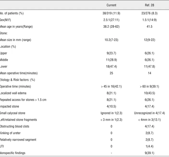
In this analysis, higher risk of emergency stenting was noted among males with larger and more proximal ureteric stones. The impact of stone location and size might be explained by increased manipulation, repeated access and development of wall edema. Increased risk in males is, however, difficult to justify. Comparable Intraoperative risk factors were reported in a similar recent study by Tanriverdi et al. (28). Summary of variables is listed in Table-3. In their analysis, about 2/3 of cases had prolonged procedure, repeated access and ureteral
Table 3 - Summary of data: current and reference no. 28 studies.
Current Ref. 28
No. of patients (%) 38/319 (11.9) 23/276 (8.3)
Sex(M:F) 2.5:1(27:11) 1.5:1(14:9)
Mean age in years(Range) 38.2 (28-62) 41.5
Stone:
Mean size in mm (range) 10.2(7-23) 12(9-22)
Location (%)
Upper 9(23.7) 6(26.1)
Middle 11(28.9) 6(26.1)
Lower 18(47.4) 11(47.8)
Mean operative time(minutes) 25 14
Etiology & Risk factors: (%)
Operative time (minutes) > 45 in 16(42.1) > 60 in 9(39.1)
Localized wall edema 8(21.1) 10(43.5)
Repeated access for stones > 1.5 cm 8(21.1) 6(26.1)
Impacted stone 4(10.5) 4(17.4)
Small calyceal stone Ignored in 1(2.3) Unrecognized in 4(17.4)
Left/retained stone fragments < 3 mm in 1(2.3) < 4mm in 3(13.1)
Obstructing blood clots 0 4(17.4)
Kinking of ureter 0 2(8.7)
Relatively narrowed segment 0 2(8.7)
UTI 0 1(4.4)
wall edema. Additional causes of postoperative obstruction were linked to residual or missed stones, blood clots, kinking or narrowed segments and UTI.
We do agree with the arguments raised against routine DJ stenting in UURS stone treat-ment (5,13-15,28,29). Insertion should better be individualized. Relative indications might include prolonged procedure (> 45 minutes), “significant” wall edema, repeated access, impacted stone, lar-ger stones (> 1.5 cm), use of access sheath, ureteric dilatation and pregnancy. Complete removal and clearance of stone(s)/fragments are highly recom-mended. The use of ureteric catheterization was not tested in our analysis.
CONCLUSIONS
Insertion of DJ stents during UURS treat-ment of stones is neither Angel nor Evil. Its role has not yet been decisively outlined. Stent inser-tion remains “opinser-tional” and a consensus is still remote. A risk-based selection may prove to be a better practice. In difficult and lengthy URS pro-cedures with significant stone burden, DJ stenting should be seriously considered.
CONFLICT OF INTEREST
None declared.
REFERENCES
1. Preminger GM, Tiselius HG, Assimos DG, Alken P, Buck AC, Gallucci M, et al.: 2007 Guideline for the management of ure-teral calculi. Eur Urol. 2007; 52: 1610-31.
2. Hosking DH, McColm SE, Smith WE: Is stenting following ure-teroscopy for removal of distal ureteral calculi necessary? J Urol. 1999; 161: 48-50.
3. Moon TD: Ureteral stenting--an obsolete procedure? J Urol. 2002; 167: 1984.
4. Netto NR Jr, Ikonomidis J, Zillo C: Routine ureteral stenting after ureteroscopy for ureteral lithiasis: is it really necessary? J Urol. 2001; 166: 1252-4.
5. Nabi G, Cook J, N’Dow J, McClinton S: Outcomes of stent-ing after uncomplicated ureteroscopy: systematic review and meta-analysis. BMJ. 2007; 334: 572.
6. Ryan PC, Lennon GM, McLean PA, Fitzpatrick JM: The effects of acute and chronic JJ stent placement on upper urinary tract motility and calculus transit. Br J Urol. 1994; 74: 434-9.
7. Lumma PP, Schneider P, Strauss A, Plothe KD, Thelen P, Ring-ert RH, et al.: Impact of ureteral stenting prior to ureterore-noscopy on stone-free rates and complications. World J Urol. 2011. Oct 29. [Epub ahead of print]
8. Denstedt JD, Wollin TA, Sofer M, Nott L, Weir M, D’A Honey RJ: A prospective randomized controlled trial comparing non-stented versus non-stented ureteroscopic lithotripsy. J Urol. 2001; 165: 1419-22.
9. Cheung MC, Lee F, Leung YL, Wong BB, Tam PC: A prospective randomized controlled trial on ureteral stenting after uretero-scopic holmium laser lithotripsy. J Urol. 2003; 169: 1257-60. 10. Pérez-Castro Ellendt E, Martínez-Piñeiro JÁ: Ureteral and renal
endoscopy. A new-approach. Eur Urol. 1982; 8: 117-20. 11. Rassweiler J: A landmark paper for endourology. Eur Urol.
2006; 50: 395.
12. Finney RP: Experience with new double J ureteral catheter stent. J Urol. 1978; 120: 678-81.
13. Pengfei S, Yutao L, Jie Y, Wuran W, Yi D, Hao Z, et al.: The results of ureteral stenting after ureteroscopic lithotripsy for ureteral calculi: a systematic review and meta-analysis. J Urol. 2011; 186: 1904-9.
14. Tang L, Gao X, Xu B, Hou J, Zhang Z, Xu C, et al.: Placement of ureteral stent after uncomplicated ureteroscopy: do we really need it? Urology. 2011; 78: 1248-56.
15. Song T, Liao B, Zheng S, Wei Q: Meta-analysis of postopera-tively stenting or not in patients underwent ureteroscopic litho-tripsy. Urol Res. 2012; 40: 67-77.
16. Keeley FX Jr, Timoney AG: Routine stenting after ureteroscopy: think again. Eur Urol. 2007; 52: 642-4.
17. Djaladat H, Tajik P, Payandemehr P, Alehashemi S: Ureteral catheterization in uncomplicated ureterolithotripsy: a random-ized, controlled trial. Eur Urol. 2007; 52: 836-41.
18. Moon KT, Cho HJ, Cho JM, Kang JY, Yoo TK, Moon HS, et al.: Comparison of an Indwelling Period Following Ureteroscopic Removal of Stones between Double-J Stents and Open-End-ed Catheters: A Prospective, Pilot, RandomizOpen-End-ed, Multicenter Study. Korean J Urol. 2011; 52: 698-702.
19. Wang CJ, Huang SW, Chang CH: Indications of stented uncom-plicated ureteroscopic lithotripsy: a prospective randomized controlled study. Urol Res. 2009; 37: 83-8.
20. Mustafa M: The role of stenting in relieving loin pain follow-ing ureteroscopic stone therapy for persistfollow-ing renal colic with hydronephrosis. Int Urol Nephrol. 2007; 39: 91-4.
21. Cheung MC, Yip SK, Lee FC, Tam PC: Outpatient ureteroscopic lithotripsy: selective internal stenting and factors enhancing success. J Endourol. 2000; 14: 559-64.
22. Akpinar H, Tüfek I, Alici B, Kural AR: Ureteroscopy and hol-mium laser lithotripsy in pregnancy: stents must be used post-operatively. J Endourol. 2006; 20: 107-10.
24. Rapoport D, Perks AE, Teichman JM: Ureteral access sheath use and stenting in ureteroscopy: effect on unplanned emer-gency room visits and cost. J Endourol. 2007; 21: 993-7. 25. El-Nahas AR, El-Assmy AM, Shoma AM, Eraky I, El-Kenawy
MR, El-Kappany HÁ: Self-retaining ureteral stents: analysis of factors responsible for patients’ discomfort. J Endourol. 2006; 20: 33-7.
26. Krambeck AE, Walsh RS, Denstedt JD, Preminger GM, Li J, Evans JC, et al.: A novel drug eluting ureteral stent: a prospec-tive, randomized, multicenter clinical trial to evaluate the safety and effectiveness of a ketorolac loaded ureteral stent. J Urol. 2010; 183: 1037-42.
27. Yakoubi R, Lemdani M, Monga M, Villers A, Koenig P: Is there a role for α-blockers in ureteral stent related symptoms? A sys-tematic review and meta-analysis. J Urol. 2011; 186: 928-34. 28. Tanriverdi O, Yencilek F, Koyuncu H, Yencilek E, Sarica K:
Emer-gent stenting after uncomplicated ureteroscopy: evaluation of 23 patients. Urology. 2011; 77: 305-8.
29. Hollenbeck BK, Schuster TG, Seifman BD, Faerber GJ, Wolf JS Jr: Identifying patients who are suitable for stentless ureteros-copy following treatment of urolithiasis. J Urol. 2003; 170: 103-6.