O R I G I N A L P A P E R
The electron transfer complex between nitrous oxide reductase
and its electron donors
Simone Dell’Acqua•Isabel Moura • Jose´ J. G. Moura•Sofia R. Pauleta
Received: 29 March 2011 / Accepted: 20 June 2011 / Published online: 8 July 2011 SBIC 2011
Abstract Identifying redox partners and the interaction surfaces is crucial for fully understanding electron flow in a respiratory chain. In this study, we focused on the interac-tion of nitrous oxide reductase (N2OR), which catalyzes the
final step in bacterial denitrification, with its physiological electron donor, either a c-type cytochrome or a type 1 copper protein. The comparison between the interaction of N2OR from three different microorganisms, Pseudomonas nautica, Paracoccus denitrificans, and Achromobacter cycloclastes, with their physiological electron donors was performed through the analysis of the primary sequence alignment, electrostatic surface, and molecular docking simulations, using the bimolecular complex generation with global evaluation and ranking algorithm. The docking results were analyzed taking into account the experimental data, since the interaction is suggested to have either a hydrophobic nature, in the case ofP. nauticaN2OR, or an
electrostatic nature, in the case of P. denitrificans N2OR
andA. cycloclastesN2OR.A set of well-conserved residues
on the N2OR surface were identified as being part of the
electron transfer pathway from the redox partner to N2OR
(Ala495, Asp519, Val524, His566 and Leu568 numbered
according to theP. nauticaN2OR sequence). Moreover, we
built a model forWolinella succinogenesN2OR, an enzyme
that has an additionalc-type-heme-containing domain. The structures of the N2OR domain and the c
-type-heme-con-taining domain were modeled and the full-length structure was obtained by molecular docking simulation of these two domains. The orientation of the c-type-heme-containing domain relative to the N2OR domain is similar to that found
in the other electron transfer complexes.
Keywords Electron transfer complexesDocking Recognition Nitrous oxide reductaseElectron transfer pathway
Introduction
Electron transfer reactions between proteins are essential for a large number of biological processes that include redox changes, such as some metabolic processes, photo-synthesis, and both aerobic and anaerobic respiration. In most bacteria, the denitrification pathway is induced by low oxygen tensions or anaerobic conditions in the presence of nitrate and involves the reduction of nitrate to nitrogen (N2)
[1,2]. This global conversion is catalyzed by a group of enzymes, nitrate reductases, nitrite reductases, nitric oxide reductases, and nitrous oxide reductases (N2ORs), that
contain transition metals as cofactors. The global process requires ten electrons that must be transferred stepwise to these enzymes through small electron donor proteins, such as c-type cytochromes or type 1 copper proteins, such as pseudoazurins and azurins [1,2].
In general, electron transfer complexes are part of a group of protein–protein complexes, the transient com-plexes, that are characterized by a short lifetime (on the
Electronic supplementary material The online version of this article (doi:10.1007/s00775-011-0812-9) contains supplementary material, which is available to authorized users.
S. Dell’AcquaI. MouraJ. J. G. Moura (&)
S. R. Pauleta (&)
REQUIMTE/CQFB, Departamento de Quı´mica,
Faculdade de Cieˆncias e Tecnologia, Universidade Nova de Lisboa, 2829-516 Caparica, Portugal e-mail: [email protected]
S. R. Pauleta
millisecond timescale) and a low binding affinity (Kdin the
millimolar to micromolar range) [3, 4]. This transient nature distinguishes the electron transfer complexes from the more stable, long-lived protein–protein complexes, such as inhibitor–enzyme complexes, antigen–antibody complexes, and signal transduction complexes.
The transient nature of these complexes, necessary for a rapid electron transfer, makes them difficult to crystallize. As a consequence, only a few three-dimensional structures of complexes of this type have been determined by X-ray crystallography [5]. An alternative to the co-crystallization is offered by protein–protein docking simulations using the coordinates of the individual proteins, coupled with experimental data that provide information on the binding interface, such as mutagenesis studies and NMR chemical shift mapping.
Several docking algorithms have been developed [Attract, BiGGER, ClusPro, 3D-DOCK, DOT, Gramm-X, HADDOCK, ICM-DISCO, Molfit, PatchDock, Rosetta-Dock, SKE-DOCK, Smooth-Rosetta-Dock, ZDOCK [6, and refer-ences therein], with HADDOCK and BiGGER being the ones mostly used to predict electron transfer complexes. In this study, we used bimolecular complex generation with global evaluation and ranking (BiGGER), which is inclu-ded in the Chemera free software package [7].
BiGGER has been used in the last decade to obtain structural models of several electron transfer and protein– protein complexes [8–10], in most cases when the three-dimensional structures of the proteins involved are avail-able and the residues at the interface are either not known or known only for one of the partners. Although the coordi-nates used are considered as ‘‘rigid bodies’’, the algorithm offers an option called ‘‘soft-docking’’ that takes into account the conformational freedom of some of the surface residue side chains, such as lysine, to assist the prediction of the mode of binding of the two proteins. However, for a complete analysis, the model structure of the complexes obtained needs to be validated by experimental data, such as from mutagenesis studies, kinetic studies of the electron transfer, or the identification of the interaction surface by chemical shift perturbation (using 2D NMR titrations).
In this work, the N2OR from different species, which
catalyzes the final step of the bacterial denitrification pathway, the two-electron reduction of nitrous oxide (N2O)
to dinitrogen (N2) [2, 11], is used as a case study. The
three-dimensional structures of N2OR from Pseudomonas nautica [12], Paracoccus denitrificans [13], and Achro-mobacter cycloclastes[14] were solved recently, revealing the presence of two multicopper centers: a binuclear electron transfer center, CuA center, and a tetranuclear catalytic center, CuZ center. The large distance between the CuA and CuZ centers within the same monomer imposes the dimeric conformation of the enzyme, which is
thus a functional homodimer, in which the two subunits are oriented ‘‘head to tail’’, bringing the CuA and CuZ centers to approximately 10 A˚ from each other, a distance appro-priate for an efficient electron transfer [15].
The binuclear CuA center of N2OR is located in a
cupredoxin-like domain similar to that found in cyto-chrome coxidase and which in both cases constitutes the proposed docking and electron entry site from the small electron donor and enables transfer to the catalytic center [11,16, 17]. The CuZ center is located in the N-terminal domain, which adopts a seven-bladeb-propeller fold.
Recently, the physiological electron donor ofP. nautica
N2OR was identified to be cytochrome c-552 [18].
A steady-state kinetic study demonstrated that the inter-action between the two proteins is mainly hydrophobic in nature and that mitochondrial cytochromecis not a com-petent electron donor to this enzyme [18]. On the other hand, N2OR isolated from Paracoccus pantotrophus, an
organism closely related to P. denitrificans, can accept electrons from cytochrome c-550 and pseudoazurin [19–21], and also from the mitochondrial cytochrome
c[22]. A structural model for the electron transfer complex betweenP. denitrificansN2OR and eitherP. panthotropus
cytochrome c-550 or P. panthotropus pseudoazurin has been proposed on the basis of a theoretical docking study [23]. In the case of A. cycloclastes N2OR, its electron
donor was shown to be only pseudoazurin [24], since no small cytochrome c was identified in the periplasm of the bacteria growing under denitrifying conditions [25]. Nevertheless, it was shown that bovine heart cytochrome
cwas also able to reduce the CuA center [26].
In Wolinella succinogenes, a host-associated organism from the Epsilonproteobacteria group, an N2OR was
identified and isolated that exhibits a unique structural feature with an additional C-terminal domain containing a
c-type heme, which is not present in any other N2OR that
has been isolated [27]. This N2OR receives electrons from
a small periplasmic c-type cytochrome isolated from the same organism [28].
The purpose of this study was to analyze the electron transfer complexes formed between P. nautica N2OR, P. denitrificansN2OR, andA. cycloclastesN2OR and their
physiological electron donors using a molecular docking approach, and to compare the results obtained with those for the nonphysiological redox partners. The ab initio calculated docked solutions were filtered using the prop-erties of the electron transfer complexes derived from the kinetic studies. The putative model structures are discussed in terms of selectivity of binding and the electron transfer pathway. Moreover, a model for W. succinogenes N2OR
Methods
Molecular docking simulation
Molecular docking simulations were performed using the algorithm BiGGER developed by Palma et al. [7]. The target protein was the functional dimer of N2OR and
the probes were each putative electron donor proteins. The coordinates for the P. nautica N2OR (1QNI [12]), P. denitrificansN2OR (1FWX [13]),A. cycloclastesN2OR
(2IWF [14]), P. nautica cytochrome c-552 (1CNO [29]),
P. denitrificans cytochrome c-550 (1COT [30]), P. pant-hotropus pseudoazurin (3ERX [31]), A. cycloclastes
pseudoazurin (1BQR (reduced) [32]), horse heart cyto-chrome c (1HRC [33]), and bovine heart cytochrome c
(2B4Z [34]) were obtained from the RCSB Protein Data Bank (http://www.rcsb.org).
The BiGGER algorithm provides a complete and sys-tematic search of the rotational space of one protein rela-tive to the other, generating a large number of putarela-tive docking geometries based on the complementarity of the molecular surfaces. The 5,000 best generated solutions were evaluated and ranked according to a combination of additional interaction criteria that included electrostatic energy of interaction, relative solvation energy, and the relative propensity of side chains to interact. For each solution, this evaluation process produces a ‘‘global score’’. The solutions can also be ranked according to each indi-vidual criterion, such as the electrostatic score or the hydrophobic score. The top solutions were analyzed using PISA (http://www.ebi.ac.uk/msd-srv/prot_int/pistart.html) to determine the size of the interface area of the complex and its hydrophobicity.
Analysis of the electrostatic surface potential
The electrostatic potential of the small electron donor proteins used in this study was generated in Chimera using the Coulombic law and partial charges from the Amber 99SB force field for all residues except for hemes, where the charges were calculated by the Gasteiger method [35]. The electrostatic potential of N2ORs used in this study was
generated using the PDB2PQR server and the Adaptative Poisson–Boltzmann Solver plug-in in PyMOL (http://www. pymol.org).
Analysis of the electron transfer pathways
The donor–acceptor coupling constant and the most prob-able electron transfer pathway were predicted using the PATHWAYS algorithm [36, 37], which is included in the HARLEM molecular modeling program (http://www. kurnikov.org/harlem_manual/html/index.html). The electronic
coupling matrix element (HAB) depends strongly on the
distance between the donor and the acceptor, since cova-lent bonds, and also hydrogen bonds to a lesser extent, produce a much stronger electronic coupling than a through-space connection [38].
Sequence analysis and alignment
Sequence alignment was carried out using the program ClustalW [39] on the EBI Web site. The WHISCY program [40] was used to predict the N2OR residues involved in
protein–protein interfaces. This program is based on sequence conservation and also takes into account struc-tural information.
Model building forW. succinogenesN2OR
The model of the N-terminal domain of W. succinogenes
N2OR was obtained through the Web-based Protein
Homology/analogY Recognition Engine [41] (PHYRE; http://www.sbg.bio.ic.ac.uk/phyre/), whereas the C-terminal domain was modeled using both PHYRE and SWISS-MODEL [42]. Putative model structures ofW. succinoge-nes N2OR were predicted by analysis of the complexes
obtained from the docking, performed with the BiGGER algorithm [7], between the model structure of the N2OR
domain andc-type-heme-containing domain.
Results and discussion
Surface homology analysis of partner proteins
N2OR is a homodimer, with each monomer being
com-posed of two domains. The N-terminal domain has a seven-blade b-propeller fold, which is named the catalytic domain, since it holds the CuZ center, whereas the C-ter-minal domain, containing the CuA center, has a cupredoxin fold and is the electron transferring domain. In Fig.1a, c, and e, the structures of the three N2OR used in this study
(fromP. nautica,P. denitrificans, andA. cycloclastes) are represented as backbones, evidencing their functional homodimeric structure, whereas in Fig. 1b, d, and f their surfaces are shown colored by electrostatic potential.
The comparison of the electrostatic surface of these N2ORs reveals that the region around the CuA center,
which is the proposed electron entry point, has a negative patch, which differs in size depending on the enzyme. In the case ofP. nauticaN2OR, the electrostatic surface is the
least negative, whereasP. denitrificansN2OR has the most
negative surface. Although the net charge of P. nautica
N2OR dimer, -38, is similar to that of A. cycloclastes
CuA center are quite different, withA. cycloclastes N2OR
being more negative and with a negative patch more sim-ilar to that of P. denitrificans N2OR dimer (which has a
global charge of-62).
As already suggested in previous studies [23, 43], the sequence alignment shows a high homology between
A. cycloclastes N2OR andP. denitrificansN2OR,
consist-ing of 89% identity, whereasP. nauticaN2OR has a lower
sequence identity relative to the other two enzymes, with 59% identity withP. denitrificansN2OR and 60% identity
withA. cycloclastes N2OR (Fig. S1). Moreover, the
map-ping of these conserved residues onto the structure of
Fig. 1 Structures and electrostatic surface potentials of Pseudomo-nas nautica nitrous oxide reductase (N2OR) (a, b),
Achromobac-ter cycloclastes N2OR (c, d), and Paracoccus denitrificans N2OR (e, f).a,c,eThe CuA and CuZ centers of the same monomer are coloredblueandlight pink, respectively. One of the N2OR monomers
is colored by secondary structure and the other is gray. The electrostatic surface potential is represented between-3 and 3kT/e
P. nautica N2OR identifies highly conserved regions,
which include the interface between the two subunits, the residues coordinating the CuA and CuZ centers, and the surface near the CuA domain (Fig.2a) [13].
The small electron carrier proteins that were used in the docking simulations are represented in Fig.3. These
proteins are either c-type cytochromes or type 1 copper proteins, and play the role of electron shuttles in the respiration and/or denitrification pathways.
Similarly to the analysis presented before for the N2ORs
used in this study, the global charges and the electrostatic surfaces of the small proteins were compared. Their global charges are quite different, ranging from positive, for mitochondrial cytochrome c (?8) and P. nautica cyto-chromec-552 (?3), to neutral in the case ofA. cycloclastes
pseudoazurin and negative forP. denitrificanscytochrome
c-550 (-2) andP. panthotropuspseudoazurin (-5). However, even though they differ in their global charge, they share some common features. Indeed, all these small electron carrier proteins have a ring of positive residues around the proposed electron entry/exit point, which is located in a region composed mainly of hydrophobic residues (Fig. 3). The only exception is P. nautica cyto-chrome c-552, in which the number of charged residues around the exposed heme edge is clearly lower than in the other proteins (Fig.3a). This cytochrome also differs from the other small proteins by being a homodimer, whereas the others are monomers (mitochondrial cytochromecand
A. cycloclastespseudoazurin) or there is a monomer–dimer equilibrium dependent on the redox state (P. denitrificans
andP. panthotropuscytochromec-550 and pseudoazurin) [44].
In general, electrostatic interactions are proposed to be instrumental in the preorientation of the partners for the formation of the encounter complex and less important in the interface of the competent electron transfer complex. The presence of a large number of opposite charges in the interface of the complex can be detrimental for an efficient electron transfer, as one of the requirements to maintain the electron flow in a pathway is fast dissociation of the part-ners after electron transfer [4]. Indeed, it has been proposed that the reason for the presence, in the electron carriers, of salt bridges between the residues that compose the positive ring and nearby negatively charged residues is to attenuate the formation of strong electrostatic interactions at the interface of the electron transfer complex [44].
In the surface of the donor protein, besides this region of charged residues, there is a hydrophobic patch that sur-rounds the exposed entry/exit site of the electron [45]. This hydrophobic patch includes the exposed heme edge in the case of c-type cytochromes or an exposed histidine side chain that coordinates the copper center in the case of pseudoazurins [46,47] (Fig.3).
Another characteristic of the small electron transfer proteins is their ‘‘pseudo-specificity’’, which is the property that allows these small electron transfer proteins to func-tion as electron donors to different enzymes in an electron transfer pathway, as the denitrification or aerobic electron transfer chain. In the case of pseudoazurin and cytochrome
Fig. 2 aThe conserved residues ofP. nautica,P. denitrificans, and
A. cycloclastesN2ORs mapped onto theP. nauticaN2OR surface are coloredred.bThe conserved residues ofP. nautica,P. denitrificans,
A. cycloclastes, andWolinella succinogenesN2ORs mapped onto the
c-550 from P. panthotropus, it has been shown that they can both donate electrons to enzymes expressed in the periplasm of this organism when the bacterium is grown under microaerophilic or anaerobic conditions [48,49].
In this study, it was also possible to assess the inter-species pseudo-specificity by determining whether some of
the small electron carriers could potentially substitute the physiological donor of N2OR in in vitro kinetic assays.
The comparison of the electrostatic surface of the small electron transfer proteins and of the enzymes from the different organisms shows that there are pairs of proteins that could be putative electron donors, whereas there are others that would be more difficult to conceive as redox partners. One of these examples is the interaction of mitochondrial cytochrome candP. nauticaN2OR, which
is actually not kinetically competent [18]. Molecular docking simulation
General analysis
A docking analysis of the complex formed between
A. cycloclastes N2OR, P. denitrificansN2OR, andP. nau-ticaN2OR and the corresponding physiological or a group
of nonphysiological electron donors was performed. In each case, the first stage of the BiGGER algorithm pro-vided a set of 5,000 solutions chosen from all the possible orientations generated by rotating the small electron donor (probe) around the surface of each N2OR (target) in steps
of 1 A˚ and with a translation step of 15. In the second
stage, the top solutions ranked by global score and either hydrophobic score or electrostatic score, depending on the nature of the electron transfer complex (see ‘‘Methods’’ and vide infra), were analyzed taking into account the distance between the redox centers.
Although the primary sequence of P. panthotropus
N2OR is not known, it is expected to have a high identity
with the P. denitrificans enzyme considering the high sequence identity that is found in other proteins from these two organisms (92 and 95% sequence identity for cyto-chrome c-550 and pseudoazurin, respectively). This high homology justifies not only the use of theP. panthotropus
pseudoazurin structure in the docking studies, but also the use of the biochemical properties ofP. panthotropusN2OR.
It was reported that the increase in ionic strength decreases the activity of P. panthotropus N2OR in the presence of
horse heart cytochrome c, which is an indication that the complex formed has an electrostatic nature [22]. The
Fig. 3 Structures and electrostatic surface potentials of P. nautica
cytochrome c-552 (a, b), P. denitrificanscytochrome c-550 (c, d),
Paracoccus panthotropuspseudoazurin (e,f),A. cycloclastes pseudo-azurin (g,h), and horse heart cytochrome c(i, j). a,c,e,g,iThe backbone is colored according to secondary structure and the heme group is colored black, whereas the copper atom is blue. The electrostatic surface potential is represented between-4 and 4kT/e
(see ‘‘Methods’’). The imageson therightare rotated by 90 with respect to those on the left, so the proposed electron entry site is facing the reader. The images were prepared using the USCF Chimera program [35]
electrostatic character has also been suggested for other electron transfer complexes involving pseudoazurin and/or cytochromec-550 from these organisms, andP. panthotr-opus cytochrome c peroxidase [48] and P. denitrificans
non-heme-iron hydroxylamine oxidase [50].
In the case of P. nautica N2OR, the complex formed
with cytochromec-552, the physiological electron donor, is hydrophobic [18]. Although there is no report in the liter-ature about the nliter-ature of the interaction ofA. cycloclastes
N2OR with its electron donor, pseudoazurin, there are
several studies that have shown that the A. cycloclastes
copper-containing nitrite reductase and A. cycloclastes
pseudoazurin form an electrostatic complex [51,52]. Thus, we propose that the electron transfer complex between
A. cycloclastes pseudoazurin and A. cycloclastes N2OR
has a similar nature.
Therefore, in the second stage of the docking, the putative complexes were analyzed taking into account the properties of these complexes as presenting an electrostatic or a hydrophobic character: the top 200 solutions were
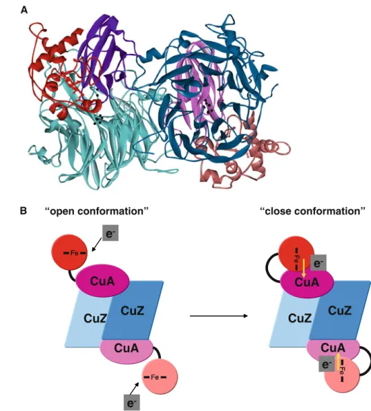
ranked by global, electrostatic, or hydrophobic score (their representations are shown in Figs. 4, S2–S4).
Previously, direct electron transfer studies have shown for P. panthotropus and P. nautica N2OR that the small
electron transfer proteins donate electrons directly to the binuclear CuA center of N2OR [18, 22]. Therefore, the
solutions were filtered using the condition that the distance between the redox centers, the CuA center and the iron ion of cytochromec, or the copper ion of pseudoazurin, should be less than 20 A˚ , a condition required for an efficient electron transfer [15] (Table1, and their representation is shown in Figs. S2–S4). This analysis is used to determine whether this docking program can be applied to discrimi-nate between effective and noneffective electron donors.
In the case ofP. nautica N2OR, the complex with the
electron donor cytochromec-552 shows a higher number of putative effective electron transfer complexes for N2OR
compared with any of the other nonphysiological electron carrier proteins (Figs.4, S4; Table1). The number of solutions with an appropriate distance between the redox
Fig. 4 The electron transfer complexes between N2OR and its physiological electron donors. a The 200 top docking solutions ranked by hydrophobic score ofP. nautica N2OR with P. nautica cytochrome c-552. b The 200 top docking solutions ranked by electrostatic score of A. cycloclastes N2OR with A. cycloclastes pseudoazurin.cThe 200 top docking solutions ranked by electrostatic score ofP. denitrificansN2OR withP. denitrificanscytochrome c -550.dThe 200 top docking solutions ranked by electrostatic score of
centers (heme–Fe and CuA) is maximum considering the hydrophobic score, as predicted taking into account the kinetic studies [18]. Moreover, it was shown that the pos-itively charged horse heart cytochromecwas not able to donate electrons toP. nauticaN2OR, a result that
corrob-orates the docking analysis, which does not present any solution with the appropriate orientation for electron transfer (Fig. S4.3).
On the other hand, this mitochondrial cytochrome was shown to be a competent electron donor toP. denitrificans
N2OR [22], and indeed the analysis of the docking
simu-lation by the different scores gives rise to several top solutions in an orientation that is predicted to enable an effective electron transfer (Table1). The number of these top orientations for electron transfer is similar whatever scoring function was used to rank them, even if the for-mation of the competent electron transfer complexes is proposed to be mainly driven by electrostatic forces.
The physiological electron donors of P. denitrificans
N2OR have long been established to be cytochromec-550
and pseudoazurin [20, 21]. In the first case, the docking simulation gave several solutions in the top 200 of each ranking with a favorable orientation for electron transfer (Fig.4c, d; Table1). In the case of pseudoazurin, this protein has a lower number of probable solutions towards
P. denitrificans N2OR compared with cytochrome c-550
(vide infra).
Our results on P. denitrificans N2OR are widely in
agreement with a previous docking analysis performed with a different algorithm, FTDOCK [23], which strengthens both the use of the docking algorithm BiGGER and the method employed in this work for the analysis of the 5,000 solutions obtained from the ab initio soft-docking calculation.
In the last case study, the molecular docking simulation between A. cycloclastes N2OR and its physiological
elec-tron donor, pseudoazurin, there are several putative dock-ing solutions with a short distance between the redox center especially when the solution were ranked by the electro-static score (Fig.4b; Table1).
The lower number of putative complexes obtained when pseudoazurins, from eitherA. cycloclastes or P. panthotr-opus, are used as probes can be attributed to the smaller size of the expected pseudoazurin surface interacting with N2OR, when compared with that of thec-type cytochromes
studied. Since there is a surface contact cutoff value below which BiGGER rejects all models, the number of ‘‘puta-tively competent’’ solutions that were rejected is expected to be higher in the docking calculations when pseudoazurin is a probe rather than in those in which cytochromes were used as probes.
Table 1 Analysis of the molecular dockings between nitrous oxide reductase (N2OR) from different microorganisms and the electron donors
performed using the BiGGER algorithm [7]
N2OR source Electron donor Best 200 Gb
?d\20 A˚a
Best 200 electrostatic
?d\20 A˚b
Best 200 hydrophobic
?d\20 A˚c
Pseudomonas nautica P. nauticacytochromec-552 23 41 139
P. nautica P. denitrificanscytochromec-550 0 0 3
P. nautica Paracoccus panthotropuspseudoazurin 0 4 4
P. nautica A. cycloclastespseudoazurin 1 4 8
P. nautica Horse heart cytochromec 0 0 0
Paracoccus denitrificans P. nauticacytochromec-552 4 34 66
P. denitrificans P. denitrificanscytochromec-550 9 83 80
P. denitrificans P. panthotropuspseudoazurin 5 20 35
P. denitrificans A. cycloclastespseudoazurin 1 27 39
P. denitrificans Horse heart cytochromec 12 92 108
Achromobacter cycloclastes P. nauticacytochromec-552 8 79 62
A. cycloclastes P. denitrificanscytochromec-550 12 126 42
A. cycloclastes P. panthotropuspseudoazurin 9 26 40
A. cycloclastes A. cycloclastespseudoazurin 6 85 55
A. cycloclastes Horse heart cytochromec 0 26 27
A. cycloclastes Bovine heart cytochromec 1 44 37
The complexes that have been studied experimentally are highlighted inbold
a
Number of solutions in the top 200 in the global score (Gb) ranking with a CuA–Fe/Cu distance shorter than 20 A˚ b
Number of solutions in the top 200 in the electrostatic ranking with a CuA–Fe/Cu distance shorter than 20 A˚ c
It is interesting to note that the mitochondrial bovine heart cytochromec, which was shown to be able to donate electrons to the CuA center of A. cycloclastes N2OR,
indeed behaves as a potential electron donor, with a larger number of putative solutions being found when the com-plexes were analyzed considering the interaction driven mainly by electrostatic forces (Fig.S2.1). Similarly,
P. denitrificans cytochrome c-550 was also predicted, by BiGGER, to function as a putative electron donor to this enzyme (considering that the interaction has an electro-static nature). This can be explained by the fact that the surfaces of these two electron shuttle proteins share a similar charge distribution (Fig.3).
A summary of the analysis of the docking simulations combined with the previous experimental data is presented in Table S1.
Analysis of the top complexes
The interface-accessible area of the top solutions for each of the physiologic docking results was evaluated. These complexes have an interface area between 943 and 1,368 A˚2(Table2), which is typical for small, short-lived complexes following the criteria of Lo Conte et al. [3]. The docking models for the P. nautica cytochrome c-552–
P. nautica N2OR complex have a higher percentage of
apolar residues in the interface area (with an average value of 79%), confirming the hydrophobic nature of the inter-action. For A. cycloclastes N2OR and P. denitrificans
N2OR complexes, the average values of apolar residues in
the interface are 64 and 69%, respectively. This result shows that although the formation of these complexes is being driven by electrostatic forces, the area of contact
Table 2 Parameters of the top model complex obtained by docking simulation of N2OR with the respective electron donor
Complex ID Gb (rank) Elect. (rank) Hydroph. (rank)
Da(A˚ ) Coupling constantb
Pathwayc Interface area (A˚2)
Apolar residuesd (%)
A. cycloclastes
N2OR–bovine heart cytochromec
4142 1.9 (387) -151.7 (56) -6.0 (440) 15.7 2.8910-5 Fe–Ala554 1,366 66
A. cycloclastes
N2OR–A. cycloclastes pseudoazurin
3203 2.2 (102) -91.5 (49) -5.4 (918) 16.9 1.1910-4 Cu–Leu627 1,090 63
A. cycloclastes
N2OR–A. cycloclastes pseudoazurin
3167 2.1 (140) -78.4 (162) -6.6 (266) 18.9 9.4910-6 Cu–Leu627 1,115 64
P. denitrificans
N2OR–P. panthotropus pseudoazurin
3077 2.3 (71) -22.7 (298) -6.9 (150) 15.7 1.3910-5 Cu–Leu593 1,277 68
P. denitrificans
N2OR–P. denitrificans cytochromec-550
4851 2.2 (62) -101.4 (152) -5.5 (1,385) 14.7 4.7910-4 Cu–His635 1,368 67
P. denitrificans
N2OR–horse heart cytochromec
1712 1.9 (86) -175.1 (121) -6.7 (38) 15.5 2.8910-4 Fe–His635 1,289 70
P. nautica
N2OR–P. nautica cytochromec-552
105 10.4 (35) -22.7 (3,545) -6.1 (13) 16.3 3.0910-5 Fe–Asp519 1,222 75
P. nauticaN2OR–
P. nauticacytochrome
c-552
579 9.2 (51) -50.7 (312) -7.0 (2) 17.5 3.0910-5 Fe–Gln497 1,211 79
P. nauticaN2OR–
P. nauticacytochrome
c-552
2181 10.4 (36) -39.6 (1,118) -4.8 (175) 17.9 2.1910-6 Fe–His566 943 84
Gbglobal score,Elect.electrostatic score,Hydroph.hydrophobic score,rankranking of the solution with that ID in the respective score a
Distance between CuA in N2OR and heme–Fe or Cu of cytochromecor pseudoazurin, respectively b
The coupling constant is the value for the best electron transfer path from Cu or Fe atoms to CuA centers as analyzed using PATHWAYS c
The residue number is according to the primary sequence of N2OR from each organism d
between the redox partners is hydrophobic, as expected in order to promote the electron transfer and provide more specificity to the complex [45].
The electron transfer complexes betweenA. cycloclastes
N2OR andP. denitrificansN2OR and their electron donors
can be ascribed to a docking scenario in which the elec-trostatic forces play the role of the long-range recognition between the donor and the acceptor and drive the formation of the complex; however, at short range, the hydrophobic patch also plays an important role in the fine-tuning of formation of the electron transfer complex.
Electron transfer pathway
To propose an electron pathway from the small electron donor protein to the CuA center and then to the catalytic center of N2OR, the CuZ center, the top complexes for
each of the docking calculations were further analyzed using the program PATHWAYS. The top complexes that were chosen had to obey the following criteria (Table2):
• They had to be part of the top 200 solutions ranked by
global score.
• They had to be part of the top solutions ranked by
hydrophobic score for P. nauticaN2OR or by
electro-static score for P. denitrificans N2OR and A. cyclocl-astesN2OR docked complexes.
• The distance between the redox center of the small
electron donor and the CuA center of N2OR had be
shorter than 20 A˚ .
Combining the analysis of the electron transfer pathway of the top complexes, we identified a conserved histidine (A. cycloclastes His625, P. denitrificans His635, and
P. nauticaHis566) in the electron transfer pathway as the entry point in three of the complexes analyzed (Table2), a leucine (A. cycloclastes Leu627, P. denitrificans Leu637, andP. nauticaLeu568) in two complexes, and an aspartate (A. cycloclastes Asp578, P. denitrificans Asp588, and
P. nautica Asp519), an alanine (A. cycloclastes Ala554,
P. denitrificans Ala564, and P. nautica Ala495), a gluta-mine (P. nautica Gln497, which corresponds to A. cyc-loclastesSer556 andP. denitrificansSer566), and a leucine (A. cycloclastes Leu583 and P. denitrificans Leu593, which corresponds toP. nauticaVal524) each just in one identified electron pathway (Table2).
The sequence alignment shows that all these residues are conserved in the primary sequence of these N2ORs,
sug-gesting that these residues constitute a conserved region located near the CuA center that functions both as the binding site for the electron donor and as the electron entry point (Figs.2a, 5a).
This conserved patch involved in complex formation and favorable electron transfer was also identified by the
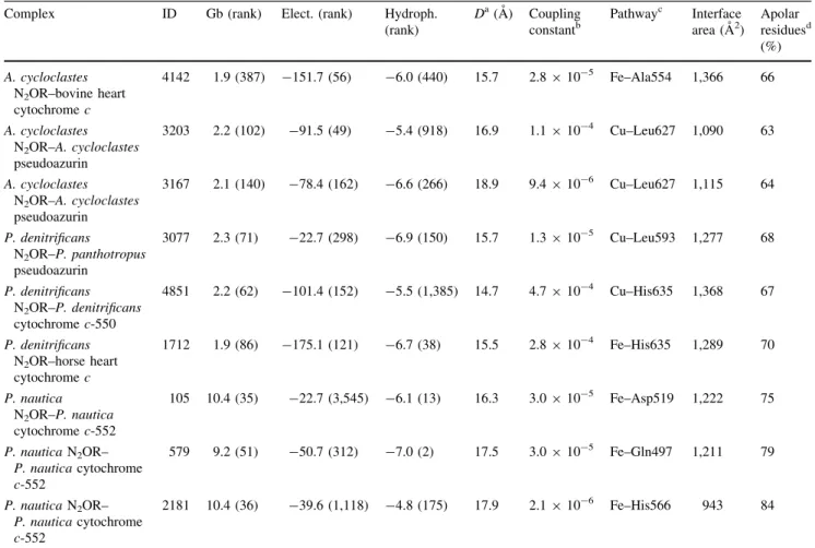
Fig. 5 Proposed electron transfer pathways in N2OR.aThe electron transfer pathway between cytochrome c-552 and the CuA center of P. nautica N2OR is represented. Two possible orientations of cytochrome c-552 are shown (for clarity only the heme group of cytochromec-552 is displayed). The residues involved in the electron transfer from the heme group to the CuA center (Ala495, Asp519, Val524, His566, Leu568) are represented inyellow. The ligands of the CuA center (His526, Cys561, Cys565, His569, Met572) are colored
WHISCY program [40] as to constitute a putative surface involved in protein–protein interaction (Fig.2c—only the
P. nauticaN2OR surface is shown).
The coupling constants for this electron transfer path-way are between 4.7910-4and 2.1910-6(values cal-culated by PATHWAYS), in which the routes involving the surface conserved histidine (A. cycloclastes His625,
P. denitrificansHis635, and P. nautica His566) show the highest coupling constant (4.7910-4
and 2.8910-4
for
P. denitrificansN2OR complexes, Table2).
The electron transfer pathway from the CuA center to the CuZ center was also analyzed with the PATHWAYS program. The most probable electron transfer pathway (with a coupling constant of 1.1910-4) implies that the electron is transferred from the CuA center to the tryp-tophan (P. nautica Trp563, P. denitrificans Trp632,
A. cycloclastes Trp622) that is bound to the copper through the main-chain carbonyl group. Then, the electron is transferred to the neighbor phenylalanine (P. nautica
Phe564, P. denitrificans Phe633, A. cycloclastes Phe623) and subsequently to the oxygen of a water molecule or a hydroxyl group, which is bound between two copper atoms of the CuZ center (CuI and CuIV) (Fig.5b— pathway represented in purple), the proposed substrate binding site [53].
Although the catalytic mechanism of N2OR is only
partially known, theoretical calculations coupled with spectroscopic studies and enzymatic assays have shown that the most favorable state of the CuZ center to bind the substrate, N2O, is the fully reduced state (4Cu
?
) [54]. The electron transfer pathway identified above is in agreement with this mechanism, in which the reduction of the CuZ center occurs prior to substrate binding. After the first step of the reaction (the N–O bond cleavage), the CuZ center is proposed to be in the [2Cu2?
–2Cu?
] redox state and has to undergo two one-electron reductions to be again in the catalytically active redox state 4Cu?
[55]. During this reduction process, the oxygen bound to the CuZ center (between CuI and CuIV) has to be protonated to be released as a water molecule.
An alternative electron pathway (with a coupling constant of 4.3910-5
) involves one of the histidine ligands of the CuA center (P. nautica His569, P. deni-trificans His638, A. cycloclastes His628), then the neigh-bor methionine (P. nautica Met570, P. denitrificans
Met639,A. cycloclastes Met629), and then transfer of the electron to the histidine (P. nautica His128, P. denitrifi-cansHis194,A. cycloclastesHis184), which is a ligand of one of the copper atoms of the CuZ center (CuII) (Fig.5b—pathway represented in blue). This electron transfer route is analogous to that proposed for the elec-tron transfer from the CuA center to the heme a in cytochromec oxidase [56].
Model structure for N2OR fromW. succinogenes
The N2OR fromW. succinogeneshas an additional domain
in its C-terminal with the canonical c-type-heme binding motif –CXXCH– [27,28].
The structure of this protein has not yet been solved and can be proposed to resemble the electron transfer complex between an N2OR and a c-type cytochrome. A protein
sequence homology search shows that there are a few organisms that have this type of N2OR, although none of
these other enzymes have been isolated. These organisms are from the Campylobacter, Sulfurimonas, and Denitro-vibrio genera, with the first two being host-associated organisms from the Epsilonproteobacteria group, as
W. succinogenes.
The model structure ofW. succinogenesN2OR was built
in two steps: first a model of the N-terminal N2OR domain
and of the C-terminalc-type-heme-containing domain were modeled, and afterwards these two domains were docked to obtain a model structure of the complete enzyme.
The N-terminal N2OR domain, composed of the CuA
and CuZ domains, shows sequence homology with all the N2ORs with known structure, and the surface mapping of
the conserved residues shows that there is a surface patch of conserved residues near the CuA center (Fig.2b), a region also identified by WHISCY as involved in protein– protein interactions. However, sinceP. nauticaN2OR has
the highest primary sequence identity with W. succinoge-nes N2OR (34%), its structure (Protein Data Bank ID
1QNI) was used to model the N-terminal domain of
W. succinogenesN2OR using the PHYRE program.
As to the C-terminal domain, proposed to contain a
c-type heme, different online programs and c-type cyto-chromes or c-type-heme-containing domains were used to model its structure (Fig.6a) on the basis of sequence homology. In particular, four models were created using the structures of the following c-type-heme-containing templates: Rhodothermus marinus caa3 cytochrome c domain (1W2L [57]), C-terminal domain of quinohe-moprotein alcohol dehydrogenase from Pseudomonas putida HK5 (1KV9 [58]), the monohemic cytochrome c2
from Rhodopila globiformis (1HRO [59]), and the mono-hemic cytochrome c from R. marinus (3CP5 [60]). The latter has the highest sequence identity (25%) and the model obtained gave the best results in the docking study. This model was obtained using SWISS-MODEL [42] (Fig.6b), and shows that this domain has a surface with positive and negative patches (Fig. 6c, d), which is con-sistent with a solvent-exposed surface.
In the second stage, these two model structures, the N2OR domain and the c-type cytochrome domain, were
(100 residues) between the N-terminal N2OR-type domain
and the C-terminalc-type-heme-containing domain which has very low homology with known protein structures. For this reason we cannot provide a realistic linker connecting the N- and the C-terminal domains. However, an accept-able docking geometry can be selected taking into account that for an effective electron transfer the distance between the CuA center and the heme iron of the heme in the C-terminal domain should be less than 20 A˚ .
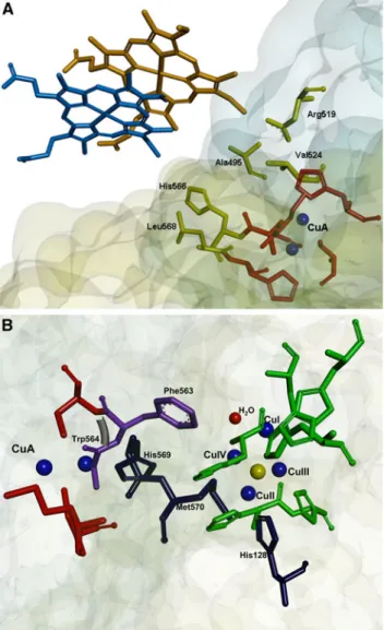
The analysis of the docking results shows that there is one solution that fulfils this criterion, providing us with a possible model structure ofW. succinogenesN2OR (Fig.7a).
The PATHWAYS analysis of these two models deter-mined a distance of 17.8 and 18.8 A˚ between the heme iron and the CuA center, with Arg557 and Cys627 in the N2OR
domain as the key residues for the electron entry. Arg557 corresponds to the conserved alanine proposed to be part of the electron transfer pathway in the other N2OR complexes
studied (A. cycloclastes Ala554, P. denitrificans Ala564, and P. nautica Ala495), and Cys627 is the neighboring residue of Ser628, which corresponds to the conserved histidine residue proposed before to be the electron entry point (A. cycloclastesHis625,P. denitrificansHis635, and
P. nauticaHis566).
The analysis of the electrostatic surface of the cyto-chrome c domain modeled on the basis of R. marinus
cytochromecreveals a prevalently charged surface, which means that this domain might not be tightly bound to the N2OR domain.
Therefore, a mechanism for the electron transfer reaction in W. succinogenes N2OR can be proposed in which the c-type-heme-containing domain can assume different orien-tations. In particular, the W. succinogenes N2OR might
present an ‘‘open conformation’’ to allow thec -type-heme-containing domain to receive electrons from an electron donor (Fig.7b), which is proposed to be a periplasmic cytochrome
c [28]. After the reduction of the c-type-heme-containing domain, the enzyme can rearrange into a ‘‘close conforma-tion’’ in which the heme group assumes the appropriate ori-entation to donate electrons to the CuA center (Fig.7b).
Conclusion
In conclusion, this docking study predicted the interacting geometries between N2OR and its physiological electron
donors. These models are corroborated by experimental data and in the case of P. denitrificans N2OR are in
agreement with the previously proposed electron transfer complexes [23].
A set of well conserved residues were identified as being involved in the electron transfer pathway, suggesting the
-CXXCH-1QNI 1QNI
2IWF 2IWF
1FWX 1FWX Pseudomonas nautica Wolinella succinogens Achromobacter cycloclastes Paracoccus denitrificans 3CP5
Rhodothermus marinuscyt. c
1W2L
Rhodothermus marinus caa3cyt. cdomain
1KV9
Pseudomonas putidaalcohol dehydrogenase
1HRO
Rhodopila globiformiscyt. c2
-CXXCH-1QNI 1QNI
2IWF 2IWF
1FWX 1FWX 3CP5 3CP5 1W2L 1W2L 1KV9 1KV9 1HRO 1HRO
-CXXCH-1QNI 1QNI
2IWF 2IWF
1FWX 1FWX 3CP5 1W2L 1KV9 1HRO
-CXXCH-1QNI 1QNI
2IWF 2IWF
1FWX 1FWX
CuZ CuA c-heme
3CP5 3CP5 1W2L 1W2L 1KV9 1KV9 1HRO 1HRO 180º D C B A Fig. 6 The domains ofW.
succinogenesN2OR and of other N2ORs with known structure. The Protein Data Bank IDs of the structures used to model the C-terminal domain are shown in
red(a). Structures 1QNI and 3CP5 were the ones that gave the highest scores for the N-terminal domain and the and C-terminal domain,
respectively. The N-terminal domain ofW. succinogenes
N2OR is composed of the CuZ domain (blue) and the CuA domain (purple), and the C-terminal domain containing thec-type-heme binding motif is represented as ared box. The model of the C-terminal domain ofW. succinogenesN2OR is shown as a backbone (b) and its surface is shown colored by electrostatic potential (c,d). The electrostatic surface potential is represented between-4 and 4
presence of a specific region in both the donor and the acceptor proteins that enables the molecular recognition, formation of the encounter complex, and efficient electron transfer. However, it was not possible to identify a single electron transfer route, and instead a patch of surface res-idues was identified as being instrumental for partner rec-ognition and complex formation, and involved in the electron transfer pathway.
Moreover, the electron transfer pathway between the CuA center and the CuZ center was investigated, sug-gesting two alternative routes for the reduction of the cat-alytic center, which agree with the accepted catcat-alytic mechanism of this enzyme: full reduction of the CuZ center is required prior to N2O binding.
A model structure for theW. succinogenesN2OR, which
has an additional C-terminal domain containing a c-type heme, was proposed. This model presents the heme iron at a distance of 18–19 A˚ from the CuA center of the N2OR
domain, forming a putative efficient electron route.
This work shows that BiGGER algorithm can be used to identify putative electron transfer partners that could be used as artificial electron donors in in vitro assays, or to propose the physiologic redox partner of an enzyme when there are several candidates in the same organism. The information presented here and the model structures can also be used as starting points for mutagenesis studies to unambiguously identify residues that are involved in electron transfer and/or partner recognition.
Acknowledgments This research was supported by Fundac¸a˜o para a Cieˆncia e Tecnologia grants PTDC/QUI/64638/2006 (to I.M.) and SFRH/BD/30414/2006 (to S.D.).
References
1. Zumft WG (1997) Microbiol Mol Biol Rev 61:533–616 2. Tavares P, Pereira AS, Moura JJG, Moura I (2006) J Inorg
Biochem 100:2087–2100
A
CuZ
CuA
e
-e
-CuZ
CuA
e
-CuZ
CuA
CuZ
CuA
FeFe
e
-Fe
Fe
CuZ
CuA
e
-e
-CuZ
CuA
e
-B “open conformation” “close conformation”
CuZ
CuA
CuZ
CuA
FeFe Fe
Fe Fe Fe
e
-FeFe
FeFe
Fig. 7 aProposed model for theW. succinogenesN2OR, which has an additional C-terminal domain with ac-type heme. The C-terminal domain (redorpink) is shown to interact with the surface of the N2OR domain surrounding the CuA domain, the proposed entry site for the electron. The two copper atoms of the CuA center and the catalytic CuZ center are colored
blackand the CuA and CuZ domains of N2OR are colored
purpleandblue, respectively.
bProposed scheme for the electron transfer mechanism in
W. succinogenesN2OR. The CuA and CuZ domains are represented inpurpleandblue, respectively, and the
cytochromecdomain isredand
pink. To receive the electron from the electron donor,
3. Lo Conte L, Chothia C, Janin J (1999) J Mol Biol 285:2177–2198 4. Prudencio M, Ubbink M (2004) J Mol Recognit 17:524–539 5. Sukumar N, Chen Z-w, Ferrari D, Merli A, Rossi GL, Bellamy
HD, Chistoserdov A, Davidson VL, Mathews FS (2006) Bio-chemistry 45:13500–13510
6. Moreira IS, Fernandes PA, Ramos MJ (2010) J Comput Chem 31:317–342
7. Palma PN, Krippahl L, Wampler JE, Moura JJG (2000) Proteins 39:372–384
8. Xu X, Schurmann P, Chung J, Hass M, Kim S, Hirasawa M, Tripathy J, Knaff D, Ubbink M (2009) J Am Chem Soc 131:17576–17582
9. Fantuzzi A, Meharenna YT, Briscoe PB, Guerlesquin F, Sadeghi SJ, Gilardi G (2009) Biochim Biophys Acta 1787:234–241 10. Almeida RM, Pauleta SR, Moura I, Moura JJG (2009) J Inorg
Biochem 103:1245–1253
11. Zumft WG, Kroneck PM (2007) Adv Microb Physiol 52:107–227 12. Brown K, Tegoni M, Prudencio M, Pereira AS, Besson S, Moura
JJG, Moura I, Cambillau C (2000) Nat Struct Biol 7:191–195 13. Haltia T, Brown K, Tegoni M, Cambillau C, Saraste M, Mattila
K, Djinovic-Carugo K (2003) Biochem J 369:77–88
14. Paraskevopoulos K, Antonyuk SV, Sawers RG, Eady RR, Hasnain SS (2006) J Mol Biol 362:55–65
15. Winkler JR (2000) Curr Opin Chem Biol 4:192–198
16. Witt H, Malatesta F, Nicoletti F, Brunori M, Ludwig B (1998) J Biol Chem 273:5132–5136
17. Maneg O, Ludwig B, Malatesta F (2003) J Biol Chem 278:46734–46740
18. Dell’Acqua S, Pauleta SR, Monzani E, Pereira AS, Casella L, Moura JJG, Moura I (2008) Biochemistry 47:10852–10862 19. Berks BC, Baratta D, Richardson J, Ferguson SJ (1993) Eur J
Biochem 212:467–476
20. Moir JWB, Ferguson SJ (1994) Microbiology 140:389–397 21. Koutny M, Kucera I, Tesarik R, Turanek J, Van Spanning RJ
(1999) FEBS Lett 448:157–159
22. Rasmussen T, Brittain T, Berks BC, Watmough NJ, Thomson AJ (2005) Dalton Trans 3501–3506
23. Mattila K, Haltia T (2005) Proteins 59:708–722
24. Fujita K, Ijima F, Obara Y, Hirasawa M, Brown DE, Kohzuma T, Dooley DM (2009) J Biol Inorg Chem 14(Suppl 1):S11–S20 25. Liu MY, Liu MC, Payne WJ, Legall J (1986) J Bacteriol
166:604–608
26. Fujita K, Chan JM, Bollinger JA, Alvarez ML, Dooley DM (2007) J Inorg Biochem 101:1836–1844
27. Teraguchi S, Hollocher TC (1989) J Biol Chem 264:1972–1979 28. Zhang CS, Hollocher TC (1993) Biochim Biophys Acta
1142:253–261
29. Brown K, Nurizzo D, Besson S, Shepard W, Moura J, Moura I, Tegoni M, Cambillau C (1999) J Mol Biol 289:1017–1028 30. Benning MM, Meyer TE, Holden HM (1994) Arch Biochem
Biophys 310:460–466
31. Najmudin S, Pauleta SR, Moura I, Romao MJ (2010) Acta Crystallogr Sect F Struct Biol Cryst Commun 66:627–635 32. Inoue T, Nishio N, Suzuki S, Kataoka K, Kohzuma T, Kai Y
(1999) J Biol Chem 274:17845–17852
33. Bushnell GW, Louie GV, Brayer GD (1990) J Mol Biol 214:585–595
34. Mirkin N, Jaconcic J, Stojanoff V, Moreno A (2008) Proteins 70:83–92
35. Pettersen EF, Goddard TD, Huang CC, Couch GS, Greenblatt DM, Meng EC, Ferrin TE (2004) J Comput Chem 25:1605–1612 36. Betts JN, Beratan DN, Onuchic JN (1992) J Am Chem Soc
114:4043–4046
37. Regan JJ, Risser SM, Beratan DN, Onuchic JN (1993) J Phys Chem 97:13083–13088
38. Onuchic JN, Beratan DN, Winkler JR, Gray HB (1992) Annu Rev Biophys Biomol Struct 21:349–377
39. Thompson JD, Higgins DG, Gibson TJ (1994) Nucleic Acids Res 22:4673–4680
40. de Vries SJ, van Dijk ADJ, Bonvin AMJJ (2006) Proteins 63:479–489
41. Kelley LA, Sternberg MJ (2009) Nat Protoc 4:363–371 42. Bordoli L, Kiefer F, Arnold K, Benkert P, Battey J, Schwede T
(2008) Nat Protoc 4:1–13
43. Prudencio M, Pereira AS, Tavares P, Besson S, Cabrito I, Brown K, Samyn B, Devreese B, Van Beeumen J, Rusnak F, Fauque G, Moura JJG, Tegoni M, Cambillau C, Moura I (2000) Biochem-istry 39:3899–3907
44. Pauleta SR, Guerlesquin F, Goodhew CF, Devreese B, Van Beeumen J, Pereira AS, Moura I, Pettigrew GW (2004) Bio-chemistry 43:11214–11225
45. Williams PA, Fulop V, Leung YC, Chan C, Moir JW, Howlett G, Ferguson SJ, Radford SE, Hajdu J (1995) Nat Struct Biol 2:975–982
46. Kukimoto M, Nishiyama M, Ohnuki T, Turley S, Adman ET, Horinouchi S, Beppu T (1995) Protein Eng 8:153–158
47. Pelletier H, Kraut J (1992) Science 258:1748–1755
48. Pauleta SR, Cooper A, Nutley M, Errington N, Harding S, Guerlesquin F, Goodhew CF, Moura I, Moura JJG, Pettigrew GW (2004) Biochemistry 43:14566–14576
49. Pearson IV, Page MD, van Spanning RJM, Ferguson SJ (2003) J Bacteriol 185:6308–6315
50. Moir JW, Wehrfritz JM, Spiro S, Richardson DJ (1996) Biochem J 319:823–827
51. Kukimoto M, Nishiyama M, Tanokura M, Adman ET, Horinouchi S (1996) J Biol Chem 271:13680–13683
52. Kataoka K, Yamaguchi K, Kobayashi M, Mori T, Bokui N, Suzuki S (2004) J Biol Chem 279:53374–53378
53. Ghosh S, Gorelsky SI, Chen P, Cabrito I, Moura JJ, Moura I, Solomon EI (2003) J Am Chem Soc 125:15708–15709 54. Dell’Acqua S, Pauleta SR, Moura I, Moura JJG (2011) J Biol
Inorg Chem 16:183–194
55. Gorelsky SI, Ghosh S, Solomon EI (2006) J Am Chem Soc 128:278–290
56. Wang K, Geren L, Zhen Y, Ma L, Ferguson-Miller S, Durham B, Millett F (2002) Biochemistry 41:2298–2304
57. Srinivasan V, Rajendran C, Sousa FL, Melo AM, Saraiva LM, Pereira MM, Santana M, Teixeira M, Michel H (2005) J Mol Biol 345:1047–1057
58. Chen ZW, Matsushita K, Yamashita T, Fujii TA, Toyama H, Adachi O, Bellamy HD, Mathews FS (2002) Structure 10:837–849 59. Benning MM, Meyer TE, Holden HM (1996) Arch Biochem
Biophys 333:338–348
![Table 1 Analysis of the molecular dockings between nitrous oxide reductase (N 2 OR) from different microorganisms and the electron donors performed using the BiGGER algorithm [7]](https://thumb-eu.123doks.com/thumbv2/123dok_br/16502435.734091/8.892.75.819.122.476/analysis-molecular-dockings-reductase-different-microorganisms-performed-algorithm.webp)