Leandro Silva MARQUES(a)
Carlos Eduardo Pinto ALCÂNTARA(b) Luciano José PEREIRA(c)
Maria Letícia RAMOS-JORGE(a)
(a)Universidade Federal dos Vales do Jequitinhonha e Mucuri – UFVJM, School of Biologic and Health Sciences, Department of Dentistry, Diamantina, MG, Brazil.
(b)Universidade Federal de Juiz de Fora – UFJF, Department of Dentistry, Governador Valadares, MG, Brazil.
(c)Universidade Federal de Lavras – UFLA, Department of Health Sciences, Human Physiology Area, Lavras, MG, Brazil.
Down syndrome: a risk factor for
malocclusion severity?
Abstract: The aims of the present study were to compare aspects
related to malocclusion between individuals with Down syndrome (DS) and a control group, establish malocclusion severity, and identify determinant factors. A total of 120 individuals (60 with DS and 60 with no physical or mental impairment), were included in the study. Data were collected through interviews, analyses of the medical charts, and oral examinations. The criteria of the Dental Aesthetic Index were used for the diagnosis of malocclusion. Chi-square test (p ≤ 0.05) and
multivariate logistic regression were used for comparisons between the two groups and to determine the association between the dependent (malocclusion severity) and independent variables. Statistically
signiicant differences were found between the two groups for the
following variables: missing teeth, diastema, overjet, mandibular protrusion, anterior open bite, posterior crossbite, facial type, lip
incompetence, and Angle classiication. DS, a history of premature
birth, and long face pattern were found to be associated with malocclusion severity. Individuals with DS exhibited more occlusal problems than those in the control group.
Keywords: Malocclusion; Down Syndrome; Orthodontics.
Introduction
Down syndrome (DS), also known as Trisomy 21, is the most common chromosomal disorder in humans, affecting 1–2 individuals out of every 1000 live births.1,2,3 Patients with this syndrome exhibit cognitive impairment as well as bone growth disorders and generalized muscle hypotonia.4,5 Moreover, oral health problems, such as inadequate oral hygiene, periodontal disease, caries, malocclusion, and tooth loss, are more prevalent in this group of individuals than in the general population.6,7,8,9 Malocclusion, in particular, exerts a considerable negative impact on the quality of life, causing problems related to the performance of daily activities, such as speech, swallowing, and chewing, and discrimination based on physical appearance.10,11
Patients with DS exhibit alterations such as an abnormal positioning of the tongue, craniofacial deformities (reduction in maxilla and mandible size, and narrow oropharynx), dental alterations (number and size of teeth), and muscle disorders.12,13,14 All these factors contribute toward the development of transversal and vertical alterations in the occlusion, such
Declaration of Interests: The authors certify that they have no commercial or associative interest that represents a conflict of interest in connection with the manuscript.
Corresponding Author:
Leandro Silva Marques
E-mail: [email protected]
DOI: 10.1590/1807-3107BOR-2015.vol29.0044
Submitted: Jul 09, 2014
as anterior open bite, posterior or anterior crossbite, and the proclination of the anterior teeth.2,3
The prevalence of malocclusion and associated factors have been assessed in individuals with DS.8,12,13 However, a critical reading of the literature reveals that there is a gap in knowledge concerning the determinant factors associated with the malocclusion establishment and severity. Many studies have failed to address certain individual, social, and behavioral aspects, such as age, birth weight, a history of premature birth, breathing pattern, and facial type.5,14,15 Moreover, methodological limitations,
including insuficient sample size, inadequate data
acquisition instruments, study designs with various sources of bias, and the use of only descriptive and comparative statistical analyses, have been encountered in the past. Thus, studies that can
provide evidence with greater scientiic weightage
are of particular importance for the development of public policies aimed at preventive strategies and oral health promotion.
The aims of the present study were to compare aspects related to malocclusion between individuals with DS and a control group, establish malocclusion severity, and identify the determinant factors.
Methodology
The present study involved a sample made up of 120 individuals; 60 with DS (37 males and 23 females, average age of 14.73 years) and 60 with no physical or mental impairment but affected by different types of malocclusion (control group: 19 males and 41 females, average age of 12.18 years). The patients in the control group were randomly selected from individuals awaiting orthodontic treatment in the Course of Orthodontics specialization at the School of Dentistry in Itaúna, Brazil. Only those individuals who had not yet been submitted to orthodontic intervention were included in the study.
Data were collected through interviews, assessments
of medical charts for the conirmatory diagnosis of DS,
and oral examinations. Information pertaining to age, gender, mother’s schooling, weight, and a history of premature birth were collected from children’s mothers during the interview. Oral examinations were performed by an orthodontist. Prior to the assessment and diagnosis
of malocclusion based on the criteria of the Dental Aesthetic Index (DAI), the orthodontist participated in a calibration exercise involving 12 individuals who did not belong to the main study and achieved high agreement values (maximal and minimal kappa values of 1.00 and 0.81, respectively). The DAI provides four outcome possibilities: mild malocclusion or absence of abnormality, for which treatment is not necessary
(DAI ≤ 25); deined malocclusion, for which treatment
is elective (DAI = 26-30); severe malocclusion, for which
treatment is highly desirable (DAI = 31–35); and very
severe or debilitating malocclusion, for which treatment
is fundamental (DAI ≥ 36).16 Because all participants exhibited some type of malocclusion, the DAI score
was dichotomized as moderate (DAI ≤ 30) and severe
(DAI > 30) malocclusion. Malocclusions were clinically categorized as Class I, Class II, or Class III, based on the
Angle classiication. During the clinical examination,
posterior crossbite, facial type, and dentition (mixed or permanent) were also determined.
A lip competence examination was performed based on the method described by Ballard;17 the mandible was in the physiological resting posture and lips were in the juxtaposition (sealed) with no contraction of the orbicular muscles of the mouth or mentalis. Lip incompetence was recorded when the individual required vigorous contractions of the orbicular muscles of the mouth and mentalis to achieve a lip seal. In children, lip incompetence and breathing patterns (nasal or mouth) were assessed during the clinical examination and by interviewing the mothers when the child was not aware of being observed, thereby revealing the inherent behavior.
Data analysis was performed using the Statistical Package for Social Sciences (SPSS for Windows, version 17.0, SPSS Inc., Chicago, USA) and included frequency distribution and association tests. Associations between the dependent (malocclusion severity) and independent variables (gender, age, birth weight, a history of premature birth, mother’s schooling, breathing pattern, dentition type, and facial type) were determined using the chi-square test (p ≤ 0.05).
Variables with a p-value of ≤ 0.20 were incorporated
0004.0.380.000–09. Parents/guardians signed the terms of informed consent authorizing their child’s participation in the study.
Results
Individuals with DS had more number of missing teeth and a greater occurrence of mandibular protrusion, anterior open bite ( mm), posterior crossbite, and lip incompetence. As shown in Table 1, individuals in the control group had a greater occurrence of
diastema (≥ 2 mm) and overjet (≥ 4 mm). Differences
were also found with regard to facial type and Angle
classiication between the two groups. Short face
pattern and Angle Class III malocclusion were more frequent among the individuals with DS, whereas long face pattern and Angle Class II malocclusion were more frequent among the individuals in the control group (Table 1).
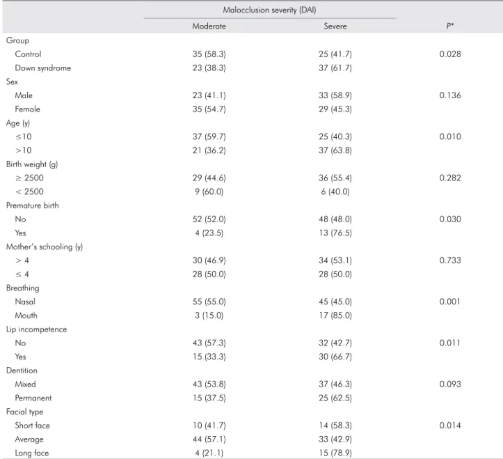
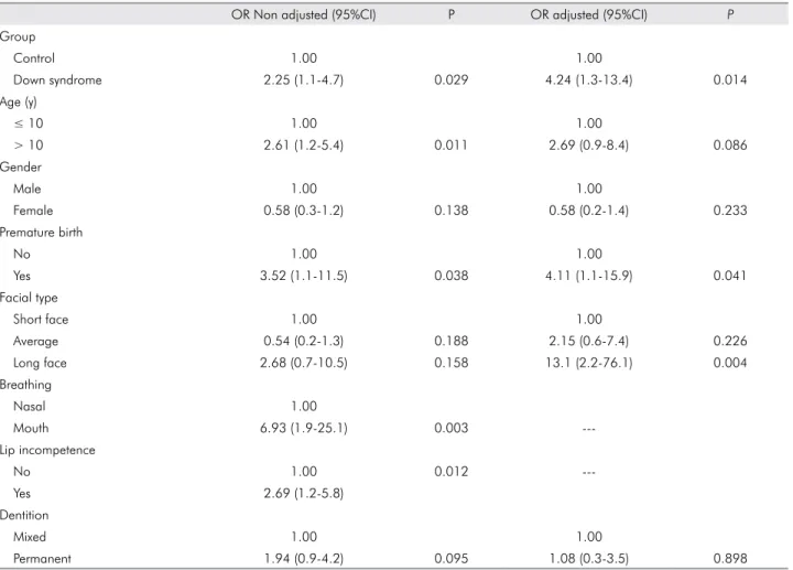
Malocclusion severity was greater among the individuals with DS than among those in the control group (p = 0.028). Considering individual, social, and behavioral factors, there was a greater frequency of severe malocclusion among individuals aged > 10 years and among those with a history of premature birth, lip incompetence, mouth breathing pattern, and long face pattern (Table 2). After the adjustment of the model, logistic regression revealed that DS, a history of premature birth, and facial type (long face) were associated with a greater malocclusion severity (Table 3), regardless of the age or gender.
Discussion
The majority of studies assessing factors associated with malocclusion in individuals with DS only provide descriptive and comparative statistical analyses and fail to determine malocclusion severity in these individuals.8,12,13 For this reason, the control group was comprised of individuals without DS and seeking orthodontic treatment, rather than individuals unaffected by malocclusion or those with similar malocclusions.
The most frequent malocclusions among the individuals with DS were mandibular protrusion, anterior open bite, and posterior crossbite. These findings confirm the results of previous studies reporting high prevalence values for malocclusions
stemming from vertical and transversal occlusal alterations.2,3,14,18 Such alterations are associated with insufficient bone development, orofacial muscle hypotonia, and the positioning of the tongue.2,14,19,20 Muscle hypotonia associated with a reduced volume of the oral cavity and characterized by a deep, atresic palate may lead to a tendency toward habitually projecting the tongue against the teeth or outside the mouth.5,18,21 Abnormal function and the position of the tongue can affect tooth eruption causing anterior open bite, tooth alignment, and the arch shape.12,22 However, these variables were not assessed in the present study. A pervious study23 using the same
control group identiied that patients with cerebral
palsy also had more anterior open bite, suggesting that muscle impairment in both disorders contributes toward malocclusion.
Patients with DS generally have a short face pattern and a reduced development of the middle third of the face, resulting in a Class III occlusal relation.8,24,25 The
present study corroborates these indings. However,
patients with long face pattern also exhibited a greater chance of developing severe malocclusion. The long face pattern is associated with muscle hypotonia and a tendency toward the downward rotatation of the mandible, favoring the development of Angle Class II malocclusion.26
Altered breathing pattern is strongly associated with malocclusion.20 Góis et al.27 found that children with mouth breathing patterns have a 10-fold greater chance of exhibiting malocclusion compared with those with nasal breathing patterns. In the present study, mouth breathing was associated with both
malocclusion prevalence and severity because 85%
of the mouth breathing patients exhibited severe malocclusion. In the logistic regression analysis, however, this variable did not adjust to the model because most individuals with DS also exhibited mouth breathing patterns. Mouth breathing patterns are more frequent due to orofacial muscle hypotonia and an absence of the lip seal.12
Children with a history of premature birth had a greater chance of developing severe malocclusion. A
recent systematic review reported scientiic evidence
for altered palatal morphology among children born
Table 1. Univariate analysis considering aspects related to malocclusion in the Down syndrome group and control group without mental impairment.
Group
Control n (%) Down syndrome n (%) P*
Missing teeth
None 59 (60.2) 39 (39.8) < 0.001
At least one 1 (4.5) 21 (95.5)
Crowding
None 31 (44.9) 38 (55.1) 0.196
1 or 2 segments 29 (56.9) 22 (43.1)
Spacing
None 34 (48.6) 36 (51.4) 0.711
1 or 2 segments 26 (52.0) 24 (48.0)
Diastema (mm)
< 2 47 (46.1) 55 (53.9) 0.041
≥ 2 13 (72.2) 5 (27.8)
Maxillary irregularity (mm)
< 2 48 (50.0) 48 (50.0) 1.000
≥ 2 12 (50.0) 12 (50.0)
Mandibular Irregularity (mm)
< 2 50 (48.1) 54 (51.9) 0.283
≥ 2 10 (62.5) 6 (37.5)
Overjet (mm)
< 4 46 (44.2) 58 (55.8) 0.001
≥ 4 14 (87.5) 2 (12.5)
Mandibular protrusion
No 59 (56.7) 45 (43.3) < 0.001
Yes 1 (6.3) 15 (93.8)
Anterior open bite (mm)
< 2 57 (56.4) 44 (43.6) 0.001
≥ 2 3 (15.8) 16 (84.2)
Posterior crossbite
Absent 53 (65.4) 28 (34.6) < 0.001
Present 7 (17.9) 32 (82.1)
Facial type
Short face 1 (4.2) 23 (95.8) <0.001
Average 45 (58.4) 32 (41.6)
Long face 14 (73.7) 5 (26.3)
Lip incompetence
No 44 (58.7) 31 (41.3) 0.014
Yes 16 (35.6) 29 (64.4)
Angle classification
Class I 31 (70.5) 13 (29.5) < 0.001
Class II 25 (61.0) 16 (39.0)
Class III 4 (11.4) 31 (88.6)
Dentition
Mixed 44 (55.0) 36 (45.0) 0.121
Permanent 16 (40.0) 24 (60.0)
Table 2. Univariate analysis of association between malocclusion severity and individual, social, and behavioral variables.
Malocclusion severity (DAI)
Moderate Severe P*
Group
Control 35 (58.3) 25 (41.7) 0.028
Down syndrome 23 (38.3) 37 (61.7)
Sex
Male 23 (41.1) 33 (58.9) 0.136
Female 35 (54.7) 29 (45.3)
Age (y)
≤10 37 (59.7) 25 (40.3) 0.010
>10 21 (36.2) 37 (63.8)
Birth weight (g)
≥ 2500 29 (44.6) 36 (55.4) 0.282
< 2500 9 (60.0) 6 (40.0)
Premature birth
No 52 (52.0) 48 (48.0) 0.030
Yes 4 (23.5) 13 (76.5)
Mother’s schooling (y)
> 4 30 (46.9) 34 (53.1) 0.733
≤ 4 28 (50.0) 28 (50.0)
Breathing
Nasal 55 (55.0) 45 (45.0) 0.001
Mouth 3 (15.0) 17 (85.0)
Lip incompetence
No 43 (57.3) 32 (42.7) 0.011
Yes 15 (33.3) 30 (66.7)
Dentition
Mixed 43 (53.8) 37 (46.3) 0.093
Permanent 15 (37.5) 25 (62.5)
Facial type
Short face 10 (41.7) 14 (58.3) 0.014
Average 44 (57.1) 33 (42.9)
Long face 4 (21.1) 15 (78.9)
*Chi-square test (p ≤ 0.05)
another well-designed controlled study suggested that prematurely born children exhibit more malocclusion characteristics and have greater requirement for orthodontic treatment than full-term born children.29
These indings suggest that preterm children should be
more closely monitored by orthodontists, who should be aware of the tendency toward severe malocclusion and the orthodontic treatment needs among such children.29 Further longitudinal studies are required to address the issue as to whether premature births result in dentofacial alterations.
A recent study used the DAI to determine the degree of malocclusion in patients with DS and
found that 83.2% of them had severe and very severe
1. Dzurova D, Pikhart H. Down syndrome, paternal age and education: comparison of California and the Czech Republic.
BMC Public Health. 2005 Jun 17;5:69.
2. Venail F, Gardiner Q, Mondain M. ENT and speech disorders in children with Down’s syndrome: an overview of pathophysiology, clinical features, treatments, and current management. Clin Pediatr (Phila). 2004 Nov-Dec;43(9):783-91.
3. Kaye PL, Fiske J, Bower EJ, Newton JT, Fenlon M. Views and
experiences of parents and siblings of adults with Down Syndrome regarding oral healthcare: a qualitative and quantitative study. Br
Dent J. 2005 May 14;198(9):571-8, discussion 559.
4. Suri S, Tompson BD, Cornfoot L. Cranial base, maxillary and mandibular morphology in Down syndrome. Angle Orthod.
2010 Sep;80(5):861-9. DOI: 10.2319/111709-650.
References
Table 3. Univariate and multivariate logistic regression analyses considering associations between malocclusion severity and independent variables.
OR Non adjusted (95%CI) P OR adjusted (95%CI) P
Group
Control 1.00 1.00
Down syndrome 2.25 (1.1-4.7) 0.029 4.24 (1.3-13.4) 0.014
Age (y)
≤ 10 1.00 1.00
> 10 2.61 (1.2-5.4) 0.011 2.69 (0.9-8.4) 0.086
Gender
Male 1.00 1.00
Female 0.58 (0.3-1.2) 0.138 0.58 (0.2-1.4) 0.233
Premature birth
No 1.00 1.00
Yes 3.52 (1.1-11.5) 0.038 4.11 (1.1-15.9) 0.041
Facial type
Short face 1.00 1.00
Average 0.54 (0.2-1.3) 0.188 2.15 (0.6-7.4) 0.226
Long face 2.68 (0.7-10.5) 0.158 13.1 (2.2-76.1) 0.004
Breathing
Nasal 1.00
Mouth 6.93 (1.9-25.1) 0.003
---Lip incompetence
No 1.00 0.012
---Yes 2.69 (1.2-5.8)
Dentition
Mixed 1.00 1.00
Permanent 1.94 (0.9-4.2) 0.095 1.08 (0.3-3.5) 0.898
and the professionals who treat such individuals are most often ill prepared to offer better care. Thus, the treatment of malocclusion for individuals with DS can lead to a considerable improvement in their quality of life.18,20
Conclusions
Vertical and transversal alterations in the occlusion, such as mandibular protrusion, anterior open bite, and
posterior crossbite were signiicantly more frequent
among the individuals with DS than those in the control group. The determinant factors associated with malocclusion severity were DS, a history of premature birth, and long face pattern.
Acknowledgments
5. Korbmacher H, Limbrock J, Kahl-Nieke B. Orofacial
development in children with Down’s syndrome 12 years
after early intervention with a stimulating plate. J Orofac Orthop. 2004 Jan;65(1):60-73.
6. Khocht A, Janal M, Turner B. Periodontal health
i n Dow n s y nd r ome: cont r ib ut ion s of me nt a l disability, personal, and professional dental care.
Spec Care Dentist. 2010 May-Jun;30(3):118-23. DOI: 10.1111/j.1754-4505.2010.00134.x.
7. Amaral Loureiro AC, Oliveira Costa F, Eustáquio da Costa
J. The impact of periodontal disease on the quality of life of
individuals with Down syndrome. Downs Syndr Res Pract.
2007 Jul;12(1):50-4.
8. Winter K, Baccaglini L, Tomar S. A review of malocclusion a mong i nd iv idua l s w it h me nt a l a nd physic a l
disabilities. Spec Care Dentist. 2008 Jan-Feb;28(1):19-26. DOI: 10.1111/j.1754-4505.2008.00005.x.
9. Anders PL, Davis EL. Oral health of patients with intellectual disabilities: a systematic review. Spec Care Dentist.
2010 May-Jun;30(3):110-7. DOI: 10.1111/j.1754-4505.2010.00136.x.
10. Sto el- Ga m mon C. Dow n sy nd rome phonolog y: developmental patterns and intervention strategies. Downs Syndr Res Pract. 2001 Oct;7(3):93-100.
11. Musich DR. Orthodontic intervention and patients with
Down syndrome. Angle Orthod. 2006 Jul;76(4):734-5.
12. Oliveira AC, Pordeus IA, Torres CS, Martins MT, Paiva SM. Feeding and nonnutritive sucking habits and prevalence of open bite and crossbite in children/adolescents with
Down syndrome. Angle Orthod. 2010 Jul;80(4):748-53.
doi: 10.2319/072709-421.1.
13. Bäckman B, Grevér-Sjölander AC, Bengtsson K, Persson
J, Johansson I. Children with Down syndrome: oral
development and morphology after use of palatal plates
between 6 and 48 months of age. Int J Paediatr Dent. 2007 Jan;17(1):19-28.
14. Quintanilla JS, Biedma BM, Rodríguez MQ, Mora MT,
Cunqueiro MM, Pazos MA. Cephalometrics in children
with Down’s syndrome. Pediatr Radiol. 2002 Sep;32(9):635-43. Epub 2002 Apr 5.
15. Hennequin M, Allison PJ, Veyrune JL. Prevalence of oral health
problems in a group of individuals with Down syndrome in France. Dev Med Child Neurol. 2000 Oct;42(10):691-8.
16. Jenny J, Cons NC. Establishing malocclusion severity levels
on the Dental Aesthetic Index (DAI) scale. Aust Dent J.
1996 Feb;41(1):43-6.
17. Ballard CF. The effect of lip morphology on the incisors
following treatment. Orthod Fr. 1969;40:181-95. French.
18. Oliveira AC, Paiva SM, Campos MR, Czeresnia D. Factors associated with malocclusions in children and adolescents
with Down syndrome. Am J Orthod Dentofacial Orthop.
2008 Apr;133(4):489.e1-8. DOI: 10.1016/j.ajodo.2007.09.014. 19. Waldman HB, Perlman SP, Swerdloff M. Orthodontics and
the population with special needs. Am J Orthod Dentofacial Orthop. 2000 Jul;118(1):14-7.
20. Oliveira AC, Paiva SM, Martins MT, Torres CS, Pordeus IA. Prevalence and determinant factors of malocclusion
in children with special needs. Eur J Orthod. 2011
Aug;33(4):413-8. DOI: 10.1093/ejo/cjq094. Epub 2010 Oct 18. 21. Zavaglia V, Nori A, Mansour NM. Long term effects of
the palatal plate therapy for the orofacial regulation
in children with Down syndrome. J Clin Pediatr Dent.
2003 Fall;28(1):89-93.
22. Chawla HS, Suri S, Utreja A. Is tongue thrust that develops during orthodontic treatment an unrecognized potential
road block? J Indian Soc Pedod Prev Dent. 2006 Jun;24(2):80-3.
23. Miamoto CB, Ramos-Jorge ML, Pereira LJ, Paiva SM, Pordeus
IA, Marques LS. Severity of malocclusion in patients
with cerebral palsy: determinant factors. Am J Orthod Dentofacial Orthop. 2010 Oct;138(4):394.e1-5; discussion 394-5. DOI: 10.1016/j.ajodo.2010.03.025.
24. Shyama M, al-Mutawa SA, Honkala S. Malocclusions and traumatic injuries in disabled schoolchildren and adolescents
in Kuwait. Spec Care Dentist. 2001 May-Jun;21(3):104-8. 25. Alio JJ, Lorenzo J, Iglesias C. Cranial base growth in
patients with Down syndrome: a longitudinal study. Am
J Orthod Dentofacial Orthop. 2008 May;133(5):729-37.
DOI: 10.1016/j.ajodo.2006.03.036.
26. Marques LS, Ramos-Jorge ML, Araujo MT, Bolognese AM.
Class II Division 1 malocclusion with severe overbite: cephalometric evaluation of the effects of orthodontic
treatment. World J Orthod. 2008 Winter;9(4):319-28.
27. Góis EG, Ribeiro-Júnior HC, Vale MP, Paiva SM, Serra-Negra
JM, Ramos-Jorge ML, et al. Influence of nonnutritive sucking
habits, breathing pattern and adenoid size on the development
of malocclusion. Angle Orthod. 2008 Jul;78(4):647-54.
DOI: 10.2319/0003-3219(2008)078[0647:IONSHB]2.0.CO;2. 28. Paulsson L, Bondemark L, Söderfeldt B. A systematic review of
the consequences of premature birth on palatal morphology, dental occlusion, tooth-crown dimensions, and tooth maturity and eruption. Angle Orthod. 2004 Apr;74(2):269-79. 29. Paulsson L, Söderfeldt B, Bondemark L. Malocclusion
traits and orthodontic treatment needs in prematurely
born children. Angle Orthod. 2008 Sep;78(5):786-92.
DOI: 10.2319/083007-402.1.
30. Abdul Rahim FS, Mohamed AM, Nor MM, Saub R. Malocclusion and orthodontic treatment need evaluated among subjects with Down syndrome using the Dental