Mediterranean Journal of Chemistry2014, 2(5), 620-631
*Corresponding author:
E-mail address:cosnalab@yahoo.fr
DOI: http://dx.doi.org/10.13171/mjc.2.5.2013.01.12.23
NMR Investigation of the complexation of (S)-2-isopropyl-
1-(o-nitrophenyl)sulfonyl)aziridine with
-cyclodextrin
Assia Keniche1 , Mohamed Z. Slimani2, José I. Miranda3, Jesus M. Aizpurua3, and Joseph Kajima Mulengi1,*
1
Laboratoire de Chimie Organique, Substances Naturelles et Analyses (COSNA). University of Tlemcen. P.O. Box 119 Tlemcen 13000, Algeria.
2
Donostia International Physics Center. San Sebastián, Spain.
3
Departamento de Química Orgánica-I. Universidad del País Vasco UPV/EHU. Joxe Mari Korta R&D Center. Avda. Tolosa-72. 20018 San Sebastián, Spain.
Abstract: Aziridines are known to undergo hydrolysis in the presence of cyclodextrins, whereas the latter are largely investigated as potential vectors of biologically active compounds. Despite this easy cyclodextrin-induced cleavage of aziridines in aqueous medium, it was of interest to find out a model aziridine derivative that would be sufficiently water-stable and form a stable complex with -cyclodextrin in aqueous medium, so that it could be used as a reference in future formulations or vectorization work. Among compounds we have investigated, we found out that only (S)-2-isopropyl-1-(o-nitrophenyl)sulfonyl)aziridine complied with the above-mentioned solubility and stability requirements. NMR studies of the inclusion complex of this derivative with -cyclodextrin provided useful parameters related to the stoichiometry of the complex and the association constant Ka. The geometry of the complex was assessed by 2D-ROESY experiments, suggesting a deep insertion of the aziridine into the cavity of -cyclodextrin.
Keywords:1H NMR, 2D-ROESY, Job’s method, -cyclodextrin, aziridine solubility.
Introduction
Up to now, major interest in cyclodextrin (CDs) research has been fuelled by commercial targets, and many of the published reports on NMR studies about interactions of small molecules with cyclodextrin have mainly focused on pharmaceuticals1. This is because formulations with cyclodextrins proved to be efficient in providing a way to increase the solubility, the stability and/or other relevant physico-chemical properties of drugs at the same time2.
withstand the induced hydrolysis catalysed by CD, so that it could serve in future investigations related to formulation and transport of biologically active aziridines of interest within our laboratory 12.
A number of investigations can be found in the literature about CD complexes with different compounds, the majority being of pharmaceutical interest13. The results and conclusions can hardly be extended to other classes of compounds, however. Thus, complexes of epoxides and N-tosylaziridines have been oxidised in the presence of 2-iodoxybenzoic acid in water by Rao and co-workers to access -hydroxyketones and -aminoketones14. Previously, the same group also observed that cyclodextrins catalyse aziridine ring opening by means of supramolecular catalysis15. Other groups focused on modified cyclodextrin-induced host-guests effects16. An attempt to determine parameters of those complexes was made by Beijnen and co-workers, but resulted in the formation of amino-alcoholsbecause of the CD-induced hydrolysis of aziridines17.
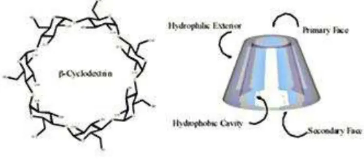
To the best of our knowledge, there are no previous studies about the encapsulation of N -sulfonyl aziridines by cyclodextrins to provide complexes with accessible NMR and physico-chemical characteristics, as those provided in this paper. In view of this, the major challenge to us was to prepare an aziridine with a well-balanced structural compromise between hydrophobic and hydrophilic groups in order to provide both enough solubility and ring-stability in water. In this work, we have used commercial -cyclodextrin (Fig.1) as a host model, based on the fact that its internal diameter allowed an efficient complexation of medium-sized aromatic compounds and heterocyclic derivatives.
Figure 1: Structure of -cyclodextrin.
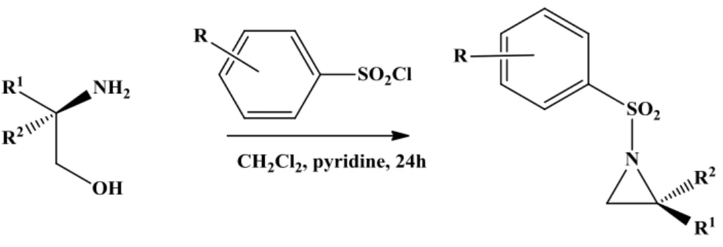
Scheme 1: Synthesis of aziridines
Furthermore, we assumed that the stereochemistry of those known chiral aziridines could promote diastereomeric interactions with the CD glucopyranose unit stereocenters to ensure a unique inclusion pattern of guest compound versus the host.
Table 1. Solubility and stability of aziridines (Az) in water: + stable / or soluble; -not soluble/or not stable/ Preparation references
Az R R1 R2 Solubility
Stability
minutes References
1 o-NO2 H i-C3H7 + 120 5a
2 p-NO2 H i-C3H7 - 60 6
3 p-Me H i-C3H7 - 30 7
4 p-Me H Me + 60 7b, 8
5 p-NO2 H Me + 30 6a, 6c
6 o-NO2 H Me + 30 9
7 p- NO2 H Ph - 180 6a
8 o- NO2 H Ph - 180 10
Before the complexation studies, all of the compounds were checked for their stability in aqueous medium (Table 1), also because the complexation and transport of drugs in water is of interest in living organisms. Therefore, a survey conducted in water would provide enough information for future formulation or studies in this field. The stability of each complex was monitored by NMR analysis of an aliquot of the corresponding solution, carried out every five minutes, searching for the appearance of a hydroxyl signal in the region of 3-3.5ppm. When the latter was observed, this gave evidence for the opening of the aziridine.
Figure 1: (S)-2-Isopropyl-1-(o-nitrophenyl)sulfonyl)aziridine 1 (AZ1).
Results and Discussion
Stoichiometry and binding constants
Stoichiometry and binding constants are the usual quantitative descriptors of the binding of a guest compound to a host, and NMR techniques are routinely used to measure these parameters as far as guest-host complexes formation is concerned. The terms of reference for such studies are either fast-exchange or slow-exchange phenomena. In the fast exchange regime, which compares well with our work, the observed NMR frequencies are defined as the mole fraction weighted average between the frequencies in the native form and the bound form of the observed molecule. For example, when the proton to be analysed is located on the host molecule, the chemical shift is given by the following equation:
obs =XH XHGG
Wherein obs is the chemical shift of the observed nucleus in the experiment, H the chemical shift of the unbound host molecule and HG is the chemical shift of the host in the complex. Besides, XH and XHG are the mole fractions of host molecule in the bound and the unbound states. In this case, the analysis was more complicated than for the slow exchange regime, because the chemical shifts in the complex could not be observed. This problem could be overcome by NMR titration, obtaining information on chemical shifts over a wide range of solutions of different compositions18.
One of the first methods used for the determination of the stoichiometry of inclusion
complexes was Job’s method, also known as the continuous variation method19,20. For the
experiment, we used stock solutions with equimolecular concentrations of Host (CD) and Guest (AZ1), according to the protocol of this method. The samples were prepared by mixing various volumes of these solutions in such a way that the total concentration [CD] + [AZ1] remained constant and the molar fraction of AZ1, varied in the range from 0 to 1. Using NMR analysis, the experimental data for the characterization of complexes were the observed variations of chemical shifts of both CD and AZ121.
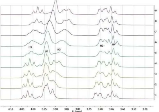
Figure 3: Expanded regions of 1H NMR spectra (500 MHz, D2O): chemical shift variations of H-3, H-6, H-5, H-2 and H-4 protons of CD in the complex.
This selective and exclusive effect of complexation on the CD internal protons is generally taken as good evidence for an interaction, where AZ1 is encapsulated in the hydrophobic core of the CD. It can be attributed to the magnetic anisotropy associated with the aromatic ring of the nitrophenyl group inside the hydrophobic cavity of CD21. No distinct chemical shifts were observed for free and bound states of the host and guest molecules, but only one set of signals was found for each species when the AZ1/CD ratio was modified. This observation was in good agreement with previously described systems regarding NMR fast-exchange limits 23,24.
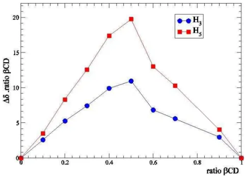
The stoichiometry of the complex was determined by using the method of continuous variation25,26. Chemical shifts differences for CD H-5 protons were used to draw the plot, and the stoichiometry was determined by plotting ratios of CD.(CD H-5) against ratios of CD (Fig. 4) to afford a maximum at ratio CD = 0.5, which meant a 1:1 complex ratio between AZ1 and CD.
Figure 4: Job’s Plot for the complex
Calculation of Ka.
The quantitative measurement of the complexation-induced shifts (CIS) of the AZ1-CD complex at 25°C is represented by the value of the equilibrium constant governing the formation of the inclusion complex (Table 2).
There are many ways to calculate the value of Ka such as Graphical (or linearization) or Curve fitting methods. The first one was designed to produce a linear relationship between obs and Ka, so that NMR data could be treated graphically. The four methods
Benesi-Hildebrand’s (Hanna-Ashbaugh) treatment27, Scatchard’s (Foster-Fyfe)28, Scott’s plot29 and
Rose-Drago’s method30 are well known for the graphical treatment of data. Curve fitting methods do not require any approximation for the calculation of Ka. We used the CLAK program that allowed a good assessment of Ka (382 mol–1)31.
Table 2: CIS = Observed increments induced on chemical shifts of CD H3 and H5 as a result of complexation with aziridine. CIS are related to concentrations of both CD and AZ1.
CD (mmol) mmol) obsH3 obs H5
7.89 0.0 0.0 0.0
7.101 0.789 3.3 4.5
6.312 1.578 10.8 14.6
5.523 2.367 8.0 14.7
4.734 3.156 11.4 21.7
3.945 3.945 21.9 39.5
3.156 4.734 24.8 43.5
2.367 5.523 24.8 41.9
1.578 6.312 26.4 41.6
0.789 7.101 25.8 35.0
The inclusion complex structure.
CD H-3 and H-5 protons provide good evidence for the formation of an internal AZ1-CD complex.
More detailed information concerning the geometry of these inclusion complexes could be derived from the evidence of the spatial neighbourhood between AZ1 and CD protons. This was achieved by investigating dipolar interactions using a 2D ROESY NMR experiment32. This technique proved to be the most sensitive for the structural analysis of inclusion complexes of CD formed in solutions33. As was done in many other situations related to inclusion complexes in water, a 300 milliseconds (300 ms) mixing time was selected to provide reliable dipolar cross-peaks with a minimal contribution of scalar transfer34,35.
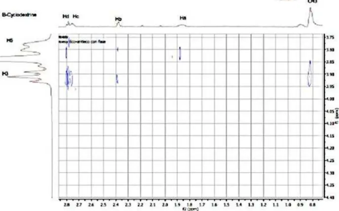
The 2D-ROESY spectrum of the AZ1/CD mixture (1:1) is depicted in Figure 5a. It shows strong 1H-1H cross peaks of the aromatic and aliphatic protons of AZ1 with CD H-3 and/or H-5 protons. These cross peaks along with the lack of correlation peaks with the protons on the outer-surface (H-2 and H-4), confirm the encapsulation of AZ1 inside the cavity of CD.
Figure 2a. Partial contour plots of ROESY experiment (mixing time: 300 ms, 500 MHz); inclusion complexCD-AZ1 (5mmol-1) in D2O, at 298 °K: areas of Az1 aliphatic (a) and
aromatic protons (b).
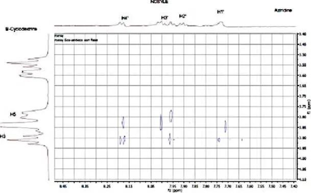
The 2D spectrum showed several intermolecular cross-peaks between CD H-3 and H-5 protons and protons of the aromatic ring of (nitrophenyl)sulfonyl group of AZ1 (H1’, H2’,
H3’and H4’), giving evidence for the inclusion of this group inside the hydrophobic cavity
Figure 3b. Partial contour plots of ROESY experiment (500 MHz, mixing time: 300 ms), inclusion complexCD/AZ1 (5mM-1) in D2O, at 298K: areas of aliphatic protons (a) and
aromatic protons (b) of aziridine.
Additional dipolar correlations were found between protons on the aziridine ring (Hb, Hd and Hc) with CD H-3 and H-5 protons. Finally, methyl protons presented a correlation only with the CD H-3 proton. This could accounted for by the spatial vicinity of this section of the aziridine with the (nitrophenyl)sulfonyl group, confirming that AZ1 is deeply inserted into the cavity of CD by means of its (nitrophenyl)sulfonyl group. We also observed intermolecular cross peaks only with the inner CD protons, and this observation established intracavity binding without giving evidence for outside contributions.
Therefore, and taking into account the attributions of the NMR analysis, we suggest the following geometry for the structure of our complex (Fig. 6).
Conclusion
We have carried out the first NMR complexation study between a stable aziridine and CD. The stoichiometry and association constant were determined by Job’s method and CLAK program. The spatial relationship between CD and AZ1 was assessed using ROESY experiments. Furthermore, NMR studies provided evidence that the interaction was an inclusion phenomenon, but not an association phenomenon, since the changes obtained for CD signals involved protons that were oriented towards the cavity of the host molecule.
The way parameters were determined in this work can serve for future investigations related not only to design new aziridines of biological interest, but also to formulation of existing aziridine-containing drugs.
Acknowledgements
We are indebted to Gobierno Vasco (ETORTEK-nanoIKER IE-11/304) and Universidad del País Vasco for financial support, and SGIker UPV/EHU for NMR facilities. A grant from Ministry of Higher Education and Scientific Research, General Directorate for Scientific Research and Technological Development (Algeria) is gratefully acknowledged.
Experimental Section
All the reactions needing anhydrous conditions were performed with dry solvents and carried out under nitrogen. Solvents were purified and dried according to standard procedures. Reagents for synthesis were used as received. I.R spectra were collected from a Mattson Genesis II FTIR. NMR spectra were recorded at 25 °C in CDCl3 (0.004mmol.L-1) on a Bruker 500, 400 or 300 MHz instrument, using tetramethylsilane (TMS) as an internal standard. Chemical shifts are given in (ppm) and coupling constant (J) values in Hertz (Hz). ROESY spectra were recorded using 500 ms mixing times and computer processing was performed using Mestrenova Software (version 6.2). Column chromatography was performed on silica gel 230-270 mesh (Merck) using CH2Cl2, MeOH or mixture of both according to thin layer chromatography (TLC) results.
Preparation of aziridines.
General procedure
Tosyl-, p-Nitrobenzensulfonyl- or o-Nitrobenzensulfonyl chloride (0.799g, 3.60 mmol) was added in one portion to a suspension of (S)-amino alcohol (0.124g, 1.20 mmol) in dry 2:1 CH2Cl2/pyridine (3mL) at 0 °C, and the resulting mixture was stirred at room temperature for 4 - 6h. The mixture was then diluted with CH2Cl2 (30 mL) and washed with 2 M HCl (3×12 mL). The aqueous layer was extracted with CH2Cl2 (20 mL), and the organic portions were combined and carefully shaken (CAUTION: an undesirable emulsion is produced under vigorous shaking) with 2M KOH aqueous solution (6×30 mL) and the aqueous layer was extracted with CH2Cl2 (20 mL). The organic layer was dried over MgSO4 and the solvent was removed to afford aziridines. Purification was carried out by column chromatography, using CH2Cl2 according to previous TLC results. The optical purity was retained in the final product and elemental analysis matched those of the literature.
(S)-2-isopropyl-1-(o-nitrophenyl)sulfonyl)aziridine1 (from L-valinol). (CAS registry number 111 0273-57-8).
(S)-2-isopropyl-1-(p-nitrophenyl)sulfonyl)aziridine2 (from L-valinol).
White solid; mp 74 °C; Rf= 0.50 (CH2Cl2) []D20 = +11.2 (c =1.0 CHCl3). 1H NMR (300 MHz, CDCl3) 0.85(d, J = 6.9Hz, 3H, CH3), 0.94(d, J = 7.0Hz, 3H, CH3), 1.39-1.53(m, 1H, CHMe2), 2.16-2.23(m, 1H, H-Az), 2.64-2.76(m, 2H, NC2-H, H-Az), 8.16(d, J = 9.0Hz, 2H, Ar-H), 8.38(d, J = 9.0Hz, 2H, Ar-H). 13C NMR (126 MHz, CDCl3) δ 19.5(CH3), 30.1(CHMe2), 33.6(Az-C3), 47.0(Az-C2), 124.2(Ar-C3), 129.3(Ar-C2), 144.1(Ar-C-SO2-), 150.6(Ar-C-NO2).. Elemental analysis: calcd for C11H14N2SO4, C 48.9; H 5.3; N 10.3. Found C 49.0; H 5.2; N 10.4.
(S)-2-isopropyl-1-Tosylaziridine 3 (from L-valinol). 1
H NMR (300 MHz, CDCl3) 0.80(d, J = 6.9Hz, 3H, CH3), 0.90(d, J = 6.8Hz, 3H, CH3), 1.37-1.46(m, 1H, CHMe2), 2.43(s, 3H, H3C-Ar), 2.49-2.56(m, 1H, Az-H1), 2.62(d, J =
7.0Hz, 1H, ), 7.33(d, J = 8.1Hz, 2H, Ar-H), 7.85(d, J = 8.1Hz, 2H, Ar-H). ). 13C NMR (126MHz, CDCl3) 19.5(CH3), 21.4(CH3-Ar), 30.0(CHMe2), 32.9(Az-C3), 46.2(Az-C2), 128.0(Ar-C2), 129.7(Ar-C3), 135.0(Ar-C-CH3), 144.5(Ar-C-SO2-). Elemental analysis: calcd for C12H17NSO2: C 60.3; H 7.2; N 5.9. Found C 60.4; H 7.1; H 5.8.
(S)-2-methyl-1-tosylaziridine 4 (from L-alaninol).
Transparent liquid. Rf = 0.58 (CH2Cl2/MeOH 9:1). 1H NMR (400 MHz, CDCl3) 1.20(d, J = 6.8Hz, 3H, CH3), 2.09(d, J= 5.8Hz, 1H, Az-H), 2.44(s, 3H, CH3), 2.62(d, J = 6.9Hz, 1H,
Az-H), 2.79(ddq, J = 7.0, 6.0, 5.0Hz, 1H, NCHMe), 7.34(d, J = 8.3Hz, 2H, Ar-H), 7.83(d, J = 8.3Hz, 2H, Ar-H). 13C NMR (126 MHz, CDCl3) δ 19.8(Ar-CH3), 21.5(CH3), 34.7(Az-C2), 36.1(Az-C3), 127.9(Ar-C2), 129.8(Ar-C3), 135.1(Ar-C-CH3), 144.6(Ar-C-SO2-). Elemental analysis: calcd for C10H13NSO2: C 56.9; H 6.2; N 6.6. Found C 57.0; H 6.3; N 6.5
(S)-2-methyl-1-(p-nitrophenyl)sulfonyl)aziridine 5 (from L-alaninol).
White solid; mp 87 °C, []D20 = + 24.5 (c = 1.0 CHCl3). 1H NMR (300 MHz, CDCl3) dJ = 6.0Hz, 3H, CH3), 2.13(d, J = 5.0Hz, 1H, Az-H), 2.75(d, J = 7.0Hz, 1H, Az-H), 2.89-3.05(m, 1H, NC2-H), 8.15(d, J = 8.9Hz, 2H, Ar-H), 8.39(d, J = 8.9Hz, 2H, Ar-H). 13C NMR (126 MHz, CDCl3) δ 19.4 CH3), 36.1(CH2N), 125.1(Ar-C-3), 130.9(Ar-C-2), 143.9(Ar-C-SO2-), 146.4(Ar-C-NO2). Elemental analysis: calcd for C9H10N2SO4 C 44.6; H 4.1; N 11.6. Found: C 44.8; H 4.0, N 11.7.
(S)-2-methyl-1-(o-nitrophenyl)sulfonyl)aziridine 6 (from L-alaninol).
Oil. 1H NMR (300 MHz, CDCl3) 1.32(d, J = 5.6Hz, 3H, CH3), 2.83(d, J = 7.0Hz, Az-CH2), 3.00-3.04(m, 1H, Az-CHMe), 7.68-8.25(m, 4H, Ar-H). 13C NMR (126 MHz, CDCl3) δ 20.1(CH3), 24.0(Az-C2), 38.1(Az-C3), 125.1(C-CNO2), 130.0(Ar-C6), 132.9(Ar–C4), 134.2(Ar-C1), 136.9(Ar-C5), 146.4(Ar-C-NO2). Elemental analysis: calcd for C9H10N2SO4 C 44.6; H 4.1; N 11.6. Found: C 44.7; H 4.2, N 115.
(S)-1-((p-nitrophenyl)sulfonyl)-2-phenylaziridine 7 (from L-(+)--phenylglycinol).
(S)-1-((o-nitrophenyl)sulfonyl)-2-phenylaziridine 8 (from L-(+)--phenylglycinol). 1
H NMR (300 MHz, CDCl3) 2.51(d, J= 5.0 Hz, 1H, NCH), 3.11(d, J= 7.0Hz, 1H, Az-H), 3.92(d, J=5Hz, 1H, Az-H), 5.30(m, 5H, Ph), 7.68-8.2(m, 4H, Ar). 13C NMR (126 MHz, CDCl3) 37.64(Az-C3), 42.83(Az-C2), 124.23(Ar-C3), 126.47(Ph-C4), 127.5(Ph-C2), 128.1(Ar-C6), 128.61(Ph-C3), 130.85(Ar-C4), 131.93(Ar-C5), 134.19(Ar-CSO2-),
148.57Ar-CNO2). Elemental analysis: calcd for C14H12N2SO4. C 55.3; H 3.9; N 9.2. Found C 55.0; H 4.1; N 9.5.
Inclusion procedure: aziridine 1- CD.
Solutions for NMR titration were prepared by mixing 7.89 mM mother solutions of AZ1
and CD to provide a series of solutions with a ratio ranging from 1 to 0. The solutions to be analyzed were kept at a constant concentration (7.89 mM) with a total volume steady at 500µl. This solution was sonicated for 10 min and subsequently analyzed by NMR. All NMR data were collected at 25 °C on a Bruker 500 MHz. The 2D data were obtained with a flip angle (90° pulse = 9.7 μs), and acquisition time of 16 s and a relaxation delay of 1s. Conditions for 1H NMR data were flip angle (90° pulse = 9.7 μs), acquisition time (AT) = 4 s,
numbers of scans (NS) = 16 and a relaxation delay of 1s.
References
1- (a) J. Szejtli J. Chem. Rev., 1998, 98, 1743-1753.
(b) M. Schirra, G. Delogu, P. Cabras, A. Angioni, G. D’Hallewin, A. Veyrat, J. Agric. Food Chem., 2002, 50, 6790-6797.
2- M. E. Davis, M.E. Brewster, Nature Rev. Drug Discov., 2004, 3, 1023-1035
3- A. A. Desai, H. Ren, M. Mukherjee, W. D. Wulff, Org. Process Res. Dev., 2011, 15, 1108-1115.
4- A. Keniche, A. Mezrai, J. Kajima Mulengi, The Open Conf. Proceed., J. 2011, 2, 28-35. 5- (a) D. Kurmich, J. R. Regan, D. Disalvo PCT Int. Appl., 2009. WO 2009015067 A2
20090129. US20100048950. Application Number: 12/521005/ Publication: 02/25/2010
(b) M. D’hoodge, I. Kerkaert, M. Rottiers, N. De Kimpe Tetrahedron, 2004, 60,
3637-3641.
6- (a) J. Farras, X. Giniesta, P. W. Sutton, J. Taltavull, F. Egeler, P. Romea, F. Urpi, J. Vilarrasa Tetrahedron, 2001, 57, 7665-7674.
(b) B. Moon, S. Mog So, H. Jin Choi Org. Lett., 2002, 4, 949-952.
(c) F. Crestey, M. Witt, K. Frydenvang, D. Stærk, J. W. Joroszewsky, H. Franzyk, J. Org. Chem., 2008, 73, 3566-3569.
(d) B. M. Chanda, R. Vyas, A. Bedekar J. Org. Chem., 2001, 66, 30-34.
7- (a) H. Xu, H. Tian, L. Zheng, Q. Liu, L. Wang, S. Zhang Tetrahedron Lett., 2011, 52, 2873-2875.
(b) H. Rubin, J. Cockrell, J. B. Morgan, J. Org. Chem., 2013, 78, 8865-8871.
8- (a) V. G. Nenajdenko, A. S. Karpov, E. S. Balenkova Tetrahedron Asymmetry, 2001, 12, 2517-2527.
(b) M. Cernerud, H. Adolfsson, C. Moberg, Tetrahedron Asymmetry, 1997, 8, 2665-2662.
(c) L. W. Bieber, M. C. F. Araujo, Molecules, 2002, 7, 902-906.
9- J. Bornholdt, J. Felding, R. P. Rasmus, J. L. Kristensen, Chemistry, A European J., 2010, 16, 12474-12480.
10- (a) H-L. Kwong, D. Liu, K-Y. Chan, C-S. Lee, K-H. Huang, C-M. Che Tetrahedron Lett.,
2004, 45, 3965-3968.
Rao J. Org. Chem., 2003, 68, 9119-9121.
15- (a) B. Srinivas, V. Pavan Kumar, R. Sridhar, K. Surendra, Y. V. D. Nageswar, K. Rama Rao, J. Mol. Catal. A: Chem., 2007, 261, 1-5.
(b) M. Somi Reddy, M. Narender, Y. V. D. Nageswar, K. Rama Rao, Tetrahedron Lett., 2005, 46, 6437-6439.
16- G. Maatz, A. Maciollek, H. Ritter Beilstein J. Org. Chem., 2012, 8, 1929-1935
17- J. H. Beijnen, S. C. van der Schoot, B. Nuijen, F. M. Flesch, A. Gore, D. Mirjovsky, L. Lenaz, Drug Devel.Indus.Pharm, 2008,34, 1130-1139.
18- K. S. Cameron, D. Fletcher, L. Fielding, Magn. Reson. Chem. 2002, 40, 251-260. 19- P. Job, Ann. Chim. 1928, 9, 113-203.
20- P. Job, Compt. Rend. Acad. Sci. Paris 1925, 180, 928-930.
21- L. Fielding, S. McKellar, J. F. Alastair, Magn. Reson. Chem. 2011, 49, 405-412. 22- (a) S. M. Ali, S. K. Upadhyay, Magn. Reson. Chem., 2008, 46, 676-679;
(b) G. Wenz, Beilstein J. Org. Chem. 2012, 8, 1890-1895.
23- N. Funasaki, S. Ishikawa, S. Neya, J. Phys. Chem. B, 2003, 107, 10094-10099. 24- K. Hirose, J. Incl. Phenom. Macrocycl. Chem. 2001, 39, 193-209.
25- (a) H. A. Benesi, J. H. Hildebrand, J. Am Chem. Soc. 1949, 71, 2703-2707. (b) M. W. Hanna, A. L. Ashbaugh, J. Phys. Chem. 1964, 68, 811-816. 26- R. Foster, C. A. Fyfe, Trans. Faraday Soc., 1965,61, 1626-1631. 27- R. Foster, C. A. Fyfe, J. Chem. Soc., Chem. Commun. 1965, 642. 28- R. L. Scott, Rec. Trav. Chim. Pays-Bas 1956, 75, 787-789. 29- G. Scatchard, Ann. N. Y. Acad. Sci. 1949, 51, 660-672.
30- N. J. Rose, R. S. Drago, J. Am. Chem. Soc. 1959, 81, 6138-6141. 31- D. Salvatierra, C. Díez, C. Jaime, J. Incl. Phenom., 1997, 27, 215-231. 3 - A. Bax, D. G. Davies, J. Magn. Reson., 1985, 65, 355-360.
33- V. Laine, A. Coste-Sarguet, A. Gadelle, J. Defaye, B. Perly, F. Djedaïni-Pilard, J. Chem. Soc. Perkin Trans, 1995, 2, 1479-1481.
34- F. Djedaïni-Pilard, N. Azaroual-Bellanger, M. Gosnat, D. Vernet, B. Perly, J. Chem. Soc. Perkin Trans, 1995, 2, 723-730.