R E S U M O A formação de trombos intraventriculares é um complicação frequente nos doentes com miocardiopatia dilatada e enfarte agudo do miocárdio, sendo o risco de embolização sistémica muito variável em função da patologia subjacente e características subjacentes dos trombos. Os autores descrevem dois casos clínicos relativos a dois doentes internados no mesmo dia com trombos intraventriculares, volumosos, protuberantes e muito móveis, no contexto de miocardiopatia dilatada e enfarte agudo do miocárdio, que embolizaram para os membros inferiores. Salientam a importância da ecocardiografia no diagnóstico, caracterização morfológica inicial e controlo evolutivo dos trombos intraventriculares, aspectos fundamentais na avaliação do risco embólico. Na ausência de recomendações específicas quanto às opções terapêuticas – anticoagulação, trombólise ou remoção cirúrgica, os autores sublinham a necessidade de avaliação individualizada, «caso a caso», tendo em conta o risco embólico, hemorrágico, e cirúrgico.
Trombos Intraventriculares
com Embolização Sistémica:
A Propósito de dois Casos Clínicos
[18]PEDROMAGNO,ANTÓNIOFREITAS,PEDROCUNHA,JOSÉLOUREIRO,JOSÉFRAGATA,RAFAELFERREIRA
Serviço de Cardiologia, Hospital Fernando Fonseca, Amadora, Portugal Serviço de Cirurgia Cardiotorácica, Hospital de Santa Marta, Lisboa, Portugal
A B S T R A C T
Intraventricular Thrombi with Systemic Embolization: Two Clinical Cases Ventricular thrombi are a frequent
complication of dilated cardiomyopathy and acute myocardial infarction, with variable risk of embolization according to the clinical setting and thrombus morphology.
The authors report two cases of patients admitted on the same day for acute myocardial infarction and dilated cardiomyopathy respectively, with high embolic risk left ventricular thrombi that embolized to the lower limbs after admission. Echocardiography, considered the gold standard diagnostic tool for intraventricular thrombi, is also the mainstay of embolic risk evaluation, for characterizing their size, mobility and echostructure. In the absence of evidence-based guidelines, management options – anticoagulation therapy, thrombolysis or thrombectomy – must be decided according to each specific case, taking account of embolic, bleeding and surgical risk.
Recebido para publicação: Julho de 2005 • Aceite para publicação: Janeiro de 2006 Received for publication: July 2005 • Accepted for publication: January 2006
Rev Port Cardiol 2006; 25 (2) :207-213
Palavras-Chave Trombos intraventriculares; Embolização sistémica; Trombectomia
Key words
Left ventricular thrombus; Systemic embolization; Thrombectomy
INTRODUÇÃO
A
formação de trombos intraventriculares (TIV) é uma complicação frequente nos doentes com miocardiopatia dilatada (MCD)(1, 2)e enfarte agudo do miocárdio (EAM)(3). A
incidência de trombo-embolismo sistémico é de um modo geral baixa, pelo que a atitude terapêutica é geralmente conservadora, na expectativa de que a resolução do trombo ocorra sem embolização sistémica(4).
Existem, no entanto, subgrupos de doentes em que o risco de embolização sistémica é elevada. Têm sido descritos vários marcadores de risco embólico, tais como a morfologia, as dimensões e a mobilidade dos TIV. Nos casos de trombos volumosos, protuberantes e muito móveis, o risco de embolização sistémica é par-ticularmente elevado, segundo alguns autores superior a 50 %(5). A informação disponível
na literatura sobre a atitude terapêutica mais adequada nestes casos é escassa, por vezes contraditória(8, 9, 11), não havendo recomendações
específicas no que se refere à indicação para anticoagulação oral, trombólise ou remoção cirúrgica.
A ecocardiografia é a técnica de imagem por excelência para o diagnóstico e controlo evolutivo dos TIV, nomeadamente dimensões, procidência na cavidade ventricular esquerda, mobilidade e modificações da ecoestrutura – aspectos fundamentais na avaliação do risco embolígeno(7).
Os autores descrevem dois casos clínicos de doentes internados simultaneamente no serviço de cardiologia com TIV volumosos, protuberantes e muito móveis, que embolizaram para os membros inferiores. A evolução clínica motivou reflexão e revisão da literatura no que se refere à atitude terapêutica mais adequada para prevenir a embolização sistémica.
CASO CLÍNICO 1
Doente do sexo feminino, 56 anos, admitida por insuficiência cardíaca congestiva – classe
IV da NYHA. Nos antecedentes pessoais há a salientar: hemorragia subaracnoideia há 16 anos, sem sequelas; e carcinoma da mama submetida a cirurgia conservadora e quimioterapia há cinco anos, sem recidivas.
Na observação à entrada apresentava polipneia, com ortopneia, ingurgitamento jugular a 45º; taquicardia sinusal, 105
208
INTRODUCTION
V
entricular thrombi are a frequent complication in patients with dilated cardiomyopathy (DCM)(1, 2) or acute myocardialinfarction (MI)(3). The incidence of systemic
thromboembolism is generally low and the therapeutic approach is accordingly conservative, in the hope that the thrombus will resolve without systemic embolization(4).
There are, however, subgroups of patients at high risk of systemic embolization. Various markers of embolic risk have been described, including the echostructure, size and mobility of the thrombus. In cases of large, protuberant, highly mobile thrombi the risk of systemic embolization is particularly high, over 50 % according to some authors(5). What little
information exists in the literature concerning the most appropriate therapeutic approach in such cases is at times conflicting(8, 9, 11), and
there are no specific guidelines on indications for oral anticoagulation, thrombolysis or thrombectomy.
Echocardiography is the gold standard technique for diagnosis and monitoring of such thrombi, particularly for characterization of their size, projection into the left ventricle (LV), mobility and changes in echostructure, which is essential for assessment of embolic risk(7).
The authors report the cases of two patients admitted on the same day to the Cardiology Department with large, protuberant, highly mobile ventricular thrombi that embolized to the lower limbs. Their clinical evolution prompted reflection and a review of the literature on the most appropriate therapeutic approach to prevent systemic embolization. CASE REPORT 1
A 56-year-old female patient was admitted for congestive heart failure (NYHA class IV). Her personal history included subarachnoid hemorrhage 16 years previously, without sequelae, and breast cancer treated by conservative surgery and chemotherapy 5 years before, with no recurrence.
Examination on admission showed tachy-pnea, orthotachy-pnea, jugular distension at 45º; sinus tachycardia, with heart rate of 105 bpm, and blood pressure 140/100 mmHg; S3 on cardiac auscultation, with no murmurs; rales at the lung bases on pulmonary auscultation; and slight
batimentos por minuto; pressão arterial 140/100 mmHg; à auscultação cardíaca: S3, sem sopros; auscultação pulmonar: fervores de estase nas bases; e edemas ligeiros dos membros inferiores.
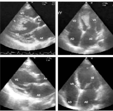
O ecocardiograma transtorácico, mostrava dilatação do ventrículo esquerdo (VE), com disfunção sistólica grave e presença de trombo volumoso, protuberante e móvel preenchendo a metade apical do VE(Fig.1).
Além da terapêutica standard para a insuficiência cardíaca, foi medicada com levosimendan em perfusão durante 24 h, e com heparina EV e varfarina, com melhoria funcional e hemodinâmica significativa, mantendo no entanto TIV volumoso com modificações da ecoestrutura – ecolucência, estreitamento do pedículo e maior mobilidade, sugestivas de maior risco embólico, o que motivou a discussão da situação clínica com a cirurgia cardiotorácica, com vista a possível trombectomia cirúrgica. Optou-se por uma atitude conservadora, atendendo ao elevado risco cirúrgico conferido pela disfunção VE, e ao contexto clínico. Ao vigésimo segundo dia de internamento, ainda sob anticoagulação e antiagregação, a doente iniciou um quadro súbito de isquemia aguda do membro inferior esquerdo, tendo sido submetida a arteriotomia femural com extracção de abundante material embólico, fragmentado.
O Ecocardiograma (Fig. 1) realizado na altura, mostrou desaparecimento do trombo intraventricular.
A doente teve alta ao vigésimo oitavo dia de internamento, em classe I NYHA. O ecocardiograma de controlo não mostrou recorrência de trombos ventriculares.
CASO CLÍNICO 2
Doente do sexo masculino, 29 anos, com antecedentes de dislipidemia e tabagismo (20 cigarros/dia), praticante de culturismo, com consumo habitual de esteróides anabolizantes e com hábitos toxifílicos esporádicos de consumo de cocaína. Transferido para o nosso hospital no terceiro dia de evolução de EAM de localização anterior, após internamento inicial noutro hospital, onde foi realizada angioplastia primária com colocação de stent no segmento médio da artéria descendente anterior.
Na admissão apresentava-se em classe 1 de Killip, normotenso, sem recorrência de angor. O
lower limb edema.
The transthoracic echocardiogram showed left ventricular dilatation, with severe systolic dysfunction, and a large, protuberant, mobile thrombus filling the apical half of the LV (Fig. 1). Besides standard therapy for heart failure, the patient was medicated with levosimendan in perfusion for 24 hours, as well as iv heparin and warfarin, with significant functional and hemodynamic improvement. However, the thrombus persisted, showing changes in echo-structure with echolucency, narrowing of the pedicle and increased mobility, suggesting higher embolic risk. A discussion of the clinical situation with the cardiothoracic surgery department followed, with a view to possible thrombectomy, but a conservative approach was
decided on given the high surgical risk associated with LV
dysfunction and the clinical setting. On the 22nd day of hospitalization, the patient, still under anticoagulation and antiplatelet therapy, presented acute ischemia in the left lower limb, and underwent femoral arteriotomy with removal of a large quantity of fragmented embolic material.
The echocardiogram (Fig. 1) performed at that time showed no sign of the ventricular thrombus.
The patient was discharged on the 28th day, in NYHAclass I. The follow-up echocardiogram showed no recurrence of ventricular thrombi. CASE REPORT 2
A 29-year-old male patient with a history of dyslipidemia, smoking (20 cigarettes/day) and body-building, with habitual use of anabolic steroids and occasional use of cocaine, was transferred to our hospital on the third day of evolution of an anterior MI after initial admission to another hospital, where he had undergone primary angioplasty, with stenting of the mide left anterior descending artery.
On admission, he presented in Killip class 1, normotensive, with no recurrence of chest pain. The transthoracic echocardiogram revealed no
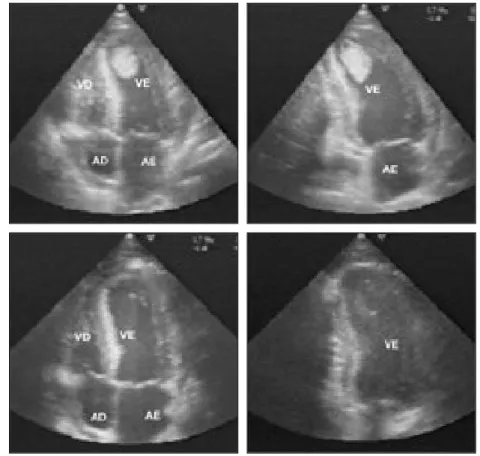
LV dilatation, slightly impaired global systolic function (ejection fraction 40 %), and akinesia of the apical segments, with a large, protuberant, mobile thrombus filling the entire apex (Fig. 2A).
ecocardiograma transtorácico na admissão revelou VE não dilatado, com função sistólica global ligeiramente comprometida (FE-40 %), e acinesia dos segmentos apicais, com volumoso trombo protuberante e móvel preenchendo todo o apex (Fig. 2).
Foi iniciada terapêutica com heparina endovenosa.
Os ecocardiogramas subsequentes mostraram ligeira redução das dimensões do trombo, modificação da eco-estrutura e aumento da mobilidade. Nesta altura foi contactada a Cirurgia Cardiotorácica, tendo em conta eventual indicação para remoção cirúrgica do trombo, mas atendendo ao contexto clínico e não havendo indicação concomitante para revascularização miocárdica, foi decidido manter apenas heparina endovenosa.
No segundo dia de internamento, iniciou quadro súbito de isquemia aguda bilateral dos membros inferiores, tendo o ecocardiograma mostrado o desaparecimento do trombo previa-mente identificado (Fig. 2). O doente foi submetido a tromboembolectomia de urgência da bifurcação aórtica, com extração de múltiplos e volumosos fragmentos de trombo.
210
Subsequent echocardiograms showed a slight reduction in the size of the thrombus, changes in its echostructure and increased mobility. At this point the cardiothoracic surgery department was contacted in view of the possible indication for thrombectomy, but given the clinical setting and absence of concomitant indication for myocardial revascularization, it was decided merely to continue with iv heparin. On the second day after admission, the pa-tient suddenly developed acute bilateral lower limb ischemia, with the echocardiogram show-ing that the previously identified thrombus had disappeared (Fig. 2B). An urgent thromboembolectomy was performed at the aortic bifurcation, with removal of several large thrombus fragments.
The procedure was uneventful and was followed by full functional recovery of the lower limbs.
DISCUSSION
Ventricular thrombi have been most frequently reported in three clinical situations –
DCM, MI and left ventricular aneurysm(1, 2, 3).
Although the pathophysiological mechanisms Fig. 1 Ecocardiograma
transtorácico:
Em cima (A), incidências paraesternal longitudinal (à esquerda) e apical quatro câmaras (à direita) que mostram um trombo (Tr) volumoso, móvel, fazendo procidência na cavidade ventric-ular esquerda.
Em baixo (B), é possível observar o desaparecimento do trombo intraventricular, após início de quadro de isquemia aguda do membro inferior esquerdo. (Ao: aorta; AD: aurícula direita; AE: aurícula esquerda; Tr: trombo; VD: ventrículo direito; VE: ventrículo esquerdo)
Fig. 1 Transthoracic echocardiogram: top (A): longitudinal parasternal view (left) and 4-chamber apical view (right), showing a large, mobile thrombus, projecting into the left ventricle; bottom (B), showing that the ventricular thrombus had disappeared following development of acute ischemia of the left lower limb. Ao: aorta; AD: right atrium; AE: left atrium; Tr: thrombus; VD: right ventricle; VE: left ventricle
O procedimento decorreu sem complicações, com total recuperação funcional dos membros inferiores.
DISCUSSÃO
A formação de TIV tem sido descrita mais frequentemente em três situações clínico-pa-tológicas – a MCD, o EAM e o aneurisma do ventrículo esquerdo(1, 2, 3). Apesar de não serem
totalmente conhecidos os mecanismos fisiopatológicos que levam à formação de TIV, é geralmente aceite que o relentamento circulatório, alterações da superfície endocárdica e o estado de hipercoagulabilidade são factores preponderantes para a sua formação.
A prevalência de TIVnos doentes com MCD
e EAM é muito variável consoante as séries, estimando-se que cerca de um terço dos doen-tes com MCD apresentem TIV ao longo da sua evolução(1, 2). Em séries de doentes com EAMde
localização anterior estão descritas prevalências de TIV até cerca de 40 %, sendo muito menos frequentes nos EAM inferiores(3).
A detecção de TIV assume especial relevância, tendo em conta a sua história natural e o risco de embolização sistémica, com
behind their formation are not fully understood, it is generally agreed that slowing of circulation, changes in the endocardial surface and hypercoagulability are predisposing factors. The prevalence of such thrombi in patients with DCM or MI varies considerably between series, but it is estimated that around a third of patients with DCM will present them at some stage(1, 2). A prevalence of up to 40 % has been
reported in series of patients with anterior MI, but they are much less common in inferior MI(3).
Detection of ventricular thrombi is particularly important given their natural history and the risk of systemic embolization, with high morbidity and mortality, particularly stroke and acute limb ischemia. Available data in the lit-erature indicate considerable variation in embo-lic potential, between 5 and 50 %(2, 5),
which reflects the heterogeneity of the populations studied in terms of underlying pathology and, more importantly, of thrombus morphology, particularly size, protrusion into the LVand intracavitary mobility.
Echocardiography, besides having high sensitivity and specificity in detecting thrombi, enables initial morphological characterization Fig. 2 Ecocardiograma transtorácico: imagens em apical 4 câmaras (à esquerda) e apical duas câmaras modificado (à direita) que mostram trombo volumoso, pediculado e móvel ocupando o ápex do ventrículo esquerdo. Na imagem de baixo (B), observa-se o desaparecimento do referido trombo intraventricular, após início de quadro de isquemia aguda bilateral dos membros inferiores. (Ao: aorta; AD: aurícula direita; AE: aurícula esquerda; Tr: trombo; VD: ventrículo direito; VE: ventrículo esquerdo).
Fig. 2 Transthoracic echocardiogram: top (A): 4-chamber apical view (left) and modified 2-chamber apical view (right), showing a large mobile pedunculated thrombus occupying the left ventricular apex; bottom (B), showing that the ventricular thrombus had disappeared follow-ing development of acute bilateral ischemia of the lower limbs. Ao: aorta; AD: right atrium; AE: left atrium; Tr: thrombus; VD: right ventricle; VE: left ventricle.
elevada morbilidade e mortalidade, particularmente os acidentes vasculares cerebrais e a isquemia aguda dos membros. Os dados actualmente disponíveis na literatura, apontam para um risco embolígeno muito variável, entre 5 a 50 %(2, 5), reflectindo a
heterogeneidade das populações estudadas, no que se refere à patologia subjacente e, sobretudo, às características morfológicas dos
TIV, nomeadamente as dimensões, a protuberância na cavidade VE e a mobilidade intra-cavitária .
A ecocardiografia, além da elevada sensibilidade e especificidade para a identificação de TIV, permite a caracterização morfológica inicial e controlo evolutivo, aspectos muito importantes na avaliação do risco cardio-embólico(5, 7). O diagnóstico
diferencial com outras massas, nomeadamente tumores cardíacos primários ou secundários, é geralmente possível com base nas caracteristicas morfológicas, embora em situações extremas só a anatomia patológica permita tal distinção.
A anticoagulação é consensual como terapêutica inicial para a maioria dos casos de
TIV, com menor risco embólico(6, 8). Quando
apesar da anticoagulação há persistência de
TIV, ou ocorrem fenómenos embólicos, está geralmente indicada a terapêutica trombolítica ou a remoção cirúrgica(9, 10).
Também os casos de TIV volumosos, protuberantes e móveis, como os descritos anteriormente, atendendo ao seu elevado potencial embólico, poderão justificar atitudes terapêuticas iniciais mais «agressivas», como a trombólise ou a remoção cirurgica, para prevenir a embolização sistémica(9, 10, 11).
Embora estejam descritos casos de fibrinólise com sucesso no contexto de TIV(10),
alguns autores alertam não só para o maior risco hemorrágico inerente a esta estratégia, mas também para uma possível promoção do fenómeno de embolização resultante da lise do ponto de fixação do trombo à parede do VE.
A remoção cirúrgica do trombo como profilaxia de embolização, tem sido descrita na literatura mas geralmente não constitui o primum movens da cirurgia, sendo, na maioria dos casos, reservada para doentes que apresentam indicação concomitante para terapêutica de revascularização miocárdica. Estão no entanto descritos casos em que a
212
and monitoring of their evolution, which is essential for assessment of embolic risk(5, 7).
Differential diagnosis with other types of mass such as primary or secondary cardiac tumors is usually possible on the basis of morphological characteristics, although in extreme situations only anatomic pathology will be able to make the distinction.
It is generally agreed that anticoagulation should be the initial therapy in most cases of ventricular thrombi, as it is associated with lower embolic risk(6, 8). If the problem persists
despite anticoagulation or when embolization occurs, thrombolysis or thrombectomy are generally indicated(9, 10).
Cases of large, protuberant, mobile thrombi, as described above, may require more aggressive initial therapies given their high embolic potential, such as thrombolysis or thrombectomy in order to prevent systemic embolization(9, 10, 11).
Although cases of successful fibrinolysis of ventricular thrombi have been reported(10), some
authors have pointed out the higher hemorrhagic risk involved in this strategy, as well as the possible increased risk of embolization arising from lysis of the point where the thrombus is attached to the LV wall. Prophylactic thrombectomy to prevent embolization has been described in the literature but generally this is not the primary reason for surgery; it is mainly reserved for pa-tients who have concomitant indication for myocardial revascularization. There have, how-ever, been cases in which prophylactic thrombectomy was the only reason for surgery
(11). Surgical risk should be assessed on an
individual basis, taking account of the likelihood of systemic
embolization; it may be acceptable not to intervene at first in patients with comorbidities or those with high surgical risk.
CONCLUSION
The two cases described above had favorable outcomes following relatively simple surgical procedures under local anesthesia, with no sequelae and disappearance of the thrombi. Embolization to the central nervous system would certainly have had more severe clinical consequences.
Echocardiography is the gold standard technique for diagnosis of ventricular thrombi
trombectomia profiláctica constituiu o único motivo da cirurgia(11). O risco cirúrgico deve ser
individualizado face à probabilidade de embolização sistémica, sendo talvez aceitável não intervir de início em doentes com co-morbilidades e risco cirúrgico elevado.
CONCLUSÃO
Os dois casos clínicos, descritos anteriormente, tiveram desfecho favorável, porque se resolveram com uma cirurgia «simples», sob anestesia local, evoluindo sem sequelas e com o desaparecimento do TIV. A embolização para o sistema nervoso central teria seguramente consequências clínicas mais gravosas.
A ecocardiografia é a técnica de imagem por excelência para o diagnóstico e estratificação do risco embolígeno dos TIV.
A anticoagulação como terapêutica para resolução do trombo e profilaxia de embolização é consensual, mas provavelmente insuficiente em situações de elevado risco embolígeno, em que a fibrinólise ou a trombectomia poderão estar indicadas. Relativamente a este ponto não há recomendações específicas, validadas
and stratification of embolic risk.
Anticoagulation is the accepted therapy to resolve thrombi and to prevent embolization, but is probably insufficient in situations of high embolic risk, in which fibrinolysis or thrombectomy may be indicated. There are, however, no specific scientifically validated guidelines as to the best therapeutic approach. Although cases have been reported in which more aggressive strategies were adopted from the start, the decision must be taken on a
case-BIBLIOGRAFIA / REFERENCES 1. Gottdiener JS, Gay JA, Van Voorhees L, DiBianco R,
Fletcher RD. Frequency and embolic potential of left ventric-ular thrombus in dilated cardiomyopathy: assessment by 2-dimensional echocardiography. Am J Cardiol 1983;52:1281-5 2. Falk RH, Foster E, Coats MH. Ventricular thrombi and thromboembolism in dilated cardiomyopathy: a prospective follow-up study. Am Heart J 1992;123:136-42.
3. Greaves SC, Zhi G, Lee RT, Solomon SD, MacFadyen J, Rapaport E, Menapace FJ, Rouleau JL, Pfeffer MA. Incidence and natural history of left ventricular thrombus following anterior wall acute myocardial infarction. Am J Cardiol 1997; 80:442-8.
4. Stokman PJ, Nandra CS, Asinger RW. Left Ventricular Thrombus. Curr Treat Options Cardiovasc Med 2001;3:515-21. 5. Visser CA, Kan G, Meltzer RS, Dunning AJ, Roelandt J. Embolic potential of left ventricular thrombus after myocardial infarction: a two-dimensional echocardiographic study of 119 patients. Am Coll Cardiol 1985;5:1276-80. 6. Mallory R, Balcezak T. Treatment of mobile left ventricular thrombus with low molecular weight heparin. N Eng J Med 1999;341:1082-3.
7. Domenicucci S, Chiarella F, Bellone P. Role of echocardi-ography in the assessment of left ventricular thrombus embolic potential after anterior acute myocardial infarction. Congest Heart Fail. 2001;7:250-5.
8. Heik SC, Kupper W, Hamm C, Bleifeld W, Koschyk DH, Waters D, Chen C. Efficacy of high dose intravenous heparin for treatment of left ventricular thrombi with high embolic risk. J Am Coll Cardiol 1994;24:1305-9.
9. Nili M, Deviri E, Jortner R, Strasberg B, Levy MJ. Surgical removal of a mobile, pedunculated left ventricular thrombus: report of four cases. Ann Thorac Surg 1988;46:396-400. 10. Rester BT, Warnock JL, Patel PB, McMullan MR, Skelton TN, Collop NA. Lysis of a left ventricular thrombus with recombinant tissue plasminogen activator. Chest 2001;120: 681-3.
11. Fortier S, Demaria RG, Pelletier GB, Carrier M, Perrault LP. Left ventricular thrombectomy in a cocaine user with normal coronary arteries. J Thorac Cardiovasc Surg 2003;125: 204-5.
Pedidos de separatas para: Address for reprints: PEDRO MAGNO
Serviço de Cardiologia do Hospital Fernando Fonseca Terreno da Via Rápida, Sintra
2720-276 AMADORA, PORTUGAL e-mail: p–magno@hotmail.com