Lectins as Biomarkers of Oral Cavity Tumors: Literature Review
Lectinas como Biomarcadores de Tumores de Cavidade Oral: uma Revisão de
Literatura
Lectinas como Biomarcadores de Tumores de la Cavidad Oral: Revisión de la
Literatura
Bruno Rocha da Silva1, Francisco Vassiliepe de Sousa Arruda2, Victor Alves Carneiro3, Theodora Thays Arruda Cavalcante4, Andréa Silvia Walter de Aguiar5, Kyria Santiago do Nascimento6, Benildo Sousa Cavada7, Edson Holanda Teixeira8
Abstract
Introduction: With the development of cancer research, a peculiar group of proteins has become the subject of great attention, namely the lectins. These proteins have the ability to bind reversibly to carbohydrates with high specificity. Because of changes in cell surface glycoprotein patterns during tumor formation, lectins have an important potential role as biomarkers of neoplastic cells. Objective: To review the literature available regarding the use of lectins as biomarkers of premalignant and neoplastic lesions, with a focus on oral cavity tumors, and to assess which groups of lectins and oral lesions have been most widely studied, with the final goal of providing a profile of publications in the field. Method: Articles were searched in the Science Direct, PubMed, and BVS databases. Inclusion criteria were: publication between 1981 and 2010; keywords “lectin” AND “binding” AND “oral” AND “tumor”; and English abstract. A total of 108 articles were selected. Articles were assessed and classified according to predetermined categories, especially number/types of lesion and number/types of lectin analyzed. Results were assessed using the chi-square test. Results: An increase throughout the years was observed in the number of articles studying the use of lectins as tumor biomarkers. Squamous cell carcinoma and Arachis hypogea (PNA) were the lesion and lectin more frequently assessed, respectively. Conclusion: It is possible to conclude that the use of lectins as a therapeutic tool in cancer research has been increasing in importance, probably as a result of its wide applicability, versatility, and reliability.
Key words: Medical Oncology; Lectins; Mouth Neoplasms
1Cirurgião-Dentista. Mestrando em Biotecnologia. Programa de Pós-graduação em Biotecnologia da Universidade Federal do Ceará – Campus Sobral.
Sobral (CE), Brasil. E-mail: brunorocha747@gmail.com.
2Biólogo. Doutorando em Biotecnologia. Rede Nordestina de Biotecnologia. Universidade Federal do Ceará. Fortaleza (CE), Brasil. E-mail: vassiliepe@gmail.com. 3Biólogo. Doutor em Bioquímica. Departamento de Bioquímica e Biologia Molecular da Universidade Federal do Ceará. Fortaleza (CE), Brasil.
E-mail: victorcarneiro@ufc.br.
4Cirurgiã-Dentista. Doutoranda em Bioquímica. Departamento de Bioquímica e Biologia Molecular da Universidade Federal do Ceará. Fortaleza (CE),
Brasil. E-mail: theodorathays@gmail.com.
5Cirurgiã-Dentista. Doutor em Odontologia. Departamento de Clínica Odontológica da Universidade Federal do Ceará. Fortaleza (CE), Brasil.
E-mail: andrea.aguiar@ufc.br.
6Engenheira de Pesca. Pós-Doutor em Biotecnologia. Departamento de Bioquímica e Biologia Molecular da Universidade Federal do Ceará. Fortaleza
(CE), Brasil. E-mail: kyriasantago@gmail.com.
7Engenheiro Agrônomo. Pós-Doutor em Bioquímica. Departamento de Bioquímica e Biologia Molecular da Universidade Federal do Ceará. Fortaleza
(CE), Brasil. E-mail: bscavada@ufc.br.
8Cirurgião-Dentista. Doutor em Bioquímica. Faculdade de Medicina da Universidade Federal do Ceará - Campus Sobral. Sobral (CE), Brasil. E-mail: edson@ufc.br.
Author for Correspondence: Edson Holanda Teixeira, Ph.D. Medicine School, Federal University of Ceará. Rua Geraldo Rangel, 100 - Campus do Derby. Sobral (CE), Brazil. CEP: 62041-040. E-mail: Edson@ufc.br.
INTRODUCTION
Malignant tumors of the oral cavity represent a major public health concern, especially in view of the increasing incidence and prevalence rates observed along the last few years1. Parkin et al. (1999) estimated about 210,000 new cases per year worldwide2. In Brazil, a total of 14,120 new cases of malignant tumors of the oral cavity have been estimated for the year 20103.
Motivated by this scenario, several studies have been conducted to investigate oral cavity malignancies, with a particular focus on cell differentiation (formation process) and cell identification methods. In particular, some studies have shown that the process of malignancy is associated with a variety of changes in cell surface carbohydrate expression, not to mention the role played by carbohydrates in determining the metastatic capabilities of neoplastic cells4-8.
Nangia-Makker et al. (2002) reported that developing cancer cells use the functional groups of carbohydrate molecules to avoid being recognized by immune cells9. During metastasis formation, carbohydrates are involved in interactions between tumor cells, between tumor cells and the extracellular membrane, or between tumor cells and endothelial cells4. However, due to the structural complexity of carbohydrates and the scarce knowledge currently available in the field of glycobiology, a better understanding of this mechanism is not yet possible6,9-10.
In an attempt to overcome these difficulties and to better understand the process of malignancy, a unique group of proteins, namely the lectins, has become the subject of special attention. Lectins are proteins of nonimmune origin comprised by several structures that have the ability to bind reversibly to carbohydrates, recognizing and agglutinating specific oligosaccharides or glycoconjugates4. Due to these specific features, lectins have been widely used in different biological and medical applications. In immunohistochemical examinations, for example, lectins recognizing different carbohydrates may provide a detection system sensitive to changes in glycosylation and carbohydrate expression at different stages of disease onset/progression, including embryogenesis, growth, and cellular pathology8. In addition, studies in the field of tumor lectinology have been able to identify differences between normal tissue and tumor cells in brain11, breast12, and oral cancer13.
The high number of lectins so far submitted to individual investigation has opened a wide variety of possibilities of studies involving these proteins. In order to facilitate and guide future studies designed to investigate the role of lectins as biomarkers of oral cavity tumors, a review of the current literature becomes extremely useful, with a focus on which lectins have already been studied
and which lesions have already been assessed in association with such lectins.
The objective of this study was to review the literature available on the use of lectins as biomarkers of premalignant and neoplastic lesions, with a focus on oral cavity tumors, and to assess which groups of lectins and which groups of oral lesions have been most widely studied, with the final goal of providing a profile of publications in the field.
METHOD
S
AmplE dElimiTATioNThe following Brazilian and international journal databases were reviewed: Science Direct, PubMed and BVS (Biblioteca Virtual em Saúde). Selection of these databases was based on the wide range of journals covered by each of them and on our goal to provide an overview of the scientific production devoted to the topic over the long timeframe under analysis.
The following inclusion criteria were considered during the review: a) articles published between January 1981 and June 2010; b) presence of the keywords “lectin” AND “binding” AND “oral” AND “tumor,” entered into the advanced search form; and c) availability of an abstract in English. A total of 108 articles were selected.
S
AmplE ClASSiFiCATioNAll the 108 abstracts were read and assessed to determine the classification of articles for subsequent quantitative analysis. In cases where the abstract was not enough to allow classification, the full article was read. Each article was classified according to the following attributes: title of article, year of publication (subcategories [quinquenniums]: first period, 1981 to 1985; second period, 1986 to 1990; third period, 1991 to 1995; fourth period, 1996 to 2000; fifth period, 2001 to 2005; sixth period, 2006 to 2010), journal name, language of publication, number and types of lesion assessed; and number and types of lectin analyzed in the study. Lesions were classified according to the World Health Organization (WHO)14 classification for oral cavity diseases; lectins were classified both in terms of kingdom (animal vs. vegetable) and family/group, based on the classifications of Peumans and Van Damme15 and Varki16.
S
TATiSTiCAl ANAlySiSData were entered into Microsoft Excel 2007 spreadsheets and systematically transferred to the Statistical Package for the Social Sciences (SPSS) version 17.0 for statistical analysis. Frequency distribution calculations were used to assess general sample characteristics, to investigate possible spelling errors in the raw data, and
to obtain an overview of the use of lectins as biomarkers of oral cavity tumors.
RESULTS
S
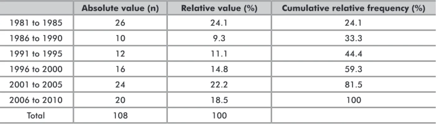
TudiES diSTRiBuTioN oVER THE yEARSAnalysis of the distribution of publications over the different quinquenniums revealed a large concentration of studies in the first period (1981 to 1985, 26 studies), followed by a significant decrease in the second period (1986 to 1990, 10 studies). In the fifth period (2001 to 2005), a return to early patterns was observed, with a total of 24 publications (Table 1).
Figure 1 shows the same distribution of studies, however divided into plant vs. animal lectins. It is possible to observe that up to the third period assessed (1981-1995), 100% of the publications focused on plant lectins, whereas studies using animal lectins started to be conducted as of 1996 and accounted for the majority of studies in the two last periods assessed (6 animal vs. 11 plant lectins in the fourth period, 17 vs. 9 in the fifth, and 16 animal vs. 4 plant lectins in the sixth period assessed) (Figure 1).
l
ANguAgE oF puBliCATioNMost of the articles reviewed were written in English (91 articles, 84.3%), followed by German (7, 6.5%), Japanese (5, 4.6%), Chinese (4, 3.7%), and French (only 1 article, 0.9%).
N
umBER oF lESioNS ANd lECTiNS ANAlyzEdOf the total of 108 studies, 47 (43.5%) analyzed only one lectin, 29 (26.9%) analyzed two, 15 (13.9%) analyzed three, and only 17 studies (15.7%) analyzed four or more lectins. In addition, 86 articles (79.6%) investigated one type of lesion, 14 (13%) investigated two lesions, and 8 articles (7.4%) investigated three or more types of lesions.
E
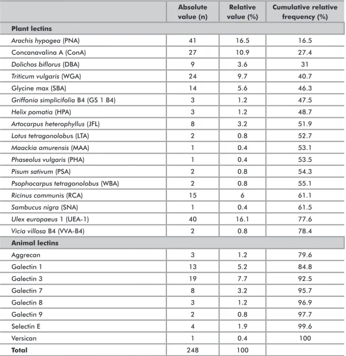
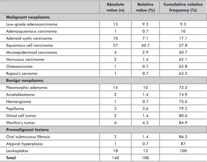
VAluATioN oF STudiEd lESioNS ANd lECTiNSThe list of lectins and types of lesions analyzed and the respective absolute and relative frequencies are shown in Tables 2 and 3. Among the lesions assessed, there was a predominance of malignant lesions (63.6%), followed by benign lesions (21.4%).
Figure 2 shows the distribution of lesions in the different quinquenniums assessed. It is possible to observe that both malignant and benign neoplasms showed a similar distribution, whereas premalignant lesions showed an important decline in the sixth/last period assessed, in which no study was found.
Absolute value (n) Relative value (%) Cumulative relative frequency (%)
1981 to 1985 26 24.1 24.1 1986 to 1990 10 9.3 33.3 1991 to 1995 12 11.1 44.4 1996 to 2000 16 14.8 59.3 2001 to 2005 24 22.2 81.5 2006 to 2010 20 18.5 100 Total 108 100
Figure 1. Distribution of publications according to kingdom of the lectin analyzed, by quinquennium
Table 1. Distribution of publications from 1981 to 2010, by quinquennium
0 10 20 30 Vegetable kingdom Animal kingdom 1981 to 1985 1986 to 1990 1991 to 1995 1996 to 2000 2001 to 2005 2006 to 2010 Quinquennium N º of P u b li c at io n s N º of P u b li c at io n s 0 10 20 Premalignant lesion Benign neoplasms 1981 to 1985 1986 to 1990 1991 to 1995 1996 to 2000 2001 to 2005 2006 to 2010 Malignant neoplasms Quinquennium
Figure 2. Distribution of the analyzed lesions according to the WHO classification, by quinquenniumnnium
In order to better assess the profile of publications, the distribution of lectins analyzed was assessed in relation to the three major groups of lesions covered by the studies (Table 4). This analysis revealed that the two lectins most commonly present in all three groups of lesions were Arachis hypogea (PNA) and Ulex europaeus 1 (UEA-1), both of vegetable origin. Moreover, in the groups of benign and malignant neoplasms, the animal lectin Galectin 3 ranked fourth among the most frequently analyzed lectins, contrasting with a predominance of plant lectins found in these groups. Conversely, in the group of
Absolute value (n) Relative value (%) Cumulative relative frequency (%) Plant lectins
Arachis hypogea (PNA) 41 16.5 16.5
Concanavalina A (ConA) 27 10.9 27.4
Dolichos biflorus (DBA) 9 3.6 31
Triticum vulgaris (WGA) 24 9.7 40.7
Glycine max (SBA) 14 5.6 46.3
Griffonia simplicifolia B4 (GS 1 B4) 3 1.2 47.5
Helix pomatia (HPA) 3 1.2 48.7
Artocarpus heterophyllus (JFL) 8 3.2 51.9
Lotus tetragonolobus (LTA) 2 0.8 52.7
Maackia amurensis (MAA) 1 0.4 53.1
Phaseolus vulgaris (PHA) 1 0.4 53.5
Pisum sativum (PSA) 2 0.8 54.3
Psophocarpus tetragonolobus (WBA) 2 0.8 55.1
Ricinus communis (RCA) 15 6 61.1
Sambucus nigra (SNA) 1 0.4 61.5
Ulex europaeus 1 (UEA-1) 40 16.1 77.6
Vicia villosa B4 (VVA-B4) 2 0.8 78.4
Animal lectins Aggrecan 3 1.2 79.6 Galectin 1 13 5.2 84.8 Galectin 3 19 7.7 92.5 Galectin 7 8 3.2 95.7 Galectin 8 3 1.2 96.9 Galectin 9 2 0.8 97.7 Selectin E 4 1.9 99.6 Versican 1 0.4 100 Total 248 100
Table 2. Lectins analyzed in the articles reviewed*
* More than one lectin was assessed in some articles, therefore the total number of lectins analyzed exceeds the total number of studies reviewed.
premalignant lesions, animal lectins were virtually absent (only one study analyzing Selectin E).
Finally, the analysis of association between lectins and the most prevalent lesions in each group (leukoplakia among premalignant lesions, pleomorphic adenoma among benign neoplasms, and squamous cell carcinoma among malignant neoplasms) revealed a predominant association between studies assessing pleomorphic adenomas and analyzing Concanavalin A (ConA) (five studies) rather than PNA, which belongs to the same group; all other lesions were associated with lectins representative of their groups of origin (Table 5).
Absolute value (n) Relative value (%) Cumulative relative frequency (%) Malignant neoplasms Low-grade adenocarcinoma 13 9.3 9.3 Adenosquamous carcinoma 1 0.7 10
Adenoid cystic carcinoma 10 7.1 17.1
Squamous cell carcinoma 57 40.7 57.8
Mucoepidermoid carcinoma 4 2.9 60.7 Verrucous carcinoma 2 1.4 62.1 Osteosarcoma 1 0.7 62.8 Kaposi's sarcoma 1 0.7 63.5 Benign neoplasms Pleomorphic adenoma 14 10 73.5 Ameloblastoma 2 1.4 74.9 Hemangioma 1 0.7 75.6 Papilloma 5 3.6 79.2
Ghost cell tumor 2 1.4 80.6
Warthin’s tumor 6 4.3 84.9
Premalignant lesions
Oral submucous fibrosis 2 1.4 86.3
Atypical hyperplasia 1 0.7 87
Leukoplakia 18 13 100
Total 140 100
Table 3. Lesions assessed (WHO classification) in the articles reviewed *
* More than one lesion was assessed in some articles, therefore the total number of lesions assessed exceeds the total number of studies reviewed.
lectins analyzed
Aggrecan
Arachis hypogea
(p
NA)
Concanavalina A (ConA) Dolichos biflorus
(d
BA)
galectin 1 galectin 3 galectin 7 galectin 8 galectin 9
Triticum vulgaris
(W
gA)
glycine max (SBA)
Griffonia simplicifolia B4 ( gS i-B4) Helix pomatia (H pA) Artocarpus heterophyllus (JF l) Lotus tetragonolobus (l TA) Maackia amurensis (m AA) Phaseolus vulgaris (p HA) Pisum sativum (p SA) Psophocarpus tetragonolobus (WBA) Ricinus communis (RCA) Sambucus nigra (SNA) Selectin E Ulex europaeus 1 ( uEA-1) Vicia Villosa B4 (VV A-B4) Total premalignant lesions N 0 13 7 2 0 0 0 0 0 9 3 2 2 4 1 0 0 2 3 0 1 12 0 0 61 % 0 3.8 2.1 0.6 0 0 0 0 0 2.7 0.9 0.6 0.6 1.2 0.3 0 0 0.6 0.9 0 0.3 3.6 0 0 18.2 Benign neoplasms N 1 12 11 4 5 9 3 2 1 7 6 1 2 2 0 0 0 0 6 0 1 12 0 0 85 % 0.3 3.6 3.3 1.2 1.5 2.7 0.9 0.6 0.3 2.1 1.8 0.3 0.6 0.6 0 0 0 0 1.8 0 0.3 3.6 0 0 25.3 malignant neoplasms N 2 29 19 5 12 17 4 1 1 16 10 3 2 8 2 1 2 3 12 1 4 31 1 2 189 % 0.6 8.7 5.8 1.5 3.6 5.1 1.2 0.3 0.3 4.8 3 0.9 0.6 2.4 0.6 0.3 0.6 0.9 3.6 0.3 1.2 9.3 0.3 0.6 56.5 Total N 3 54 37 11 17 26 7 3 2 32 19 6 6 14 3 1 2 5 21 1 6 55 1 2 335 0.9 16.1 11.0 3.3 5.1 7.8 2.1 0.9 0.6 9.6 5.7 1.8 1.8 4.2 0.9 0.3 0.6 1.5 6.3 0.3 1.8 16.4 0.3 0.6 100.0 Table 4. Distribution of lectins according to the type of lesion assessed (WHO classification) in the articles reviewed
DISCUSSION
Several experimental data on different types of malignancies have been published in recent years6,8,13,17. These studies have revealed that the process of malignancy is associated with several changes in cellular glycosylation and that tumor cells show aberrant patterns in relation to carbohydrates attached to glycoproteins and glycolipids5. In view of this wide range of studies devoted to the study of malignancies and lectins, we felt the need for conducting a review of studies published in the field along the last decades. To the authors’ knowledge, no previous review study has focused on lectins, and therefore we strongly believe that our results will provide an up-to-date overview of the topic and will better guide future efforts to improve our understanding of the role of lectins as tumor biomarkers.
Our review revealed a peak of studies on the use of lectins as markers of oral cavity tumors in the years 1981-1985, when the first lectins were isolated and their biological functions had not yet been established. Over subsequent years, the number of publications in the field decreased due to the belief that lectins did not have an important biological role6,10,15. Then, in the second half of the 1990s, with the discovery of animal lectins, a new research field emerged, encouraging authors to investigate the association between the ability of lectins to bind to carbohydrates and the tumor cell differentiation process16. This historical movement is illustrated in Figure 1.
The variable characteristics of studies found in our review, especially in terms of the numbers and types of lesions and lectins analyzed, hinders the possibility to
conduct comparisons across studies. For example, there were very few studies addressing the role of multiple lectins in one particular type of lesion, or the role of a single lectin in different lesions. As revealed in our review, most studies addressed the role of one lectin in one particular type of lesion, probably as a result of limitations regarding lectin isolation methods, as pointed out by Goto18.
An interesting feature observed in our review was a decrease in the number of studies assessing premalignant lesions (Figure 2), whereas studies assessing other groups of lesions maintained a pattern similar to the overall findings of the study (Table 1). Caldeira19 had already emphasized that the search for biomarkers that reflect the biological behavior of leukoplakias was necessary to identify predictive characteristics of malignant transformation, thus contributing to a less empirical approach to these lesions.
Among all lesions assessed in the articles submitted to review, the squamous cell carcinoma was the most prevalent one. According to Oliveira et al.20, this is the most common type of malignancy of the oral cavity, responsible for 55-75% of all cancers affecting this region of the face. This finding is in line with the frequencies observed for premalignant lesions in our review, with leukoplakia being the most frequent lesion. Finally, with respect to benign tumors, variable prevalence rates were found. According to Barbosa et al.21, for example, pleomorphic adenomas are the most prevalent type of benign neoplasm affecting the oral cavity.
Plant lectinology has evolved extensively in recent decades, in great part due to technological development. In addition, plant lectins are more easy to obtain when
lectins analyzed
Aggrecan
Arachis hypogea (PNA) Concanavalina A (ConA) Dolichos biflorus (DBA)
galectin 1 galectin 3 galectin 7 galectin 8 galectin 9
Triticum vulgaris (WGA) glycine max (SBA)
Griffonia simplicifolia B4 (GS I-B4)
Helix pomatia (HPA)
Artocarpus heterophyllus (JFL) Lotus tetragonolobus (LTA) Maackia amurensis (MAA) Phaseolus vulgaris (PHA)
Pisum sativum (PSA)
Psophocarpus tetragonolobus (WBA)
Ricinus communis (RCA) Sambucus nigra (SNA)
Selectin E
Ulex europaeus 1 (UEA-1) Vicia Villosa B4 (VVA-B4)
Total leukoplakia N 0 12 6 2 0 0 0 0 0 8 3 2 2 3 1 0 0 0 1 2 0 1 10 0 53 % 0 6.1 3.2 1 0 0 0 0 0 4.1 1.5 1 1 1.5 0.5 0 0 0 0.5 1 0 0.5 5.1 0 27 pleomorphic adenoma N 1 3 5 0 3 4 1 0 1 2 1 0 1 1 0 0 0 0 0 2 0 0 5 0 30 % 0.5 1.5 2.6 0 1.5 2.1 0.5 0 0.5 1 0.5 0 0.5 0.5 0 0 0 0 0 1 0 0 2.6 0 15.3 Squamous cell carcinoma N 2 18 9 2 9 7 3 1 1 9 3 3 2 7 2 1 1 1 2 5 1 3 20 1 113 % 1 9.3 4.6 1 4.6 3.6 1.5 0.5 0.5 4.6 1.5 1.5 1 3.6 1 0.5 0.5 0.5 1 2.6 0.5 1.5 10.3 0.5 57.7 Total N 3 33 20 4 12 11 4 1 2 19 7 5 5 11 3 1 1 1 3 9 1 4 35 1 196 % 1.5 16.7 10.3 2.1 6.1 5.6 2.1 0.5 1 9.7 3.6 2.6 2.6 5.6 1.5 0.5 0.5 0.5 1.5 4.6 0.5 2.1 17.8 0.5 100.0 Table 5. Distribution of lectins according to the most prevalent lesions in each group
compared to animal lectins, and they can be conveniently purified using simple and efficient methods such as affinity chromatography8,18. Our study confirmed the wide use of plant lectins in the study of tumors, as revealed by the high numbers obtained: 78.6% vs. only 21.4% of animal lectins (Galectin 3 was the animal lectin most frequently assessed). In the group of plant lectins, PNA, UEA-1, and ConA (all leguminous) were the lectins most frequently assessed and associated with different types of lesions.
The Leguminosae family of plant lectins has been widely described in the literature, especially because they contain ions Mn2+ and Ca2+, which are associated with a series of highly conserved amino acids involved in carbohydrate binding processes8,9,13,18. Usually the lectins in the Leguminosae family (subfamily Papilonideae) have similar molecular structures, however they can produce patterns of affinity for different carbohydrates, which directly contributes to the identification of tumor biomarkers6,10.
In the study conducted by Sobral et al.8, leguminous lectins ConA and UEA-1 were used as histochemical markers of neoplastic glandular tissue with low, intermediate, and high grade dysplasia. The authors found that ConA localization in the cytoplasm and/or plasma membrane was significantly associated with neoplastic cells of all three grades of severity, whereas UEA-1 was associated with low and intermediate grade dysplasia. Similar results were obtained by the authors in the analysis of other cell regions.
In turn, the use of PNA as a tumor biomarker has been the subject of several studies. Pillai et al.13 assessed the patterns of PNA binding in different types of lesions, including keratinizing and non-keratinizing epithelia of the oral mucosa, dysplastic and non-dysplastic oral leukoplakias, and squamous cell carcinomas. The authors found a high intensity of lectin binding in normal keratinizing tissue, erosive leukoplakias and squamous cell carcinomas at all three differentiation stages assessed.
Mori et al.17 assessed the use of several lectins as immunohistochemical markers of benign lesions affecting the jaws, including ameloblastomas and dentinogenic ghost cell tumors (GCT). ConA, PNA, and UEA-1 were significantly more present in ameloblastic cells, whereas GCT showed a high rate of binding for UEA-1 only (ConA and PNA presented low binding rates in this type of tissue).
Among animal lectins, galectins stand out because they have a permanent sequence of about 130 amino acids for the recognition of carbohydrates, in addition to a high affinity for beta-galactosides present in both normal and tumor cells. As a result of this lectin-cell interaction property, galectins are involved in several biological processes, such as cell cycle control, immune response, cell adhesion, apoptosis, and metastasis4,22-24.
Galectin 3 can present both nuclear and cytoplasmic immunohistochemical staining. Cytoplasmic staining is associated with the antiapoptotic function of this lectin, thus promoting tumor progression; however, when expressed in the nucleus, the apoptotic function of Galectin 3 is lost24.
Gillenwater et al.25 analyzed the expression of Galectins 1 and 3 in 35 cases of primary squamous cell carcinoma of the head and neck and found that Galectin 1 was usually expressed in the basal layer of adjacent normal tissue, stroma and at the periphery of invasive tumor islands. Galectin 3, in turn, was found in superficial mucosal layers and adjacent to keratin pearls in areas of invasion. The authors concluded that galectins are expressed in the tumor cell surface, where they may participate in cellular interactions, and that the expression pattern of galectins appears to be associated with tumor grade, suggesting a role of galectins as biological markers in squamous cell carcinomas of the head and neck.
The absence of studies investigating the use of animal lectins in leukoplakias was especially interesting (Table 5). However, this finding can be explained by the predominance of studies assessing these lesions in the first three time periods assessed (1981 to 1995), when animal lectins had not yet been discovered.
CONCLUSION
A relatively high number of studies has been conducted to investigate the role of lectins as biomarkers of oral cavity tumors, covering a variety of lesions and lectins. In our review, studies assessing squamous cell carcinomas and PNA and UEA-1 lectins were the most frequent ones, as well as studies focusing on one specific lectin and one lesion. Further analyses of lectins as biomarkers should be undertaken to improve our understanding of the processes involved in malignant tumor formation.
ACKNOWLEDGMENTS
The authors thank CNPq for fellowship granted to Bruno Rocha da Silva.
CONTRIBUTIONS
Bruno Rocha da Silva - from initial idea to final draft of the work; Francisco Vassiliepe de Sousa Arruda - initial drafting, assistance in literature review and translation process of the article; Victor Alves Carneiro - assistance in literature review, categorization of studies found and translation process of the article; Theodora Thays Arruda Cavalcante - reading and categorization of studies found, final editing and translation process of the article; Andréa SilviaWalter de Aguiar - from initial idea to final draft
of the work; Kyria Santiago do Nascimento - statistical analysis, review of final drafting and translation of the article; Benildo Sousa Cavada - review of the methodology, statistical analysis and final editing, and translation of the article; Edson Holanda Teixeira - initial idea of the work, review of the methodological process, review of the final draft and the translation process.
Conflicts of Interest: All authors disclose any financial and personal relationships with other people or organizations that could inappropriately influence (bias) this work.
REFERENCES
1. Leite ICG, Nunes LC, Moreira RC, Couto CA, Teixeira MTB. Mortalidade por câncer de boca e faringe em cidade de médio porte na região sudeste do Brasil, 1980-2005. Rev bras cancerol. 2010;56(1):17-23.
2. Parkin DM, Pisani P, Ferlay J. Estimates of the worldwide incidence of 25 major cancers in 1990. Int J Cancer. 1999;80(6):827-41.
3. Instituto Nacional de Câncer (Brasil). Estimativa 2010: incidência de câncer no Brasil. Rio de Janeiro: INCA; c2009. 98 p.
4. Chiang WF, Liu SY, Fang LY, Lin CN, Wu MH, Chen YC, et al. Overexpression of galectin-1 at the tumor invasion front is associated with poor prognosis in early-stage oral squamous cell carcinoma. Oral Oncol. 2008;44(4):325-34.
5. Gorelik E, Galili U, Raz A. On the role of cell surface carbohydrates and their binding proteins (lectins) in tumor metastasis. Cancer Metastasis Rev. 2001;20(3-4):245-77.
6. Pusztai A, Bardocz S, Ewen SW. Uses of plant lectins in bioscience and biomedicine. Front Biosci. 2008;13:1130-40.
7. Colombo J, Rahal P. Alterações genéticas em câncer de cabeça e pescoço. Rev bras cancerol. 2009;55(2):165-74. 8. Sobral AP, Rego MJ, Cavalacanti CL, Carvalho Júnior LB, Beltrão EI. ConA and UEA-I lectin histochemistry of parotid gland mucoepidermoid carcinoma. J Oral Sci. 2010;52(1):49-54.
9. Nangia-Makker P, Conklin J, Hogan V, Raz A. Carbohydrate-binding proteins in cancer, and their ligands as therapeutic agents. Trends Mol Med. 2002;8(4):187-92.
10. Sharon N, Lis H. Carbohydrates in cell recognition. Sci Am. 1993;268(1):82-9.
11. Beltrão EI, Medeiros PL, Rodrigues OG, Figueredo-Silva J, Valença MM, Coelho LC, et al. Parkia pendula lectin as histochemistry marker for meningothelial tumour. Eur J Histochem. 2003;47(2):139-42.
12. Korourian S, Siegel E, Kieber-Emmons T, Monzavi-Karbassi B. Expression analysis of carbohydrate antigens in ductal carcinoma in situ of the breast by lectin histochemistry. BMC Cancer. 2008;8:136.
13. Pillai KR, Remani P, Kannan S, Sujathan K, Mathew B, Vijayakumar T, et al. Lectin histochemistry of oral premalignant and malignant lesions: correlation of JFL and PNA binding pattern with tumour progression. Eur J Cancer B Oral Oncol. 1996;32B(1):32-7.
14. Barnes L, Eveson JW, Reichart P, Sidransky D, editors. Pathology and genetics of head and neck tumours. Lyon: IARC Press; 2005. 430 p. (World Health Organization Classification of Tumours).
15. Peumans WJ, Van Damme EJ. Lectins as plant defense proteins. Plant Physiol. 1995;109(2):347-52.
16. Varki A. Discovery and classification of animal lectins. In: Varki A, Cummings R, Esko J, Freeze H, Hart G, Marth J, editors. Essentials of glycobiology. Cold Spring Harbor: Cold Spring Harbor Laboratory Press; 1999. p. 1-51. 17. Mori M, Kasai T, Nakai M, Sato K, Takeuchi H, Takai Y,
et al. Dentinogenic ghost cell tumor: histologic aspects, immunohistochemistry, lectin binding profiles, and biophysical studies. Oral Oncol. 2000;36(1):134-43. 18. Goto LS. Estudos estruturais e funcionais sobre duas
lectinas: cadeia B recombinante da Pulchellina & Camptosemina [tese]. São Carlos: Universidade de São Paulo, Instituto de Física de São Carlos; 2007.
19. Caldeira PC. Leucoplasias bucais: estudo comparativo entre o grau histológico de displasia, imunoexpressão de hMLH1 e p53 e análise quantitativa de AgNOR [dissertação]. Belo Horizonte: Universidade Federal de Minas Gerais, Faculdade de Odontologia; 2010. 20. Oliveira LR, Ribeiro-Silva A, Zucoloto S. Perfil da
incidência e da sobrevida de pacientes com carcinoma epidermóide oral em uma população brasileira. J bras patol med lab. 2006;42(5):385-92.
21. Barbosa RPS, Meireles SS, Guimarães KB, Costa LJ. Neoplasias malignas de glândulas salivares: estudo retrospectivo. Rev odonto ciênc. 2005;20(50):361-66. 22. Alves PM. Avaliação imuno-histoquímica das galectinas-1,
-3, -4 e -7 em carcinoma epidermóide de língua [tese]. Natal: Universidade Federal do Rio Grande do Norte, Departamento de Odontologia, Programa de Pós-Graduação em Patologia Oral; 2009.
23. Fukumori T, Kanayama HO, Raz A. The role of galectin-3 in cancer drug resistance. Drug Resist Updat. 2007;10(3):101-8.
24. Takenaka Y, Fukumori T, Raz A. Galectin-3 and metastasis. Glycoconj J. 2004;19(7-9):543-9.
25. Gillenwater A, Xu XC, Estrov Y, Sacks PG, Lotan D, Lotan R. Modulation of galectin-1 content in human head and neck squamous carcinoma cells by sodium butyrate. Int J Cancer. 1998;75(2):217-24.
Resumo
Introdução: Com o advento de novas pesquisas abordando o processo neoplásico, um grupo peculiar de proteínas tem sido amplamente estudado, as lectinas. Estas proteínas possuem a capacidade de se ligarem de forma reversível a carboidratos com alta especificidade. Devido às alterações no padrão glicoproteico de superfície celular durante o processo neoplásico, as lectinas se tornam uma ferramenta potencial como biomarcadores de células neoplásicas. Objetivo: Investigar a produção científica da aplicação lectinas como biomarcadores de lesões neoplásicas e potencialmente neoplásicas da cavidade oral e analisar quais grupos de lectinas e lesões orais foram mais extensamente estudados, com o objetivo final de traçar o perfil dessas publicações. Método: Realizou-se uma pesquisa de artigos científicos integrando periódicos indexados na base de dados Science Direct, Pubmed e BVS. Os critérios de inclusão estabelecidos foram: Tempo – de 1981 a 2010; Descritores – “lectin” AND “binding” AND “oral” AND “tumor”; Resumo/abstract – língua inglesa. Foi obtido um total de 108 artigos. As publicações foram avaliadas e classificadas em categorias pré-estabelecidas, como número/tipos de lesões e número/tipos de lectinas analisadas. As variáveis estudadas foram correlacionadas e o teste Qui-quadrado foi aplicado. Resultados: Houve notadamente um crescimento de estudos utilizando lectinas como biomarcadores tumorais ao longo dos anos, em que a lesão mais amplamente estudada foi o carcinoma espinocelular e a lectina mais avaliada foi a Arachis hypogea (PNA). Conclusão: Pode-se concluir que a utilização de lectinas como ferramenta de diagnóstico é de crescente importância para a pesquisa em cancerologia devido à sua aplicabilidade, versatilidade e fidedignidade de resultados.
Palavras-chave: Oncologia; Lectinas; Neoplasias Bucais
Resumen
Introducción: Con el desarrollo de nuevas investigaciones sobre el proceso neoplásico, un grupo peculiar de las proteínas se ha convertido en objeto de gran atención, a saber, las lectinas. Estas proteínas tienen la capacidad de unirse de forma reversible a los carbohidratos con alta especificidad. Debido a los cambios en el patrón de glicoproteína de superficie celular durante la formación del tumor, las lectinas tienen un importante rol como marcadores biológicos de las células neoplásicas. Objetivo: Investigar la producción científica disponible sobre el uso de lectinas como marcadores biológicos de las lesiones neoplásicas y potencialmente neoplásicsa de la cavidad oral y analizar cuáles grupos de las lectinas y de las lesiones orales que han sido más estudiadas, con el objetivo final de ofrecer un perfil de las publicaciones en este campo. Método: Se llevó a cabo una investigación de artículos científicos en las bases de datos Science Direct, PubMed, y BVS. Los criterios de inclusión fueron: Tiempo - de 1981 a 2010; Descriptores - "lectin" AND "binding" AND "oral" AND "tumor"; Resumen/abstract - Inglés. Un total de 108 artículos fueron seleccionados. Las publicaciones fueron evaluadas y clasificadas en categorías predeterminadas, como número/tipos de lesión y número/tipos de lectinas analizadas. Resultados: Hubo, a lo largo de los años, un aumento notable en el número de estudios que utilizan lectinas como marcadores biológicos de tumores. La lesión más ampliamente estudiada fue el carcinoma espinocelular y la lectina mas evaluada, la Arachis hipogea (PNA). Conclusión: Es posible concluir que el uso de lectinas como herramienta de diagnóstico ha aumentado en importancia para la investigación en cancerología debido a su aplicabilidad, versatilidad y fiabilidad de resultados.