17
Case R eport
REVISTA PAULISTA DE MEDICIN ACystic struma ovarii: a rare
pre se ntation of an infre que nt tumor
Departament of Pathological Anatomy, Faculdade de Ciências Médicas,
Universidade de Campinas, Campinas, Brazil
a b s t r a c t
CO N TEX T: Struma o varii, a rare neo plasm, is a mo no phyletic terato ma co mpo sed o f thyro id tissue. It is generally co nsidered to acco unt fo r less than 5 % o f mature terato mas.
CASE REPO RT: A diag no sis o f struma o varii may be the so urce o f many diag no stic pro blems. It may be cystic and micro sco pic exami-natio n may o nly reveal a few typical thyro id fo llicles, resulting in co nfusio n with o ther cystic o varian tumo rs. Extensive sampling sho uld be undertaken and immuno histo chemistry may be decisive in estab-lishing the thyro id nature o f the epithelial lining . The autho rs repo rt two cases o f cystic struma o varii, and discuss diag no stic criteria and the limitatio ns o f fro z en bio psies in these tumo rs.
KEY W O RDS: Cystic struma o varii. Fro z en sectio n. Terato ma.
• Rita Barbo sa de Carvalho • Maria Letícia Cintra • Patrícia Sabino de Mato s • Paulo Sérg io Bueno de Campo s
INTRODUCTION
Struma o varii, a rare neo plasm, is a mo no phyletic terato ma co mpo sed o f thyro id tissue. It is generally co n-sidered to acco unt fo r less than 5% o f mature terato mas. Its intrao perative evaluatio n can be difficult and erro rs in judging fro zen sectio ns may lead to o vertreatment o f patients.1 We recently enco untered two cases o f cystic
struma that were misdiagno sed o n fro zen sectio n. It was po ssible to make the definitive diagno sis o nly after ex-aminatio n o f multiple sectio ns o n permanent material. The aim o f the present study is to discuss diagno stic cri-teria and limitatio ns o f fro zen bio psies in these tumo rs.
CASE REPORT
Case 1
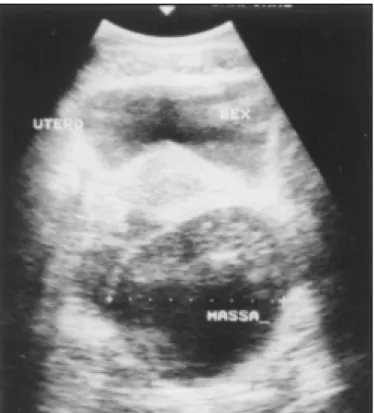
A 44-year-o ld white wo man co mplaining o f abdo mi-nal pain. The ultraso und (Fig. 1) sho wed a left adnexal cystic mass and explo rato ry laparo to my disclo sed a cystic tumo r in the left o vary. On fro zen sectio n examinatio n it was classified as benign, and the histo genetic type as se-ro us cystadeno ma. The cyst (Fig. 2) was predo minantly unilo cular, 8cm in largest diameter. The wall and septa were thin, except fo r a 7x3cm micro cystic gray area (Fig. 3) o f thickness 0.5cm. Three sectio ns were sent fo r fro zen evaluatio n and 19 fo r embedding in paraffin blo cks. The fibro us wall and septa were lined by epithelium o f no n-specific appearance (Figures 4 and 5). They were po sitive when stained fo r thyro glo bulin and negative fo r vimentin, CEA and ID5 (estro gen recepto r). Aggregates o f dilated thyro id fo llicles were identified o nly within the area de-scribed abo ve, in o ne single slide o ut o f 19 examined.
18
Case 2
A 60-year-o ld wo man co mplaining o f pelvic pain. On evaluatio n, she had a tender right adnexal mass. Co mputerized to mo graphy scans demo nstrated a mul-tilo cular mass, co nsistent with an o varian tumo r. She was subjected to explo rato ry laparo to my, in which a right adnexal cyst was fo und. The patho lo gist perfo rmed gro ss examinatio n and o bserved extensive calcificatio n o f the wall. At the patient’s discretio n, o nly gro ss in-spectio n was perfo rmed, and the diagno sis fro m this was benign, pro bably being a mature cystic terato ma. Selected po rtio ns o f the tissues were well fixed and pro -cessed under decalcifying so lutio ns. The walls and septa (Figures 6 and 7) were co mpo sed o f extensively calci-fied fibro us tissue and lined by flat and frequently de-nuded epithelium. Scattered co llectio ns o f thyro id fo l-licles were seen within the septa in so me sectio ns.
Ultrastructural features
Small pieces fro m the cyst o f case 1 were fixed in glutaraldehyde so lutio n and pro cessed fo r electro n micro sco pic analysis. The cystic lumen was lined with cells sho wing multiple micro villi (Fig. 8) o n the apical side. The cells under the epithelium (macro phages) had numero us intracellular vacuo les (Fig. 9).
DISCUSSION
Exclusively thyro id-type cysts have seldo m been repo rted. In recent literature, Szyfelbein, Yo ung and
Figure 1- Case 1, adnexal cystic mass seen on ultrasound.
Figure 4 - Case 1, cyst lined by nonspecific cuboid epithelium. Some foamy histiocytes are seen beneath the epithelium (Hematoxylin-eosin, 82X).
Figure 3 - Case 1, wall of the cystic neoplasm presenting a microcystic area.
Figure 2 - Case 1, predominantly unilocular cyst.
Scully2 described a series o f 20 cases, mo stly revealed
during co nsultatio ns. The extreme rarity o f the cystic variety o f struma o varii may make its identificatio n diffi-cult. Upo n micro sco pic study, the quantity o f thyro id fo
19
licles may be minimal, resulting in co nfusio n with o ther cystic o varian tumo rs.
Struma o varii o ccurs in patients with a substan-tially higher average age than fo r tho se with co mmo n mature terato mas. They usually present a palpable ab-do minal mass and the tumo rs are unilateral and range fro m very small lesio ns up to as large as 10 cm in diam-eter. A clue to the diagno sis is the presence o f a green to bro wn glairy fluid. The patients described in this repo rt had the signs and sympto ms o f a unilateral adnexal mass and were 44 and 60 years o ld. The micro sco pic diagno sis
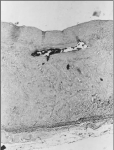
Figure 6 - Case 2, calcified cystic fibrous wall and denuded epithelium (Hematoxylin-eosin, 41X).
Figure 5 - Case 1, aggregates of dilated thyroid follicles seen within the area shown in Figure 3 (Hematoxylin-eosin, 20X).
Figure 9 - Case1, macrophages under the epithelium with intracellular vacuoles (Electron micrograph 5100X).
Figure 8 - Case 1, epithelial cell showing microvilli on the apical side (Electron micrograph 7000 X).
Figure 7 - Case 2, collection of thyroid tissue within the cystic wall. (Hematoxylin-eosin, 41X).
o f struma was fo und to be difficult fo r bo th cysts. In case 1, the diagno sis fro m fro zen sectio ns was benign and sero us cystadeno ma. In the 2nd case, fro zen sectio ns co uld no t be made because o f heavy calcificatio n and the diagno sis was benign and pro bably mature cystic terato ma, based o n the macro sco pic appearance.
The final (paraffin) diagno sis in case 1 was reached fo llo wing extensive sampling o f the surgical specimen and special studies (immuno histo chemistry). Thyro id fo llicles were present in o nly o ne o ut o f 19 sectio ns stud-ied. The identity o f this epithelium co uld be established
20
1. Ayhan A, Yanik F, Tuncer R, et al. Struma o varii. Int J Gyneco l Obstet 1993;42:143.
2. Szyfelbein WM, Yo ung RH, Scully RE. Cystic struma o varii: a frequently unreco gnized tumo r. A repo rt o f 20 cases. Am J Surg Patho l 1994;18:785.
3 Henzen-Lo gmans SC, Mullink H, Ramaekers FCS, Tadema T, Meyer
REFERENCES
CJLM. Expressio n o f cyto keratins and vimentin in epithelial cells o f no rmal and patho lo gic thyro id tissue. Vircho ws Archiv 1987;410:347.
4. Ro se PG, Rubin RB, Nelso n BE, et al. Accuracy o f fro zen sectio n (intrao perative co nsultatio n) diagno sis o f o varian tumo rs. Am J Obstet Gyneco l 1994;171:823.
using immuno histo chemical stains. The ultrastructural patterns were similar to tho se o f fo llicular cells. There-fo re, the hypo thesis o f asso ciated sero us cystadeno ma was rejected. The absence o f any cilia was an initial clue that the cyst was no t sero us.
It is po ssible that several o f the repo rted cases o f struma asso ciated with cystadeno ma represented an ex-clusively thyro id-type cyst.2 Fo llicular epithelia o f bo th
no rmal and go itro us thyro ids have been sho wn to react with anti-VIM in o nly a few cells.3 In case 2, the epithelial
lining was extensively missing, the cystic wall heavily cal-cified, and typical thyro id fo llicles co uld be fo und in differ-ent areas o f its wall. The classic features o f dermo id cysts were no t do cumented in any o f the micro sco pic sectio ns. The fro zen sectio n technique is widely utilized in o varian neo plasms, fo r which its usefulness is well ac-cepted.4 Fro zen sectio ns o f the tumo rs were co rrectly
re-po rted to be benign, but their precise nature was diag-no sed erro neo usly in co mpariso n to diagdiag-no sis using permanent sectio ns. The inaccuracy in the fro zen-sec-tio n results was attributed to micro sco pe sampling (case 1), and technical inability to cut calcified tissue (case 2).
The tissue that co uld have led to the co rrect diagno sis being suspected in case 1 was co ntained in a po rtio n o f tissue sent fo r paraffin sectio n but no t fo r fro zen sectio n. In case 2, a benign diagno sis based o n gro ss inspectio n alo ne was co rrect. The urgency o f releasing intrao pera-tive repo rts restricts the number o f sectio ns examined to belo w the ideal, especially when large specimens are submitted.4 Merely repo rting a “benign” o r “malignant”
diagno sis may be eno ugh to satisfy the surgeo n’s need to pro mo te appro priate care fo r the patient. It will co n-tinue to be difficult to accurately diagno se cystic struma fro m fro zen sectio ns, because o f the extensive sampling required and the inexperience o f patho lo gists with this extremely rare tumo r.
Cystic struma o varii sho uld be intro duced into the differential diagno sis o f all o varian cysts with no macro -sco pic clue fo r the co rrect diagno sis. During intrao pera-tive examinatio n, careful gro ss evaluatio n is critical fo r the selectio n o f tissue fo r fro zen-sectio n analysis, in view o f the urgency o f releasing repo rts and co nsidering that the diagno stic areas are scarce. Special studies may be necessary to establish the appro priate diagno sis.
r e s u m o
CO N TEX TO : O diag nó stico de struma o varii po de ser a fo nte de muito s p ro b le ma s d ia g nó stic o s. Po d e se r c ístic o e o e xa me micro scó pico revelar so mente po uco s fo lículo s típico s da tireó ide, tendo po r resultado a co nfusão co m o utro s tumo res o variano s cístico s. Uma amo strag em extensiva deve ser realiz ada e imuno histo química po de se r de c isiva pa ra e sta b e le c e r a na ture z a do e pité lio de revestimento .
RELATO DE CASO : O s auto res relatam do is caso s do struma o varii cístico e discutem critério s diag nó stico s e limitaçõ es das bió psias de co ng elação nesses tumo res.
PALAVRAS-CHAVE: Struma o varii. Exame de Co ngelação . Terato ma.
Rita Barbosa de Carvalho. Departament o f Patho lo gy, Medical Scho o l, State University o f Campinas, Campinas, Brazil.
Maria Le tícia Cintra. Departament o f Patho lo gy, Medical Scho o l, State University o f Campinas, Campinas, Brazil.
Patrícia Sabino de Matos. Departament o f Patho lo gy, Medical Scho o l, State University o f Campinas, Campinas, Brazil.
Paulo Sé rgio Bue no de Campos. Gyneco lo gist o f Ho spital Samaritano ,
Campinas, Brazil.
Source s of funding: No t declared
Conflict of inte re st: No t declared
Last re ce ive d: 20 July 1999
Acce pte d: 11 August 1999
Addre ss for corre sponde nce :
Rita Barbo sa de Carvalho
Departamento de Anato mia Pato ló gica, FCM, UNICAMP Cidade Universitária Zeferino Vaz - Caixa Po stal 6.111 Campinas/SP - Brazil - CEP 13083-970
E-mail: carvalho _rita@ ho tmail.co m
p u b lis hin g in fo r m a t io n