Article
J. Braz. Chem. Soc., Vol. 22, No. 8, 1595-1600, 2011.
Printed in Brazil - ©2011 Sociedade Brasileira de Química
0103 - 5053 $6.00+0.00
A
*e-mail: [email protected]
Quantum Coninement in PbI
2Nanodisks Prepared with Cucurbit[7]uril
Erick M. S. dos Santos, Lourivaldo S. Pereira and Grégoire J.-F. Demets*
DQ-FFCLRP, Departamento de Química, Universidade de São Paulo, 14040-901 Ribeirão Preto-SP, Brazil
Este trabalho apresenta uma rota alternativa para a preparação de nanodiscos de iodeto de chumbo (ca. 50-340 × 7Å, diâmetro × espessura) utilizando o macrocíclico cucurbit[7]urila como molde de síntese e agente estabilizante. Estas nanopartículas apresentam um deslocamento para o azul de gap óptico consistente com seu tamanho reduzido e coninamento quântico 1D. Suas
espessuras são compatíveis com a de uma camada de iodeto de chumbo esfoliado, indicando que cucurbit[7]urila impede a formação de estruturas lamelares e limita o crescimento das nanopartículas. A estrutura, a morfologia e as propriedades destes discos foram veriicadas por difratometria de raios X em pó (XRD), espectroscopia no UV-Visível, microscopia de força atômica (AFM), microscopia eletrônica de varredura com análise de luorescência de raios X por dispersão de energia (SEM-EDS) e microscopia eletrônica de transmissão de alta resolução (HRTEM).
This work presents an alternative route for the preparation of heavy metal iodide nanoparticles, particularly lead iodide nanodisks (ca. 50-340 × 7 Å, diameter × thickness), using the macrocycle
cucurbit[7]uril as a synthetic template and stabilizing agent. These nanoparticles exhibit an optical-gap blue shift consistent with their small size and 1D quantum coninement. Their thicknesses are compatible with an exfoliated single layer of lead iodide, indicating that cucurbit[7]uril, preventing the stacking and formation of tactoids, thus limiting nanoparticles growth in the z direction. The structure, morphology and properties of these disks were analyzed by X-ray powder diffractometry (XRD), UV-Visible spectroscopy, atomic force microscopy (AFM), scanning electron microscopy with analysis of energy dispersive X-ray luorescence (SEM-EDS) and high resolution transmission electron microscopy (HRTEM).
Keywords: lead iodide, cucurbit[7]uril, synthetic template, nanodisks, quantum coninement
Introduction
Lead iodide is a lamellar solid consisting of layers of metal and iodide ions united by covalent bonds. Weak van der Waals interactions hold these layers together in a three dimensional sandwich-like semiconductor. This structure can be described as a hexagonal close-packed array of iodide anions with alternate layers of octahedral interstices occupied by lead(II) cations. Therefore, each layer can be described as a I-Pb-I sequence layer.1 Very
interesting materials can be obtained by diminishing the PbI2 particle size to the nanoscale in order to favor the appearance of new properties resultant of quantum coninement, such as those observed in zero-dimensional solids, quantum dots and other N-dimensional, quantum-conined nanomaterials. Quantum dots encounter many applications in several technological ields such as sensor
devices and anti-counterfeiting systems, illumination (as high eficiency white-light emitting diodes), medicine (as cellular markers), lasers, solar cells and others.2-5 Another
important property of lead iodide is its ability to form intercalation compounds with many chemical species, such as hydrazine, ammonia, aniline, butylamine, pyperidine and quinoline, among others.1 The insertion of guest molecules
in the interlamellar gap considerably affects the chemical and physical properties of the material.
Cucurbiturils (CB[n]) are pumpkin-shaped thoroidal molecules with two distal portals separated by 9.1 Å. They are produced by condensation of glycoluril and formaldehyde in acidic medium and were synthesized for the irst time by Behrend et al. in 1905.6 However,
their structure remained uncertain until the findings of Freeman et al., 75 years later.7 Since then, many
The general reaction based on the condensation of glycoluril derivatives and formaldehyde leads to several homologues containing mainly 5, 6, 7 and 8 glycolurilic moieties (CB[5], CB[6], CB[7] and CB[8], respectively). Among these, CB[7] is the only homologue with considerable solubility in water (ca. 30 mmol L-1).12 The
portals are composed by carbonyl groups and behave as Pearson’s hard bases. Obviously, the portal sizes vary with the number of monomeric units, ranging from 2.4 to 6.9 Å in the CB[5]-CB[8] series.8,12,13 Cucurbituril cavities are
hydrophobic and can accommodate many guest molecules in a series of inclusion compounds, just like cyclodextrins and calixarenes (see molecular structure in Figure 1).11
Corma et al.14 have recently synthetized gold
nanoparticles using CB cavities as templates, obtaining almost monodisperse particle populations, a very important aspect when one aims at absolute size control. The size control in this level is even more important during the production of quantum dots, since their physical and physicochemical properties are strongly size- and shape-dependent. According to Pearson’s HSAB concept,15 Pb2+
ions should exhibit low afinity for cucurbiturils. However, other scientists have reported high formation constants for Pb2+ and decamethylcucurbit[5]uril complexes, which is
a slightly different, smaller homologue.16 There are very
few works in the literature reporting on CB[n]-containing nanostructured materials, but few others encompassing gold, palladium and cadmium sulide can be cited.17-19
In this context, we have studied the formation of colloidal lead iodide usingCB[7] acting as an external template, generating lead iodide particles with extremely reduced dimensions. We have observed the formation of randomly stacked discoid nanostructures with 1-D quantum coninement instead of small clusters.
Experimental
Cucurbit[7]uril (CB[7]) was prepared and puriied as described in the literature.20 Lead iodide/CB[7]
nanoparticles were prepared using Pb(NO3)2 0.1 mol L-1
(Synth), KI (Synth) and cucurbit[7]uril 1 × 10⁻³ mol L-1.
The cucurbituril solution (10 mL) was kept under vigorous stirring at 27 oC and received the addition of 100 µL of the
Pb(NO3)2 solution, followed by 200 µL of the KI solution.
The molar ratio between the reactants CB[7]/Pb2+/I−
was 1:1:2, respectively. The resulting mixture immediately formed a pale greenish-yellow colloid with gel-like consistence in a reproducible way, quite different from the large golden yellow crystals obtained when no CB[7] is present in the solution. This colloidal suspension was unstable and precipitated after two days. The resulting solid had the same greenish-yellow color and was washed three times with water (in order to remove the excess of KNO3)
and dried under vacuum. For comparison purposes, reference colloidal PbI2 (2H-polytype) was synthesized exactly in the
same way, except for the addition of the macrocycle. The powder X-ray diffraction (XRD) measurements were carried out on a Siemens 5005 equipment using Cu Kα radiation (λ = 1.54 Å). The diffuse relectance spectra were measured with an Ocean optics USB 4000 spectrometer at 77 K (the absorption intensities were not corrected for the powder size, which is relevant for using the Kubelka-Munk function). The luminescence experiments were registered at 77 K using a Fluorog SPEX F2121 spectrometer equipped with a 450 W Xe lamp, a cooled Hamamatsu R918 photomultiplier and double excitation and emission monochromators. The atomic force microscopy images were collected on a Shimadzu SPM-9600 scanning probe microscope (contact mode). The scanning electron microscopy and energy-dispersive X-ray spectroscopy (EDS) measurements were carried out on a Zeiss EVO 50 microscope. The samples were covered with gold by sputtering. High resolution transmission electron microscopy (HRTEM) images were obtained with a TEM-JEM 2100 ARP electron microscope. TEM sample grids were prepared by placing 1 µL of the particle suspension on a carbon-coated copper grid (300 meshes) and evaporating the solvent at room temperature.
Results and Discussion
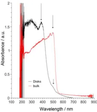
Absorption spectra of both bulk and CB-assisted PbI2
display typical semiconductor absorption proile, except for their typical absorption edge at ca. 3 eV, which is
blue-shifted by 0.5 eV (ca. 90 nm) for the hybrid material
semi-conducting materials lead to quantum coninement of excitons in one, two or three dimensions, depending on the shape and size of the particles.21-29 In fact, the size
affects the band-gap energy when the dimension becomes inferior to the bulk-exciton Bohr radius (ca. 1.9 nm for
PbI2),21,22 although literature reveals that PbI2 absorption
edge shifts can be assigned to many other factors, from intercalation of electron-donor species between the iodide layers in few atoms clusters to ripening processes, and in low-polarity solvents.1,25 In fact, all these phenomena are
interconnected and the shift is always related to quantum coninement mechanisms. Normally, low-polarity solvents tend to stabilize larger colloidal particles in detriment of the smaller ones as shown by Sandroff et al.,24 but also change
the dielectric constant of the medium. Small molecules can be easily intercalated into PbI2, once the layers are weakly
held together, essentially by van der Waals interactions. The insertion of guest species between the layers has the same effect of that produced by negative pressure on this kind of lamellar material. In other words, the intercalation increases the interlamellar distances, lowering the electronic interaction between layers and the extent of the dispersion in the valence band. This increases considerably the energy, promoting 1D quantum coninement in the axial direction.25
Using the effective mass approximation model, it is possible to estimate the crystallite size and thickness, as did Sandroff et al.24 and others.21,22 Thus, in such anisotropic
nanomaterial, the equation governing the band-gap shifts has the form:
(1)
where the effective reduced masses of the electron-hole pairs in the xy plane and in the axis perpendicular to them
are represented by µxy and µz, respectively, and Lxy and Lz
are the dimensions of the crystallites. The reduced masses have been experimentally determined as 0.32 (µxy) and
1.4 (µz) electron mass units, according to magneto-optic
experiment data.24,30 Taking into account the absorption
shift, it is reasonable to consider that almost no electronic interaction exists between the layers of PbI2 in our case,
meaning that the thickness of the nanoparticles should not be larger than a single layer of PbI2. When considering a
single layer of the material (7 Å), equation 1 becomes:
(2)
In our case, the shift observed is 3.10 − 2.56 eV = 0.54 eV. According to equation 2, this value corresponds to the shift caused by the exfoliation of the structure, independently of the size of layers, which are too large to produce any spectral shift (the irst term of such equation tends to 0 when Lz = 7 Å). Thus, the spectral shift observed for the
CB[7]/PbI2 sample is consistent with nanolakes constituting
of PbI2 monolayers stabilized by the macrocycle. No
luorescence at all could be measured for the hybrid solid CB[7]/PbI2, indicating that the electron-hole pairs in these
crystals are captured by surface traps and quenched by non-radiatively recombination processes. This is a quite common phenomenon in quantum-conined nanocrystals.26,31,32
The X-ray diffraction patterns of the pure PbI2 and
CB[7]/PbI2 sample are compared in Figure 3. Pure PbI2
exhibits very sharp and intense peaks characteristic of crystalline materials, while CB[7]/PbI2 reveals an
amorphous structure, with a total loss of structural
Figure 2. Absorption spectra (converted from diffuse relectance data) of bulk PbI2 and the CB[7]/PbI2 sample at 77 K.
coherence. In fact, the absence of the 001 peak for the hybrid structure suggests the absence of a regular ordering of the PbI2 layers and it is expected for more or less
randomly-oriented isolated nanolakes consisting of iodide monolayers. According to this, it is possible to conclude that no continuous turbostratic structure can be present, because the size of the clusters is too small for this. According to Sandroff et al.,24 the interlayer distance of lead iodide can
vary depending on the synthetic procedure, but it is never inferior to 12 Å, while the thickness of a single layer is 7 Å. The height of CB[7] molecule is 9.1 Å and should provoke an expansion of the basal distance of this magnitude if it is intercalated between the layers, acting as a pillar in the structure. An important factor that must be considered is sterical hindrance: cucurbit[7]uril is a very large molecule (ca. 9 Å) and it is geometrically impossible to keep a 1:1
Pb2+/CB[7] ratio inside a lamellar structure. Therefore, part
of the macrocycle molecules would be pushed out from the structure or the iodide would be forced to adopt a crystalline coniguration different from the lamellar one. Furthermore, K+ ions are present in large amounts in the reaction medium
and it is certain that they bind the macrocycle portals, forming large cationic species in solution. These positively charged species may interact with iodine ions by electrostatic interactions, but it was veriied that electron-donor species are better guests for intercalation in lead iodide. In other words, nothing favors the formation of lamellar structures in this case.33-38 A few and very weak signals may be observed
in the diffraction pattern of the hybrid material. Even using the best acquisition methods, it is impossible to state, with our equipment, that these signals are peaks and not noise, since they are not reproducible. Furthermore, the signal does not correspond to any known PbI2 polytype.
Medium resolution SEM micrographs (× 2,000 magniication) reveal overlaid sheets of PbI2, just as in the
case of re-crystallized matrices.33 A SEM image obtained
with high magniication (× 200,000) shows that these sheets are made of small disks measuring around 50 nm (Figure 4). EDS spectra display strong lead, iodine, carbon, oxygen and potassium signals arising from the sample (data not shown). The detected potassium is probably reminiscent from the KI starting material. This fact is easily understood once, as already mentioned, K+ ions form very stable coordination
compounds with cucurbiturils.39
The presence of the nanodisks was conirmed by AFM imaging, making clear that the sheets are composed by aggregates of regular disk-like particles. The average size was estimated to be around 34 nm (Figure 5). HRTEM images show these disks as single sheets of lead iodide that are totally exfoliated, forming a “house of cards” structure (Figure 6). This kind of structure is frequently observed in
exfoliated clay gels, for instance Laponite®. In this synthetic
smectite, the gel is formed by weak interactions between 1 × 25 nm disks.40
Conclusions
CB[7] does not act as a hollow shell template for lead iodide formation as it does in the case of metal nanoparticles. The present work indicates that the macrocycle forms a random structure composed of small PbI2 disks separated
by CB[7] and K+ ions. The hybrid material presents reduced
interlayer coupling terms, which affect its absorption spectrum when compared to pure 2H-PbI2. The mechanism
involved in the formation of this nanomaterial is complex, since it starts from an unstable colloid, which then sediments. A possible mechanism could be proposed with three major steps: the irst is the formation of K2CB[7]2+ Figure 4. SEM image of the CB[7]/PbI2 sample with aggregates under
× 200,000 magniication. The grain size estimated in 54 nm.
Figure 5. AFM micrograph of the CB[7]/PbI2 sample with CB[7]/PbI2
cations which interact with iodine ions in solution. Then, lead ions bind the halogens forming nucleation centers for lead iodide single layers. As the single layers grow, CB[7] becomes too bulky and is gradually pushed to their borders, limiting nanoparticle growth. Another possibility is the formation of adducts with water molecules and Pb2+
ions instead of potassium complexes during the nucleation process, because this kind of structure has already been observed in many cases and with several metal ions. The nanodisk formation mechanism is still being investigated with other metal halides and will be the subject of another paper.
Supplementary Information
Additional information about the exciton band of lead iodide, including the absorption spectra of solid PbI2 and
PbI2 nanodisks (Figure S1), is available free of charge at
http://jbcs.sbq.org.br as PDF ile.
Acknowledgements
We sincerely thank Dr. Ivana Aparecida Borin and Dr. Rodrigo Ferreira Silva for microscopy images, as well as Prof. Koiti Araki, Prof. Antonio Osvaldo Serra and Prof. Paulo César da Souza Filho for fluorescence measurements. This work was sponsored by the Conselho Nacional de Desenvolvimento Cientíico e Tecnológico (CNPq), Coordenação de Aperfeiçoamento de Pessoal de Nível Superior (CAPES) and Fundação de Amparo à Pesquisa do Estado de São Paulo (FAPESP). HRTEM images were obtained at Laboratório Nacional de Luz Síncrotron (LNLS, Campinas-SP, Brazil)(TEM-MSC-9054).
References
1. Coleman, C. C.; Goldwhite, H.; Tikkanen, W.; Chem. Mater. 1998, 10, 2794.
2. Michalet, X.; Pinaud, F. F.; Bentolila, L. A.; Tsay, J. M.; Doose, S.; Li, J. J.; Sundaresan, G.; Wu, A. M.; Gambhir, S. S.; Weiss, S.; Science 2005, 307, 538.
3 Xu, T.; Nikiforov, A. Y.; France, R.; Thomidis, C.; Williams, A.; Moustakas, T. D.; Phys. Status Solidi A 2007, 204, 2098. 4. Tang, J.; Sargent, E. H.; Adv. Mater. 2010, 20, 1.
5. Hendrick, E.; Frey, M.; Herz, E.; Wiesner, U.; J. Eng. Fibers Fabr.2010, 5, 21.
6. Behrend, R.; Meyer, E.; Rusche, F.; Liebigs J.; Ann. Chem.
1905, 339, 1.
7. Freeman, W. A.; Mock, W. L.; Shih, N.-Y.; J. Am. Chem. Soc. 1981, 103, 7367.
8. Day, A.; Arnold, A. P.; Blanch, R. J.; Snushall, B.; J. Org. Chem.
2001, 66, 8094.
9. Day, A. I.; Blanch, R. J.; Arnold, A. P.; Lorenzo, S.; Lewis, G. R.; Dance, I.; Angew. Chem., Int. Ed. 2001, 41, 275
10. Kim, J.; Jung, I.-S.; Kim, S.-Y.; Lee, E.; Kang, J.-K.; Sakamoto, S.; Yamaguchi, K.; Kim, K.; J. Am. Chem. Soc. 2000, 122, 540.
11. Liu, S.; Ruspic, C.; Mukhopadhyay, P.; Chakrabarti, S.; Zavalij, P. Y.; Isaacs, L.; J. Am. Chem. Soc. 2005, 127, 15959.
12. Lagona, J.; Mukhopadhyay, P.; Chakrabarti, S.; Isaacs, L.;
Angew. Chem., Int. Ed. 2005, 44,4844.
13. Demets, G. J.-F.; Quim. Nova 2007, 30, 1313.
14. Corma, A.; Garca, H.; Montes-Navajas, P.; Primo, A.; Calvino, J. J.; Trasobares, S.; Chem. Eur. J. 2007, 13, 6359.
15. Pearson, G. R.; Inorg. Chem. 1988, 27, 734.
16. Zhang, X. X.; Krakowiak, K. E.; Xue, G.; Bradshaw, J. S.; Izatt, R. M.; Ind. Eng. Chem. Res. 2000, 39, 3516.
17. Lee, T.-C.; Scherman, O. A.; Chem. Commun. 2010, 46, 2438. 18. Li, M.; Zaman, M. B.; Bardelang, D.; Wu, X.; Wang, D.; Margeson, J. C.; Leek, D. M.; Ripmeester, J. A.; Ratclife, C. I.; Lin, Q.; Yange, B.; Yu, K.; Chem. Commun. 2009, 44, 6807. Figure 6. HRTEM micrographs of (I) the CB[7]/PbI2 sample; (II) a single
PbI2 nanodisk (diameter ca. 30 nm), inset: electron diffraction pattern;
19. Cao, M.; Lin, J.; Yang, H.; Cao, R.; Chem. Commun. 2010, 46, 5088.
20. Day, A. I.; Arnold, A. P.; Blanch, R. J.; Snushall, B.; J. Org. Chem. 2001, 66, 8094.
21. Mallik, K.; Dhami, T. S.; Phys. Rev. B: Condens. Matter Mater. Phys. 1998, 58, 13055.
22. Finlayson, C. E.; Sazio, P. J. A.; J. Phys. D: Appl. Phys. 2006,
39, 1477.
23. Nozue, Y.; Tang, Z. K.; Goto, T.; Solid State Commun. 1990,
73, 531.
24. Sandroff, C. J.; Kelty, S. P.; Hwang, D. M.; J. Chem. Phys. 1986, 85, 5337.
25. Ghorayeb, A. M.; Coleman, C. C.; Yoffe, A. D.; J. Phys. C: Solid State Phys. 1984, 17, L715.
26. Artemyev, M. V.; Rakovich, Y. P.; Yablonski, G. P.; J. Cryst. Growth 1997, 171, 447.
27. Barnakov, Y. U.; Ito, S.; Dmitruk, I.; Tsunekawa, S.; Kasuya, A.; Scr. Mater. 2001, 45, 273.
28. Micic, O. I.; Zongguan, L.; Mills, G.; Sullivan, J. C.; Meisel, D.; J. Phys. Chem. 1987, 91, 6221.
29. Tubbs, M. R.; Forty, A. J.; J. Phys. Chem. Solids 1965, 26, 711. 30. Sandroff, C. J.; Hwang, D. M.; Chung, W. M.; Phys. Rev. B:
Condens. Matter Mater. Phys. 1986, 33, 5953. 31. Dag, I.; Lifshitz, E.; J. Phys. Chem. 1996, 100, 8962.
32. Sengupta, A.; Jiang, B.; Mandal, K. C.; Zhang, J. Z.; J. Phys. Chem. B 1999, 103, 3128.
33. Schaefer, R. W.; Ardelean, M.; Powder Diffr. 2001, 16, 16. 34. Agrawal, V. K.; Chadha, G. K.; Trigunayat, G. C.; Acta Cryst.
1970, A26, 140.
35. Chand, M.; Trigunayat, G. C.; Acta Cryst. 1975, B31, 1222.
36. Flahaut, E.; Sloan, J.; Friedrichs, S.; Kirkland, A. I.; Coleman, K. S.; Williams, V. C.; Hanson, N; Hutchison, J. L.; Green, M. L. H.; Chem. Mater. 2006, 18, 2059.
37. Kasi, G. K.; Dollahon, N. R.; Ahmadi, T. S.; J. Phys. D: Appl. Phys. 2007, D40, 1778.
38. Koutselas, I.; Dimos, K.; Bourlinos, A.; Gournis, D.; Avgeropoulos, A.; Agathopoulos, S.; Karakassides, M. A.;
J. Optoelectron. Adv. Mater. 2008, 10, 311.
39. Buschmann, H. J.; Cleve, E.; Schollmeyer, E.; Inorg. Chim. Acta 1992, 193, 93.
40. Kroon, M.; Vos, W. L.; Wegdam, G. H.; Int. J. Thermophys.
1998, 19, 887.
Submitted: August 21, 2010 Published online: May 24, 2011
Supplementary Information
S
I
J. Braz. Chem. Soc., Vol. 22, No. 8, S1, 2011. Printed in Brazil - ©2011 Sociedade Brasileira de Química 0103 - 5053 $6.00+0.00
*e-mail: [email protected]
Quantum Coninement in PbI
2Nanodisks Prepared with Cucurbit[7]uril
Erick M. S. dos Santos, Lourivaldo S. Pereira and Grégoire J.-F. Demets*
DQ-FFCLRP, Departamento de Química, Universidade de São Paulo, 14040-901 Ribeirão Preto-SP, Brazil
On lead iodide exciton band
The exciton band of lead iodide does not appear always as a sharp and intense peak. Its shape and intensity may vary with crystal form, polytype, temperature, light polarization, synthetic procedure, crystallite size and especially with the purity of the material.1 Many authors have reported similar spectra, for which the absorption edge is associated with the excitonic level, separated from gap transitions by few meV, since the binding energy is very small. The presence of electron donors in solution also affects exciton absorption bands, as demonstrated in other papers.2-5 It is well described in the literature that the irst excitonic band may undergo blue shifts when extremely small particles are obtained.1,6-8 In the present work, a set of optical ibers and a light source essentially designed for Vis-NIR region were used in the irst attempts to obtain absorption spectra. This could be the reason for the lack of spectral resolution. Furthermore, the Kubelka-Munk function could not be used since no information about the powder size of the samples after drying the colloid was known. The UV-Vis absorption measurements were repeated at room temperature (Figure S1), using another set of optical ibers designed for working in the UV region. This time the bands clearly appear. Using the Tauc equation, the Eg value was calculated, being 2.34 and 2.80 eV (diff = 0.46 eV) for bulk PbI2 and the nanodisks, respectively.
For pure lead iodide, the exciton band is located at the absorption edge (499 nm), which is coherent with the literature (490-500 nm). For the nanodisks, it is blue-shifted to 387 nm and a small shoulder is observable at 499 nm, which could be assigned to larger aggregates or to hetero-nuclear excitons.3 As mentioned before, these new values were obtained at room temperature, but the onset absorption energies do not vary considerably from our previous measurements at 77 K (see Figure 2 in the main text), and would not affect the presented conclusions from the effective mass model. Probably, the problem is related to our experimental apparatus.
References
1. Tubbs, M. R.; Forty, A. J.; J. Phys. Chem. Sol. 1965, 26, 711. (29 in the Article text).
2. Sengupta, A.; Jiang, B.; Mandal, K. C.; Zhang, J. Z.; J. Phys. Chem. B 1999, 103, 3128. (32 in the Article text).
3. Bhavsar, D. S.; Saraf, K. B.; J. Mater. Sci.: Mater Electron.
2003, 14, 195.
4. Koutselas, I.; Dimos, K.; Bourlinos, A.; Gournis, D.; Avgeropoulos, A.; Agathopoulos, S.; Karakassides, M. A.;
J. Optoelectron. Adv. Mat. 2008, 10, 311.
5. Nozue, Y.; Tang, Z. K.; Goto, T.; Solid State Commun. 1990,
73, 531. (23 in the Article text).
6. Kasi, G. K.; Dollahon, N. R.; Ahmadi, T. S.; J. Phys. D: Appl. Phys. 2007, 40, 1778. (37 in the Article text).
7. Artemyev, M. V.; Rakovich, Y. P.; Yablonski, G. P.; J. Cryst. Growth 1997, 171, 447. (26 in the Article text).
8. Micic, O. I.; Li, Z.; Mills, G.; Sullivan, J. C.; Meisel, D.;
J. Phys. Chem. 1987, 91, 6221.
![Figure 1. Representation of the chemical structure of cucurbit[7]uril.](https://thumb-eu.123doks.com/thumbv2/123dok_br/18995341.462020/2.892.71.411.387.552/figure-representation-chemical-structure-cucurbit-uril.webp)
![Figure 3. XRD patterns of pure PbI 2 (a) and of the CB[7]/PbI 2 sample (b).](https://thumb-eu.123doks.com/thumbv2/123dok_br/18995341.462020/3.892.486.819.753.1073/figure-xrd-patterns-pure-pbi-cb-pbi-sample.webp)
![Figure 4. SEM image of the CB[7]/PbI 2 sample with aggregates under](https://thumb-eu.123doks.com/thumbv2/123dok_br/18995341.462020/4.892.455.796.121.386/figure-sem-image-cb-pbi-sample-aggregates.webp)
![Figure 6. HRTEM micrographs of (I) the CB[7]/PbI 2 sample; (II) a single PbI 2 nanodisk (diameter ca](https://thumb-eu.123doks.com/thumbv2/123dok_br/18995341.462020/5.892.94.440.116.629/figure-hrtem-micrographs-pbi-sample-single-nanodisk-diameter.webp)