Letters to the Editor
Radiol Bras. 2016 Nov/Dez;49(6):406–413
406
0100-3984 © Colégio Brasileiro de Radiologia e Diagnóstico por Imagem
Letters to the Editor
Female urethral diverticulum containing a urothelial carcinoma
Dear Editor,
We report the case of a 63-year-old black female who pre-sented with complaints of difficulty in urinating and pollakiuria. She also reported an eight-month history of episodes of dysuria and hematuria. She had smoked for 10 years and had quit 30 years prior. She had three pregnancies, all with vaginal delivery, and had undergone total hysterectomy 17 years prior.
Magnetic resonance imaging (MRI) of the pelvis revealed, below the urinary bladder, a cystic formation involving the ure-thra, consistent with urethral diverticulum (UD), within which there was a solid component showing paramagnetic contrast en-hancement, suggesting an expansive process. Communication with the urethra was well defined after a urethral catheter had been inserted. The diverticulum was surgically resected. On the basis of histological and immunohistochemical studies of the surgical sample, the patient was diagnosed with papillary urothelial diver-ticular carcinoma.
The reported prevalence of UD is 0.6–6.0%, and the condi-tion is most common in women between 30 and 60 years of age(1– 8). Some studies have reported that the incidence of UD is higher
in black individuals(3,4,7). Typically, UD is underdiagnosed because, in most cases, the clinical profile is nonspecific(2,4–6) and up to 20% of patients are asymptomatic(4,8). The site most often affected is the middle third of the urethra, where the paraurethral glands (Skene’s glands) are typically located, and 96% of diverticular ori-fices are posterolateral(1–5,8). Most patients with UD have the ac-quired form, which probably arises from dilation/abscess in parau-rethral glands. Other causes include trauma and surgery(1–5,8). Typically measuring 0.2–1.6 cm(5), UDs can be single or mul-tiple, simple or multiloculated, and locally restricted or surround-ing the urethra (in a “horseshoe” shape), with one or more (nar-row or broad) orifices(1,2). Differential diagnoses include cervical
cysts, vaginal cysts, abscesses, tumors, urethral endometriosis, and ectopic ureterocele(2,4,5).
Clinical findings include the classic triad of dysuria (in 30– 70% of cases), dyspareunia (in 10–25%) and postmicturition dribble (in 10–30%), as well as pollakiuria or urinary urgency (in 40–100%), urinary incontinence (in 32–60%), recurrent urinary tract infections (in 30–50%), hematuria (10–25%), and bulging in the anterior vaginal wall (in 35%), accompanied by purulent urethral discharge on palpation (in 12%)(1–8). The chronic inflam-mation and urinary stasis seen in UDs result in complications(3,4), including calculi (in 1.5–10%) and malignant tumors(1–8). As for the tumors, UDs are responsible for less than 5% of all urethral neoplasms(4,8), fewer than 200 cases having been reported(1,2,7,8). Malignancies in UD include adenocarcinoma, in 49–61% of cases, transitional cell carcinoma, in 27–30%, and squamous cell carcinoma, in 10–12%(1–5).
Diagnostic imaging methods include the following: voiding cystourethrography—a technically simple method, with an accu-racy of 85%, that demonstrates the diverticulum through con-trast and identification of filling gaps suggestive of calculi or tu-mors, although it uses an iodized agent might not detect UDs with small orifices(1,4,5); double-balloon urethrography—a method with an accuracy of 90%, showing findings similar to those cystourethrography, with the disadvantage of being invasive and complex(1,4,5); ultrasound—a method with excellent accuracy (near 100%) when intraurethral (highly invasive) or translabial/ transperineal (less invasive) that characterizes the cystic forma-tions and any vascularized solid content, although examiner-dependent and limited in the evaluation of collapsed UDs(1,4,5); computed tomography—a method that is useful in identifying cal-culi and tumors (evident solid components), albeit with low sensi-tivity for small UDs(1,4,5); and MRI—the method of choice, with near 100% sensitivity, which is noninvasive, with excellent con-trast between tissues and discrimination of the complexity of the
Letters to the Editor
Radiol Bras. 2016 Nov/Dez;49(6):406–413
407
http://dx.doi.org/10.1590/0100-3984.2015.0115
Rodolfo Mendes Queiroz1, Paula Puty e Costa1, Nara Yamada
Fabril de Oliveira1, Juliana Alves Paron1, Eduardo Miguel
Febronio1
1. Documenta – Hospital São Francisco, Ribeirão Preto, SP, Brazil. Mailing address: Dr. Rodolfo Mendes Queiroz. Documenta – Centro Avançado de Diagnóstico por Imagem. Rua Bernardino de Campos, 980, Centro. Ribeirão Preto, SP, Brazil, 14015-130. E-mail: [email protected]. structures, capable of detecting small UDs and identifying
neo-plasms(1,2,4–6,8). In T-2 weighted MRI sequences, UDs show hyperintense signals, although they can be hypointense if they have thick content(1,2,4,6). Solid tumor components present as vegetative lesions with intermediate signals on T1- and T2-weighted sequences, potentially restricting the diffusion, and show significant enhancement after intravenous administration of con-trast(1,2).
REFERENCES
1. Chou CP, Levenson RB, Elsayes KM, et al. Imaging of female urethral diverticulum: an update. Radiographics. 2008;28:1917–30.
2. Chaudhari VV, Patel MK, Douek M, et al. MR imaging and US of female urethral and periurethral disease. Radiographics. 2010;30:1857–74. 3. Tines SC, Bigongiari LR, Weigel JW. Carcinoma in diverticulum of the
female urethra. AJR Am J Roentgenol. 1982;138:582–5.
4. Khati NJ, Javitt MC, Schwartz AM, et al. MR imaging diagnosis of a urethral diverticulum. Radiographics. 1998;18:517–22.
5. Hosseinzadeh K, Furlan A, Torabi M. Pre- and postoperative evaluation of urethral diverticulum. AJR Am J Roentgenol. 2008;190:165–72. 6. Dwarkasing RS, Dinkelaar W, Hop WCJ, et al. MRI evaluation of
ure-thral diverticula and differential diagnosis in symptomatic women. AJR Am J Roentgenol. 2011;197:676–82.
7. Ahmed K, Dasgupta R, Vats A, et al. Urethral diverticular carcinoma: an overview of current trends in diagnosis and management. Int Urol Nephrol. 2010;42:331–41.
8. Grimsby GM, Wolter CE. Signet ring adenocarcinoma of a urethral di-verticulum. J Surg Case Rep. 2011;2011:2.
Chronic kernicterus: magnetic resonance imaging findings
Dear Editor,
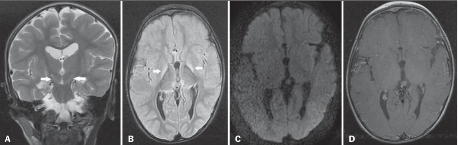
A 3-year-old male child who had developed bilirubin encepha-lopathy in the neonatal period, due to Rh incompatibility, presented with delayed neuromotor/psychomotor development and involun-tary movements. The prenatal and perinatal periods had been free of complications. Serology for cytomegalovirus, toxoplasmosis, and HIV were negative, as was the VDRL test. The results of a com-plete blood count, serum ceruloplasmin, electrolytes, and thyroid function were all within the limits of normality. Magnetic reso-nance imaging (MRI) of the brain showed bilateral, symmetrical hyperintense signals on FLAIR and T2-weighted sequences, af-fecting the globus pallidus and subthalamic nuclei, with no mass effect, with no diffusion restriction or evidence of gadolinium enhancement (Figure 1). Those imaging findings, together with the clinical and biochemical history, confirmed the suspected di-agnosis of chronic kernicterus.
Recent studies conducted in Brazil have highlighted the im-portance of MRI studies to improving the diagnosis of central ner-vous system disorders(1–5). Kernicterus, also known as bilirubin encephalopathy, is a rare complication of hyperbilirubinemia in childhood, occurring when serum bilirubin levels in the neonate are in excess of 20 mg/dL at term or even lower values in
prema-ture infants, which result in bilirubin deposition in the globus pallidus, subthalamic nuclei, hippocampus, putamen, thalamus, and cranial nerves, primarily the third, fourth, and sixth cranial nerves(6). Symptoms include drowsiness, hypotonia, opisthotonus, rigidity, and seizures. The factors involved in its pathogenesis are hyperbilirubinemia, reduced serum bilirubin binding capacity, changes in the permeability of blood-brain barrier, and neurotox-icity. Although the main causes of kernicterus are ABO and Rh mismatches, it can also be caused by sepsis and other types of hemolytic anemia such as glucose-6-phosphate dehydrogenase deficiency(7). The clinical symptoms and signs can regress com-pletely if properly treated with phototherapy and blood transfu-sions(6); without treatment, permanent damage can occur, gen-erating encephalopathy with symptoms related to the basal nu-clei, including involuntary movements, asymmetric spasticity, ri-gidity, ataxia, and hearing loss(8).
The MRI findings in kernicterus are characterized by a hyperintense signal on T1-weighted sequences in the globus pallidus, progressing chronically to a shift from a hyperintense signal on T1-weighted sequences to a bilateral, symmetrical hyperintense signal on T2-weighted and FLAIR sequences in the globus pallidus and subthalamic nuclei(7,9–11), corresponding to the areas of preferential deposition of unconjugated bilirubin, characterizing chronic kernicterus, as in the case presented here.
Figure 1. A: Coronal T2-weighted MRI sequence showing a bilateral, symmetrical hyperintense signal in the subthalamic nuclei (arrows), without a mass effect. B:
Axial FLAIR MRI sequence showing a bilateral, symmetrical hyperintense signal in the globus pallidus (arrows). C: Axial diffusion-weighted MRI sequence showing no diffusion restriction. D: Axial T1-weighted MRI sequence showing no evidence of gadolinium enhancement.