118
Mata BELF et al. Infantile myofibromatosis: report of a rare disease
Radiol Bras. 2012 Mar/Abr;45(2):118–120
Infantile myofibromatosis: report of a rare disease
*
Miofibromatose infantil: relato de uma rara doença
Bruna Emmanuelle Linhares Fonseca Mata1, Juliana Santos Bayerl de Oliveira1, Damião Ranulfo
Fernandes Soares2
Infantile myofibromatosis is a rare disease with various presentations. Usually, such condition manifests itself as subcutaneous nodules, either in association or not with visceral nodules. The diagnosis should be achieved by means of physical examination and imaging studies, with emphasis on the lesions pattern to allow the staging and classification of the disease. The treatment is still controversial.
Keywords: Myofibromatosis.
A miofibromatose infantil é uma rara doença que tem várias formas de apresentação. Habitualmente, manifesta-se com nódulos subcutâneos, que podem ou não estar associados à presença de nódulos viscerais. Deve-se estar apto a fazer o diagnóstico por meio do exame físico e de imagem, que evidenciarão o padrão das lesões para estadiar/ classificar a doença. O tratamento ainda é controverso.
Unitermos: Miofibromatose.
Abstract
Resumo
* Study developed at Hospital Infantil Nossa Senhora da Gló-ria and Hospital Universitário Cassiano Antônio de Moraes, Vitó-ria, ES, Brazil.
1. MDs, Residents in Radiology and Imaging Diagnosis, Uni-versidade Federal do Espírito Santo (UFES), Vitória, ES, Brazil. 2. Radiologist, Physician Assistant, Hospital Infantil Nossa Senhora da Glória, Vitória, ES, Brazil.
Mailing Address: Dra. Bruna Emmanuelle Linhares Fonseca Mata. Avenida Rio Branco, 1512, ap. 1003, Praia do Canto. Vitória, ES, Brazil, 29055-642. E-mail: brunaemmanuelle@ yahoo.com.br
Received September 1st, 2011. Accepted after revision No-vember 10, 2011.
Mata BELF, Oliveira JSB, Soares DRF. Infantile myofibromatosis: report of a rare disease. Radiol Bras. 2012 Mar/Abr;45(2):118–120.
0100-3984 © Colégio Brasileiro de Radiologia e Diagnóstico por Imagem
CASE REPORT
DISCUSSION
The pathogenesis of generalized fibro-matosis still remains unknown, despite re-ports on association with estrogen recep-tors(4). This disease is classified into three types as follows: solitary fibromatosis, con-genital generalized fibromatosis without visceral involvement, and congenital gen-eralized fibromatosis with both cutaneous and visceral involvement(5). In most cases, the disease is sporadic, but there are reports on familial occurrence of the three presen-tations of the disease, so a possible genetic autosomal inheritance (either dominant or recessive) is postulated, with a male preva-lence(3). The range of differential diagnoses is wide and should include other types of fibromatosis, congenital infantile fibrosa-rcoma, hemangiopericitoma, myofibro-blastic tumors, neurofibromas, leyomiomas and nodular fasciitis(6,7).
The imaging findings of the present case are compatible with those described by other reports in the literature, but one should consider the extent of the lesion, with possible central degeneration in cases of major masses. MRI imposes itself as the best method to evaluate the extent and vis-ceral involvement by the disease.
The treatment still remains controversial and includes surgery in cases of obstruction of skin nodules, particularly on the scalp
and trunk (Figure 1A).
Investigation of upper intestinal ob-struction was performed with contrast-en-hanced imaging of the esophagus, stomach and duodenum, demonstrating an appar-ently extrinsic narrowing in the pyloric re-gion. The patient was submitted to explor-atory laparotomy that demonstrated the presence of a mass adjacent to the pylorus which was biopsied. Histopathological study revealed spindle cells characteristic of muscle cells, intermingled with fibro-blasts, confirming the diagnosis of infan-tile myofibromatosis (Figure 1B).
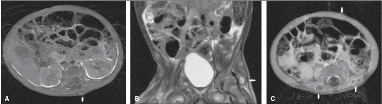
The lesions workup proceeded with ul-trasonography which demonstrated the presence of diffuse, hypoechoic nodules with a more echogenic nucleus affecting the subcutaneous tissue of the trunk, scalp (Figure 2A) and the gallbladder wall (Fig-ure 2B). Computed tomography demon-strated subtly hypodense lesions with ho-mogeneous contrast uptake on the late phase (Figure 3). Magnetic resonance im-aging (MRI) demonstrated the presence of nodules in subcutaneous, intramuscular and visceral tissues, with hyposignal on T1-weighted sequences (Figure 4A), variable hypersignal on T2-weighted sequences (Figure 4B) and peripheral paramagnetic contrast uptake (Figure 4C).
INTRODUCTION
Infantile myofibromatosis, a rare child-hood disease, was first described by Stout in 1954, as congenital generalized fibroma-tosis(1). Classically, this disease presents solitary or multicentric nodular masses aris-ing at the first months of life and involv-ing the skin, subcutaneous tissues, bones or viscus(1–3).
The present report is aimed at review-ing the findreview-ings of this rare disease, empha-sizing that the diagnosis must be sped up considering the significant morbidity of this condition.
CASE REPORT
119
Mata BELF et al. Infantile myofibromatosis: report of a rare disease
Radiol Bras. 2012 Mar/Abr;45(2):118–120
like in the present report, radiotherapy, corticotherapy and chemotherapy – all of these treatments still with limited results(6). The disease prognosis is less favorable in cases of visceral involvement(3). The pa-tient is currently undergoing chemotherapy and has presented a significant clinical and radiological improvement of the lesions and good weight gain progression. The rel-evance of imaging methods in the identifi-cation of lesions which otherwise could not be identified at physical examination, as well as their relationship with anatomical structures, should be highlighted.
Therefore, the association of imaging methods and histopathological analysis allows the staging and classification of the disease, leading to the adoption of the most appropriate treatment for this rare patho-Figure 2. Ultrasonography. A: Hypoechoic nodules in subcutaneous tissue with hyperechoic nucleus, causing skin bulging. B: Image of parietal nodule in the gallbladder with iso- to hyperechoic aspect.
A B
Figure 3. Computed tomography. A: Hypodense nodule in the left occipital region. B: Appearance of late contrast enhancement on the central region of nodules.
A B
Figure 1. Skin lesions. A: Innumerable subcutaneous nodules on the trunk and scalp. B: Biopsy slide with visceral nodule specimen demonstrating muscle cells and fibroblasts.
120
Mata BELF et al. Infantile myofibromatosis: report of a rare disease
Radiol Bras. 2012 Mar/Abr;45(2):118–120 logical entity. The authors conclude that the
knowledge of the typical radiological find-ings of this entity is critical, considering the wide range of differential diagnoses includ-ing disorders commonly found in children.
REFERENCES
1. Stout AP. Juvenile fibromatosis. Cancer. 1954;7: 953–78.
2. Dimson OG, Drolet BA, Southern JF, et al. Con-genital generalized myofibromatosis in a neonate. Arch Dermatol. 2000;136:597–600.
3. Robbin MR, Murphey MD, Temple T, et al. Imag-ing of musculoskeletal fibromatosis. Radiographics. 2001;21:585–600.
4. Chapman PR, Judd CD, Felgenhauer JL, et al. In-fantile myofibromatosis of the posterior fossa. AJR Am J Roentgenol. 2005;184:1310–2.
5. Thunnissen BT, Bax NM, Rövekamp MH, et al.
Figure 4. Magnetic resonance imaging. A: Axial T1-weighted image demonstrates dorsal nodule with iso/hyposignal. B: Coronal T2-weightd image with fat saturation demonstrating deep nodule with marked hypersignal. C: Axial T1-weighted image following gadolinium injection, demonstrating peripheral enhance-ment of subcutaneous nodules.
B
A C
Infantile myofibromatosis: an unusual presentation and a review of the literature. Eur J Pediatr Surg. 1993;3:179–81.
6. Kuo FY, Huang SC, Eng HL, et al. Solitary infan-tile myofibromatosis: report of two cases. Chang Gung Med J. 2002;25:393–8.