49
Case R eport
REVISTA PAULISTA DE MEDICIN AAnalysis of the p53 ge ne
by PCR-SSCP in te n case s of Wilms’ tumor
Department of Pediatrics, Faculdade de Medicina de Ribeirão Preto,
Universidade de São Paulo and Fundação Hemocentro de Ribeirão Preto, Riberão Preto, Brazil
a b s t r a c t
CO N TEX T: Mutatio ns o f the p5 3 tumo r suppresso r g ene are the mo st frequent alteratio ns o bserved in human neo plasias affecting adults. In pediatric o nco lo g y, ho wever, they have seldo m been identified. W ilms’ tumo r is a renal neo plasia co mmo nly o ccurring in children and is asso ciated with mutatio ns o f the W T1 g ene. The co rrelatio n between W ilms’ tumo r and alteratio ns o f the p5 3 g ene has no t been well established, with a lo w frequency o f mutatio ns having been repo rted in this type o f tumo r. Mutatio n may be asso ciated with advanced stag e disease and unfavo rable histo lo g y.
O BJECTIV E: To screen fo r mutatio ns o f the p5 3 g ene by the PCRSSCP metho d and DN A sequencing in cases o f W ilms’ tumo r sug -g estive o f mutatio n.
DESIGN : Case Repo rt.
CASE REPO RT: Evaluatio ns o f exo ns 5 -9 o f the p5 3 g ene in DN A samples extracted by PCR-SSCP fro m 1 0 W ilms’ tumo rs in children at different stag es, and DN A sequencing . Chang es in SSCP analy-sis were o bserved in exo n 8 in two samples. The pro bable muta-tio ns were no t co nfirmed by DN A sequencing . The absence o f po int mutatio ns in p5 3 g ene o bserved in the 1 0 samples o f W ilms’ tumo r studied ag rees with literature data, with DN A sequencing being o f fundamental impo rtance fo r the co nfirmatio n o f po ssible mutatio ns.
KEY W O RDS: W ilms’ tumo r. p5 3 g ene. PCR-SSCP. So lid tumo rs.
• Ricardo Defavery • Jo sé Alexandre Ro drig ues Lemo s • Simo ne Kashima • Jo sé Eduardo Bernardes Carlo s Alberto Scridelli • Dimas Tadeu Co vas • Luiz G o nz ag a To ne
INTRODUCTION
The p53 tumo r suppresso r gene is lo cated o n the sho rt arm o f human chro mo so me 17 (17p13) and it co des fo r a nuclear pho spho pro tein o f 53 kDa. The func-tio n o f wild type p53 is the negative regulafunc-tio n o f cell pro liferatio n, with a transcriptio n actio n that inhibits the G1 phase o f the cell cycle in the presence o f dam-aged DNA.1-3 p53 is the mo st intensely investigated gene
in human cancer.2 Its mutatio ns have been identified in
50% o f adult neo plasias invo lving the co lo n, lung, eso phagus, sto mach, liver, breast, and uterine cervix.4
Ho wever, mutatio ns o f the p53 gene have been little o bserved in children.5,6
Wilms’ tumo r is the renal neo plasia mo st fre-quently o ccurring in children, with an incidence o f 1:10,000 children, especially amo ng tho se yo unger than 6 years.7 Appro ximately 5 to 10% o f children with Wilms’
tumo r have bilateral invo lvement. Wilms’ tumo r is asso ciated with co ngenital abno rmalities including genito -urinary malfo rmatio ns, spo radic aniridia, mental retar-datio n, and hemihypertro phy.8
Mutatio ns o f the WT1 gene are asso ciated with Wilms’ tumo r.9 The co rrelatio n between p53 gene and
Wilms’ tumo r is no t co mpletely understo o d. There are few repo rts o n mutatio ns o f this gene in this tumo r type. A po ssible asso ciatio n with unfavo rable pro gno sis and advanced stage o f the disease has been repo rted.6,9,10
In the present study we investigated po ssible mu-tatio n in the regio ns o f the 5-9 exo ns o f the p53 gene in children with Wilms’ tumo r by the po lymerase chain re-actio n – single strand co nfo rmatio nal po lymo rphism (PCR-SSCP). Samples that presented ano malo us
50
tio n by PCR-SSCP analysis were submitted to auto matic DNA sequencing to co nfirm po ssible mutatio ns.
CASE REPORT
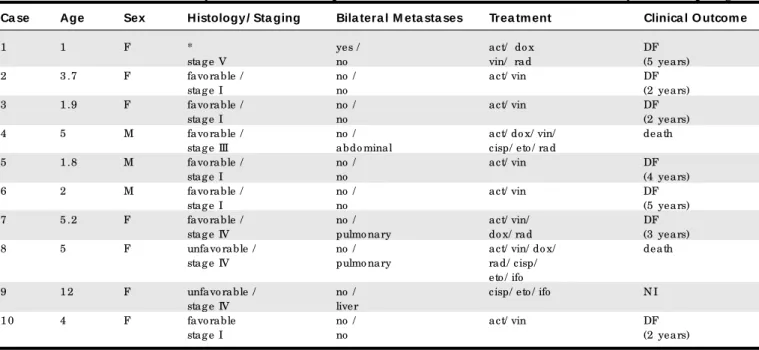
Tumo r samples fro m 10 patients with a diagno sis o f Wilms’ tumo r were studied by PCR-SSCP to determine the presence o f po ssible mutatio ns due to genetic changes in the p53 gene. The patients were seen at the Pediatric Onco lo gy Outpatient Clinic o f the University Ho spital, Faculty o f Medicine o f Ribeirão Preto , Univer-sity o f São Paulo . The clinical and histo lo gical character-istics o f the patients (7 girls and 3 bo ys) are presented in Table 1. Mean age at diagno sis was 4.1 years. Histo lo gy was favo rable in 6 cases and unfavo rable in 3. In o ne case o f bilateral Wilms’ tumo r the material was insuffi-cient fo r histo patho lo gical analysis. The tumo r was stage I in 5 cases, stage III in 1, stage IV in 3, and stage V in 1. The classificatio n o f Wilms’ tumo r was based o n the cri-teria o f the Natio nal Wilms’ Tumo r Study.11 Tumo r
samples were o btained during surgery and DNA was ex-tracted by the pheno l/chlo ro fo rm metho d.12
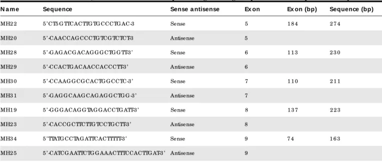
The po ssible alteratio ns o f the p53 gene in the re-gio ns co rrespo nding to exo ns 5-9 were evaluated by PCR-SSCP. Each o f the five regio ns analyzed was amplified by PCR using a pair o f co rrespo nding primers13 (Table 2).
Fo r PCR, each DNA sample (0.1 mg/ml) was added to a mixture co ntaining 2.5 mM buffer so lutio n (0.2 M Tris-HCI, 0.5 M KCl, pH 8,4); 10 mM dNTPs (dATP, dCTP, dGTP, dTTP); 1.5 mM Mg fo r exo ns 5, 6, 7 and 8; 2 mM Mg fo r exo n; a pair o f primers (10 mg/ml) co rrespo nding to the
exo n under study, and taq po lymerase (5 U/ml), to a final vo lume o f 25 ml. All samples were submitted to the fo l-lo wing amplificatio n co nditio ns: 35 successive cycles o f denaturatio n (1 minute at 94ºC fo r all exo ns), annealing (1.5 minutes at 61ºC fo r exo ns 5 and 8, 54ºC fo r exo n 6, 58ºC fo r exo n 7 and 53ºC fo r exo n 9) and extensio n (1 minute at 72ºC fo r all exo ns).
Fo r SSCP, the PCR pro ducts were diluted 1:10 in a so lutio n co ntaining 0.1% SDS and 10 mM EDTA, and an equal vo lume o f dye with 20 mM EDTA, 95% fo rmamide, 0.05% bro mo pheno l blue and 0.05% xylene cyano l was added. Denaturatio n was then carried o ut at 96ºC fo r 10 minutes. A fractio n o f this so lutio n was put into 6% no n-denaturing po lyacrylamide gel and submitted to elec-tro pho resis under the fo llo wing co nditio ns: 8 W, 40 mA and 200 V fo r 2-4 ho urs at 4ºC. The gel was stained with silver nitrate, develo ped, pho to graphed, and analyzed.
Fo r DNA sequencing o f the samples with pro bable mutatio ns screened by PCR-SSCP, a 100 ml aliquo t o f each PCR pro duct was used fo r DNA purificatio n. The purified DNA pro ducts and the primers labeled with fluo rescein13
(Table 2) were submitted to the sequencing reactio n us-ing the Thermo Sequenase kit (Amersham Pharmacia, Little Chalfo nt, Buckinghamshire, UK) acco rding to the manufacturer’s instructio ns. Electro pho resis was then car-ried o ut using a 7% no n-denaturing gel with an auto matic ALF sequencer (Amersham Pharmacia Bio tech, Uppsala, Sweden) at 1500 V, 25 mA and 60 W fo r 4-6 ho urs. The DNA sequences o btained fo r each sample were co mpared to the no rmal sequence o f the p53 gene o btained fro m the Gene Bank, sequencing access number x54156.
Table 1 - Clinical and laboratory characte ristics of patie nts with Wilms’ tumor submitte d to analysis of the p53 ge ne
Ca se Age Sex Histology / Sta ging Bila tera l M eta sta ses Trea tm ent Clinica l O utcom e
1 1 F * yes / act/ do x DF
stag e V no vin/ rad (5 years)
2 3 .7 F favo rable / no / act/ vin DF
stag e I no (2 years)
3 1 .9 F favo rable / no / act/ vin DF
stag e I no (2 years)
4 5 M favo rable / no / act/ do x/ vin/ death
stag e III abdo minal cisp/ eto / rad
5 1 .8 M favo rable / no / act/ vin DF
stag e I no (4 years)
6 2 M favo rable / no / act/ vin DF
stag e I no (5 years)
7 5 .2 F favo rable / no / act/ vin/ DF
stag e IV pulmo nary do x/ rad (3 years)
8 5 F unfavo rable / no / act/ vin/ do x/ death
stag e IV pulmo nary rad/ cisp/ eto / ifo
9 1 2 F unfavo rable / no / cisp/ eto / ifo N I
stag e IV liver
1 0 4 F favo rable no / act/ vin DF
stag e I no (2 years)
act: actino mycin; do x: do xo rubicin; vin: vincristine; cis: cisplatin; eto : eto po side; ifo : ifo sfamide; rad: radio therapy; * preo p erative chemo therapy; N I: no info rmatio n; DF: disease free
51
Table 2 - Prime rs, characte ristics and amplifie d re gions of ge ne p53 use d in the pre se nt study
N a m e Sequence Sense a ntisense Ex on Ex on (bp) Sequence (bp)
MH2 2 5 ’ CT5 G TTCACTTG TG CCCTG AC-3 Sense 5 1 8 4 2 7 4
MH2 0 5 ’ -CAACCAG CCCTG TCG TCTCT-3 Antisense 5
MH2 8 5 ’ -G AG ACG ACAG G G CTG G TT-3 ’ Sense 6 1 1 3 2 3 0
MH2 9 5 ’ -CCACTG ACAACCACCCTT-3 ’ Antisense 6
MH3 0 5 ’ -CCAAG G CG CACTG G CCTC-3 ’ Sense 7 1 1 0 2 1 1
MH3 1 5 ’ -G AG G CAAG CAG AG G CTG G -3 ’ Antisense 7
MH1 9 5 ’ -G G G ACAG G TAG G ACCTG ATT-3 ’ Sense 8 1 3 7 2 2 3
MH2 3 5 ’ -CACCG CTTCTTG TCCTG CTT-3 ’ Antisense 8
MH3 4 5 ’ TTATG CCTAG ATTCACTTTTT-3 ’ Sense 9 7 4 1 6 3
MH2 5 5 ’ -CATCG AATTCTG G AAACTTTCCACTTG AT-3 ’ Antisense 9
bp: base pair; Exo n 8
The DNA samples fro m the 10 Wilms’ tumo r speci-mens were submitted to screening by PCR-SSCP fo r po s-sible mutatio ns o f the p53 gene in the regio ns co rre-spo nding to exo ns 5-9.
Abno rmal bands were revealed by SSCP in exo n 8 in 2 samples. One was fro m a patient with bilateral in-vo lvement (case 1) (Figure 1) and the o ther fro m a pa-tient with favo rable histo lo gy (case 3). The clinical o ut-co me was go o d fo r all o f these patients. The DNA se-quencing analyses o f these 2 samples with altered mi-gratio n in exo n 8 were no rmal. Thus, po int mutatio n al-teratio ns were no t co nfirmed in the sequencing o f DNA fro m the 2 samples.
DISCUSSION
In the present study we assessed the presence o f po ssible mutatio ns due to genetic alteratio n in the re-gio ns between exo ns 5 and 9 o f the p53 gene in 10 cases o f Wilms’ tumo rs affecting children. In 2 cases, altered electro pho retic migratio n was o bserved by SSCP but was no t co nfirmed by DNA sequencing.
Fo r the screening o f samples with pro bable muta-tio ns o f the p53 gene we used PCR-SSCP, a metho d with appro ximately 90% sensitivity and specificity fo r the de-tectio n o f mutatio ns co nfirmed by DNA sequencing in PCR pro ducts with 200 base pairs o r less.14,15 Even tho ugh
the co ding regio n o f the p53 gene co nsist o f 10 exo ns (the first is no n-co ding), the analysis o f mutatio ns was perfo rmed in the regio n between exo ns 5 and 9. The rea-so n fo r this is that mo re than 98% o f the mutatio ns o f the p53 gene in human neo plasias are lo cated in these exo ns.5 In additio n, the regio n between exo ns 5 and 8
(co do ns 126 and 331 with 540 base pairs) co ntains DNA sequences that co de fo r evo lutio narily co nserved do -mains co nsidered to be functio nally impo rtant.3
The false-po sitive results o btained here by SSCP in 2 cases agree with repo rts by o ther investigato rs who have studied the p53 gene in Wilms’ tumo r, such as Waber et al.16 and Malkin et al.,9 demo nstrating that DNA
sequenc-ing is o f fundamental impo rtance in the determinatio n o f the existence o f mutatio ns in a given DNA segment.
Acco rding to several repo rts, the incidence o f po int mutatio ns o f the p53 gene in Wilms’ tumo rs is small.
1 2 3
Figure 1 - SSCP on exon 8. Numbers 1 and 3: Normal controls. Number 2: anomalous run (case 1).
52
REFERENCES
1. Fields S, Jang S. Presence o f a po tent transcriptio n activating sequence in the p53 pro tein. Science 1990;249:1046-9.
2. Levine A, Mo mand J, Finlay C. The p53 tumo r suppresso r gene. Nature (Lo nd) 1991;351:453-6.
3. Vo gelstein B, Kinzler K. p53 functio n and dysfunctio n. Cell 1992;70:523-6. 4. Chang F, Syrjanen S, Syrjanen K. Implicatio ns o f the p53 tumo r-suppresso r
gene in clinical o nco lo gy. J Clin Onco l 1995;13:1009-22.
5. Levine AJ. The p53 tumo r suppresso r gene. N Engl J Med 1992;326:1350-2. 6. Kusafuka T, Fuzukawa M, Oue T, Ko mo to Y, Yo neda A, Okada A. Mutatio n analysis o f the p53 gene in childho o d malignant so lid tumo rs. J Pediat Surg 1997;32:1175-80.
7. Breslo w N, Beckwith JB, Cio l M, Sharples K. Age distributio n o f Wilms’ tumo r: repo rt fro m the Natio nal Wilms’ Tumo r Study. Cancer Res 1988;48:1653-7. 8. Clericuzio C. Clinical pheno types and Wilms’ tumo r. Med Pediat Onco l
1993;21:182-7.
9. Malkin D, Sexsmith E, Yeger H, Williams BRG, Co ppes MJ. Mutatio ns o f the p53 tumo r suppresso r gene o ccur infrequently in Wilms’ tumo r. Cancer
Res 1994;54:2077-9.
10. Bardeesy N, Falko ff D, Petruzzi M, et al. Anaplastic Wilms’ tumo r: a subtype displaying po o r pro gno sis harbo rs p53 gene mutatio ns. Nature Genet 1994;7:91-7.
11. D’Anglio GJ, Evans A, Breslo w N, et al. Treatment o f Wilms’ tumo r: results o f the third Natio nal Wilms’ Tumo r Study. Cancer (Phila) 1989;64:349-60. 12. Sambro o k J, Fritsch EF, Maniats T. Mo lecular Clo ning: a labo rato ry manual.
2nd ed. Co ld Spring Harbo r, NY: Co ld Spring Harbo r; 1989.
13. Hsiao M, Yu AA, Yeargin J, Ku D, Haas M. No nhereditary p53 mutatio ns in t-cell acute lympho blastic leukemia are asso ciated with relapse phase. Blo o d 1994;83:2922-30.
14. Hayashi K. PCR-SSCP. A simple and sensitive metho d fo r detectio n o f mutatio ns in the geno mic DNA. PCR Meth Applicatio ns 1991;1:34-8. 15. Greenblatt M, Bennett WP, Ho llstein M, Harris CC. Mutatio ns in the p53
tum o r sup p re sso r ge ne : c lue s to c anc e r e tio lo gy and m o le c ular patho genesis. Cancer Res 1994;54:4855-78.
16. Waber PG, Chen J, Nisen PD. Infrequency o f ras, p53, WT 11 o r RB gene alteratio ns in Wilms’ tumo r. Cancer 1993;72:3732-8.
Kusafuka et al.6 studied 13 cases o f Wilms’ tumo r and
detected no mutatio n o f the p53 gene. In a study o f 38 cases o f Wilms’ tumo r, Waber et al.16 also did no t detect
any mutatio n o f the p53 gene. Malkin et al.9
detected 2 cases with mutatio ns o ut o f a to tal o f 21. One o f the pa-tients had an advanced stage tumo r with favo rable his-to lo gy and the o ther had fo cal anaplasia. Bardeesy et al.10 did no t o bserve mutatio ns o f the p53 gene in 92
cases o f Wilms’ tumo r with favo rable histo lo gy.
Based o n these findings, the absence o f detectio n
o f mutatio ns o bserved in the present study agrees with current data, despite the small number o f cases analyzed. We suggest that the number o f Wilms’ tumo r specimens to be evaluated sho uld be increased in future studies, es-pecially tho se fro m patients with unfavo rable histo lo gy and advanced stage disease, and that mo re co mprehen-sive studies sho uld be perfo rmed by analyzing the remain-ing co dremain-ing exo ns o f the p53 gene and their expressio n, in o rder to better determine any po ssible asso ciatio n be-tween alteratio ns o f p53 gene and Wilms’ tumo r.
r e s u m o
CON TEXTO: Mutações do gene supressor tumoral p5 3 são observadas como as mais freqüentes alterações em neoplasias humanas em adultos. Em oncologia pediátrica, entretanto, são pouco identificadas. O tumor de W ilms é uma neoplasia renal comum em crianças e associa-se com mutações do gene W T1 . A correlação entre tumor de W ilms e alterações do gene p5 3 não está bem estabelecida, sendo baixa a freqüência tumoral, podendo haver associação com doença em estágio avançado e histologia desfavorável.
TIPO DE ESTUDO: Relato de caso .
RELATO DE CASO : Foi realizar uma triagem para mutações do gene p5 3 pelo método de PCR-SSCP e seqüenciamento de DNA nos casos sugestivos de mutação em tumor de W ilms.Avaliação do gene p5 3 nos exons 5 -9 em amostras de DNA extraídos de 1 0 tumores de W ilms de c ria nç a s e m d ife re nte s e stá g io s p e lo mé to d o d e PC R-SSC P e seqüenciamento de DN A. Alteraçõ es na análise do SSCP fo ram observadas em duas amostras no exon 8 . As prováveis mutações não fo ram co nfirmadas pelo seqüenciamento de DN A. A ausência de mutações de ponto do gene p5 3 observada nas 1 0 amostras de tumor de W ilms estuda da s está de a c o rdo c o m a litera tura , sendo o seqüenciamento de DNA fundamental para a confirmação de possíveis mutações.
Pa la vra s-cha ve: Tumo r de W ilms. G ene p5 3 . PCR-SSCP. Tumo res só lido s.
Ricardo De fave ry, MD. Department o f Pediatrics, Faculty o f Medicine o f Ribeirão Preto , University o f São Paulo , Ribeirão Preto , Brazil.
José Ale xandre Rodrigue s Le mos, PhD. Labo rato ry o f mo lecular Bio lo gy, Department o f Pediatrics, Faculty o f Medicine o f Ribeirão Preto , University o f São Paulo , University o f São Paulo , Ribeirão Preto , Brazil.
Simone Kashima, PhD. Labo rato ry o f mo lecular Bio lo gy, Fundação Hemo centro de Ribeirão Preto , University o f São Paulo , Ribeirão Preto , Brazil.
José Eduardo Be rnarde s, MD. Department o f Pediatrics, Faculty o f Medicine o f Ribeirão Preto , University o f São Paulo , University o f São Paulo , Ribeirão Preto , Brazil.
Carlos Albe rto Scride lli, MD. Assistant Physician, Department o f Pediatrics, Faculty o f Medicine o f Ribeirão Preto , University o f São Paulo , University o f São Paulo , Ribeirão Preto , Brazil.
Dimas Tade u Covas, MD. Directo r o f Fundação Hemo centro de Ribeirão Preto , University o f São Paulo , Ribeirão Preto , Brazil.
Luiz Gonzaga Tone , MD. Department o f Pediatrics, Faculty o f Medicine o f Ribeirão Preto , University o f São Paulo , University o f São Paulo , Ribeirão Preto , Brazil.
Source s of funding: No t declared
Conflict of inte re st: No t declared
Last re ce ive d: 17 de August 1999
Acce pte d: 21 September 1999
Addre ss for corre sponde nce :
Luiz Go nzaga To ne
Departamento de Pediatria da Faculdade de Medicina de Ribeirão Preto da Universidade de São Paulo
Av. Bandeirantes, 3900
Ribeirão Preto /SP - Brasil - CEP 14049-900 E-mail: lgto ne@ fmrp.usp.br
p u b lis hin g in fo r m a t io n