Expression of Ki-67 and P16
INK4ain chemically-induced perioral
squamous cell carcinomas in mice.
Expressão KI-67 e P16INK4a em carcinomas espinocelulares periorais
quimicamente induzidos em camundongos.
Ângela valéria Farias alves1; danielle rodrigues riBeiro1; sonia oliveira liMa2; FranCisCo prado reis2; andréa Ferreira soares3;
Margarete zanardo goMes4; riCardo luiz CavalCantide alBuquerque Júnior4.
INTRODUCTION
S
quamous cell carcinomas (SCC) represent the most prevalent oral cancer, accounting for about 90% to 95% of cases, being more frequent in the lower lip, tongue and oral floor1. Amongthe etiological factors of these malignancies, there is the action of tobacco combustion prod-ucts in chronic smokers2.
One of the most used chemical carcino-gens in neoplastic dynamic study is the compound 9,10-dimethyl-1,2-benzanthracene (DMBA), which is an organic pollutant of polycyclic aromatic hy-drocarbon type, largely released into the environ-ment, especially due to human activity3. DMBA
has cytotoxic, mutagenic and immunosuppressant properties4,5.
The transformation of normal cells into ma-lignant ones is mediated by disorders in several cell
cycle regulating agents, whether positive or nega-tive. Cell cycle progression is positively regulated by multiple cyclins and cyclin-dependent kinases, and negatively by a number of cyclin-dependent kinase inhibitors6.
Ki-67 is a nuclear protein expressed in all phases of the cell cycle (G1, S, G2 and M), which, however, is absent in the G0 phase (“resting phase”). The precise function of the Ki-67 antigen is still unknown, but it has been suggested that this protein is possibly associated with the nucle-olus and fibrillar components, and also seems to play an essential role in ribosome synthesis during cell division. Studies have shown that the Ki-67 immunohistochemical expression correlates with the proliferative potential of oral malignant tu-mors7,8.
The p16INK4a (p16) is a oncosuppressor
protein encoded by the INK4a gene (also known as
1. Instituto de Tecnologia e Pesquisa, Aracaju/SE, Brasil; 2. Curso de Medicina, Universidade Tiradentes, Aracaju/SE, Brasil; 3. Universidade Federal de Sergipe, SE, Brasil; 4. Programa de Pós-Graduação em Saúde e Ambiente, Universidade Tiradentes, Aracaju/SE, Brasil.
A B S T R A C T
Objective:to evaluate the influence of Ki-67 and P16INK4a proteins immunohistochemical expressions on the clinical and
morphologi-cal parameters of perioral squamous cell carcinoma induced with 9,10-dimethyl-1,2-benzanthracene (DMBA) in mice. Methods: we topically induced the lesions in the oral commissure of ten Swiss mice for 20 weeks, determining the time to tumors onset and the average tumor volume up to 26 weeks. In histopathological analysis, the variables studied were histological malignancy grade and the immunohistochemical expression of Ki-67 and P16INK4a proteins. The correlation between variables was determined by application of the
Spearman correlation test. Results: the mean time to onset of perioral lesions was 21.1 ± 2.13 weeks; mean tumor volume was 555.91 ± 205.52 mm3. Of the induced tumors, 80% were classified as low score and 20% high score. There was diffuse positivity for Ki-67 in 100% of lesions – Proliferation Index (PI) of 50.1 ± 18.0. There was a strong direct correlation between Ki-67 immunoreactivity and tu-mor volume (R = 0.702) and a low correlation with the malignancy score (R = 0.486). The P16INK4a protein expression was heterogeneous,
showing a weak correlation with tumor volume (R = 0.334). There was no correlation between the immunohistochemical expression of the two proteins studied. Conclusion: in an experimental model of DMBA-induced perioral carcinogenesis, tumor progression was as-sociated with the tumor proliferative fraction (Ki-67 positive cells) and with tumor histological grading, but not with P16INK4a expression.
MTSI, CDK4I or CDKN2) located on chromosome 9p, locus 21, involved in cell cycle progression blocking process. It is inactive in a wide range hu-man malignancies. The loss of p16INK4a
immunoex-pression has been observed in the early stages of oral carcinogenesis and has been considered a mo-lecular event of significant value in the prognostic analysis of such tumors9,10.
This study evaluated the influence of Ki-67 and p16 proteins immunohistochemical ex-pression on morphological parameters (mean tu-mor volume and histological malignancy grade) of DMBA-induced perioral SCC in mice. In addition, it sought to verify the existence of correlation between the immunoreactivity of p16 and Ki-67 proteins.
METHODS
The development of the study had the ap-proval of the Ethics in Research Committee of the Universidade Tiradentes - Aracaju / SE, with protocol number 191208.
Animals and chemical carcinogenesis induction procedure
We used ten Swiss mice without distinction between gender, from the vivarium of the Universi-dade Tiradentes, with a body mass of about 150 ± 30g (Average age 100 days).
We induced the oral lesions in the mice left oral commissure by the topical application of 9,10-dimethyl-1,2-benzanthracene (DMBA), diluted to 0.5% in acetone, on a weekly basis in three al-ternate days for 20 weeks11. After this period, the
animals were kept under observation for six weeks, and we duly registered the time of tumor onset (clinical) of each animal.
Macroscopic analysis of DMBA-induced injuries
To determine tumor volume, we used a dig-ital caliper so that we could verify the average diam-eter of the induced lesions and apply the following formula12: V = 4/3.π.d, where: V = volume; π = 3,14;
d = average diameter.
Specimens collection and histologic processing
After 26 weeks the animals were sacri-ficed in a CO2 chamber (Insight, Ribeirão Preto, SP – continuous flow of 100% CO2 for 50 minutes). Then the tumor area was subjected to post-mor-tem removal. The tissue specimens were fixed in buffered formalin (10%, pH 7.4) for 24 hours, de-hydrated in increasing ethanol solutions and di-aphanized in xylene for subsequent impregnation and embedding in paraffin.
For each tumor we obtained 15, 5μm-thick histologic sections, subject to routine staining with hematoxylin and eosin. The lesions were morpho-logically analyzed by light microscopy (Optical Mi-croscope Olympus CX31). Two previously trained observers examined ten histological fields and clas-sified the tumors according to a histological malig-nancy grading system13. This system aims both at
the analysis of the tumor cell population, and at the host response by assessing parameters such as the degree of keratinization, nuclear pleomorphism, number of mitoses, invasion pattern, invasion stage and lymphoplasmacytic infiltrate. It has a estab-lished score between 1 and 4, as recommended by the authors. The sum of the scores was divided by six (the number of evaluated parameters) to obtain the average final score for each case. The evaluated cases were divided into two groups, based on the average final score: Group I, low score, with cases whose average value was less than 2.6; and Group II, high score, those with average values equal to or greater than 2.6.
Immunohistochemical analysis
IN-K4a and Ki-67 proteins with rabbit anti-mouse
monoclonal antibodies, types Ab-7 (Neomarkers, Fremont, CA, USA, dilution 1:100) and MIB-1 (Dako, Glostrup, Denmark, dilution 1:50), re-spectively, both for 30 minutes. The reaction was revealed using diaminobenzidine (DAB, Ventana Medical Systems, Tucson, AZ, USA) and coun-terstained with Meyer’s hematoxylin. Both steps were developed in a four minute interval each. The positive control was performed with human tonsil (for Ki-67) and dermal nevocellular nevus (for p16)14. For negative control, we substituted
primary antibody by phosphate buffered saline in the reaction.
Interpretation of the immunohistochemistry results
Cells whose nuclei and / or cytoplasm were stained brown by Ab-7 antibody (anti-p16) were considered positive, regardless of the immunostain-ing intensity. The grade of immunohistochemical expression was determined by intensity semiquan-tification (0, negative; 1, weak; 2, moderate; 3, strong) and percentage of positively stained cells (1, less than 30%; 2, between 30 and 60%;. 3, more than 60%) The final score of each tumor was cal-culated by summing the intensity and percentage scores, as previously described by Prowse et al.15.
Cells whose nuclei were stained with MIB-1 anti-body (anti-Ki-67), regardless of cytoplasmatic stain-ing, were considered positive. The gradw of immu-noreactivity was determined by the percentage of positive cells in 1,000 cells.
Statistical analysis
We applied the Spearman linear correlation test to determine the degree of correlation between mean tumor volume, malignancy grade and immu-nohistochemical expression of Ki-67 and p16INK4a
an-tigens. The correlation was stronger the closer to 1 was the R value.
To compare interobserver means, and de-termine the average values of the scores, we used the Student’s t test, with significance level set to a value of p <0.05.
RESULTS
After 26 weeks, all the animals devel-oped perioral tumor lesions, with mean and stan-dard deviation (SD) of 21.1±2.13 weeks for in-juries onset. The mean tumor volume ± SD was 555.91±205.52 mm3.
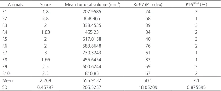
With respect to the specimens histological analysis, we observed that all visible tumors were squamous cell carcinomas. These were characterized by proliferation of keratinocytes, well to moderate-ly differentiated, with varying degrees of individual keratinization (dyskeratosis) and in group (keratin pearls), infiltrating the adjacent mucosa and skin. We also found a predominantly lymphocytic inflammato-ry reaction of intensity ranging between mild, mod-erate and severe. As shown in table 1, of the ten cas-es of lip squamous cell carcinoma, eight (80%) were classified as low-grade malignant lesions, while only two (20%) were interpreted as having high degree malignancy. There was a moderate direct correlation between the mean tumor volume and tumor malig-nancy grade (R=0.659) (Figure 1).
As shown in table 1, all the analyzed tu-mors showed nuclear staining for the Ki-67 anti-gen, although at varying grades, with a mean ± SD proliferative index (PI) of 50.1 ± 18.0. In tumors with weak immunostaining (less than 30% reac-tive cells), we observed the immunohistochemical positivity predominantly in the basal parabasal layers of the nests and neoplastic sheets, whereas tumors with moderate (between 30 and 60% re-active cells) and stronger (more than 60% rere-active cells) markings showed a quite diffuse positivity.
This antigen was also well expressed in tumor cells during all phases of mitosis.
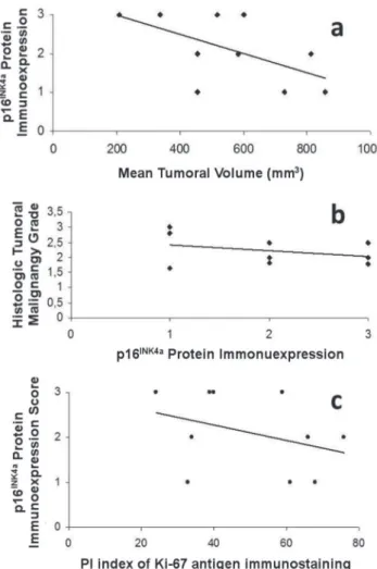
We also observed a strong direct correla-tion between the PI index of Ki-67 positive cells and the mean tumor volume (R=0.702) (Figure 2a), but a weak one between the index and histological malig-nancy grade (R=0.486) (Figure 2b).
Regarding the p16INK4a antigen
immuno-histochemical expression, there positivity was mild in 30% of cases, moderate in 30% and intense in 40% of analyzed lesions. The immunoreactivity pat-tern was quite heterogeneous, with staining some-times nuclear, somesome-times nuclear and cytoplasmic. Eminently nuclear immunostaining was most com-mon in well-differentiated tumor cells located in the surface portion of the tumor. The nuclear / cyto-plasmic positivity, on the other hand, was found in tumor cells of more central regions and rarely of the invasive front of the tumor. Keratinized areas (dyskeratosis and keratin pearl), as well as mitotic figures, were negative for this antigen.
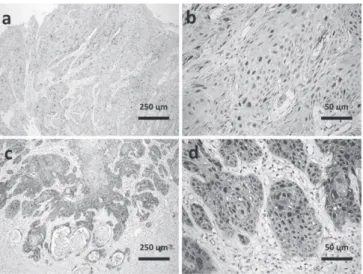
Figure 3 shows immunostaining for p16INK4a
protein and immunohistochemical positivity for Ki-67 antigen.
By comparing the expression profile of p16 IN-K4a protein and the mean tumor volume, we found
only a weak inverse correlation between these two variables (R=0.334) (Figure 4a), and no correlation
between immunohistochemical expression of this antigen and histological malignancy grade (R=0.143) (Figure 4b). Also, there was no immunoreactivity cor-relation between the p16INK4a protein and the Ki-67
antigen (R=0.124) (Figure 4c).
Table 1 - Histopathological and immunohistochemical evaluation of DMBA-induced perioral squamous cell carcinomas.
SD - Standard Deviation
DISCUSSION
In this study, there was a strong direct correlation between the mean tumor volume and tumor malignancy grade, ie, the lesions classified as high grade showed the highest rates of tu-mor growth when compared with the low-grade ones, showing that morphologically undifferen-tiated cells are genetically unstable and easily es-cape from cell cycle control mechanisms, with a tendency to have high cell proliferation rates, in agreement with other studies16,17.
The Ki-67 immunoreactivity relates to evidence of cell proliferation, so that it is ex-pressed in all cell cycle phases but G0, in which cells are quiescent18. According to Sousa et al.19,
immunohistochemical analysis of this marker is an effective method for assessing the human malignancies growth fraction, providing valuable information about prognosis.
In the studied sample, we observed that the Ki-67 immunostaining showed vary-ing grades, and in high-grade lesions there was strong expression in diffuse distribution. In some low-grade lesions, with evidence of high mean tumor volume index, we also found strong markings by said antibody, thus confirming the
strong direct correlation between Ki-67 and this clinical parameter. Several authors report said correlation, such as Balassiano20, who analyzed
the expression of Bcl-2 markers, p53, mutated p53, caspase-3 and Ki-67 as prognostic factors in proliferative lesions of the oral cavity, such as inflammatory fibrous hyperplasia, actinic chei-litis and squamous cell carcinoma of the lower lip, and found high Ki-67 expression in all le-sions.
The Ki-67 positivity kept a weak direct correlation with histological malignancy grade. This is due to the antibody’s immunoreactivity instability. According to a published study21,
Ki-67 allows inferences about the time of life of a particular cell, stating only if it is in the cell cycle21. Therefore, it is possible that a particular
neoplasm has a high proliferation rate and a low percentage of cells positive for this antibody.
Some authors evaluated the expression of PCNA, Ki-67, p53 and bcl-2 in patients with squamous skin carcinoma (n=10) and actinic keratosis (n=10), and confirmed the absence of Ki-67 expression in two cases, confirming the variability in this marker’s immunoreactivity18.
However, it is interesting to note that in several studies the Ki-67 presents a tendency to strong direct correlation with the lesion malignancy degree, being valuable as a prognostic predic-tor17,18.
The p16INK4a tumor suppressor gene,
encoding the p16 protein, is inactivated by hy-permethylation in several types of malignancies, including oral squamous cell carcinoma, consti-tuting a crucial event in the early stages of ma-lignant transformation of the affected tissue.
Using immunohistochemical techniques, it is common to highlight the absence of p16 IN-K4a protein immunoreactivity, a fact that is highly
correlated with the findings provided by molec-ular techniques, which show inactivation of the aforementioned gene22,23.
In this experimental group, there was little positivity for p16INK4a in high-grade lesions,
and moderate to intense staining in low-grade
ones. Immunoexpression was strictly nuclear in well-differentiated tumor cells, especially in sur-face areas, but also in the central areas of the tumor. These findings ar in agreement with oth-er scientific work16, which reported an
immuno-localization trend of p16 in the central and sur-face areas of the tumor mass, with progressive decrease in regions of the invasion front, which concentrates the most undifferentiated cells, with a higher degree of cell adhesion loss.
Some studies show strong direct rela-tionship between the lack of p16 immunoreac-tivity and the severity of the histological grading and clinical staging. However, we could not
es-tablish such correlation, consistent with another study16.
We also found a weak inverse correla-tion between p16INK4a expression and tumor
vol-ume, as well as no statistically significant correla-tion between the immunoreactivity of p16INK4a
and Ki-67.
According to the aforementioned au-thors, this lack of correlation of p16, with im-portant prognostic parameters, is due to the fact that the inactivation of the p16INK4a gene and its
related protein would occur in the early stages of oral carcinogenesis and therefore would be more efficient as early markers of malignant transfor-mation than as prognostic markers, being un-reliable in predicting the biological behavior of neoplastic lesions.
The results of this study show that tu-mor volume is an important clinical parameter to measure the aggressiveness of malignant neoplasms, and Ki-67 immunoreactivity was ef-fective as a marker of cell proliferation. Never-theless, such marker not always displays a sig-nificant correlation with the immunoreactivity pattern of proteins that regulate the cell cycle, due to the instability of its expression in the tu-mor parenchyma.
Moreover, when one intends to cor-relate the expression of proteins that control the cell cycle with the degree of tumor malignancy, there may be inconsistencies justified by the fact that these proteins either act by independent molecular pathways or at different stages of the cell cycle and of tumor progression, such that their expression may not reflect the proliferative potential of malignant lesions.
In conclusion, this study showed that in the perioral carcinogenesis induced by DMBA in an experimental model, tumor progression is associated with the proliferative fraction of the tumor (Ki-67 positive cells) and with tumor dif-ferentiation, but without correlation with the p16INK4a protein expression. New studies are
nec-essary to elucidate the mechanisms of action of genes and proteins involved in the cell cycle.
REFERENCES
1. Melo AUC, Albuquerque Júnior RLC, Melo MFB, Ri-beiro CF, Santos TS, Gomes ACA. Análise das esti-mativas de incidência de câncer de boca no Brasil e em Sergipe (2000 - 2010). Odontol Clín-Cient. 2012; 11(1):65-70.
2. Turati F, Garavello W, Tramacere I, Pelucchi C, Ga-leone C, Bagnardi V, et al. A meta-analysis of al-cohol drinking and oral and pharyngeal cancers: results from subgroup analyses. Alcohol Alcohol. 2013;48(1):107-18.
3. Saha D, Hait M. An ontological design: two stage mouse skin carcinogenesis induced by DMBA and promoted by croton oil. Asian J Res Pharm Sci. 2012;2(1):1-3 4. Lindhe O, Granberg L, Brandt I. Target cells for
cyto-chrome p450-catalysed irreversible binding of 7,12-di-methylbenz[a]anthracene (DMBA) in rodent adrenal glands. Arch Toxicol. 2002;76(8):460-6.
5. Buters J, Quintanilla-Martinez L, Schober W, So-balla VJ, Hintermair J, Wolff T, et al. CYP1B1 de-termines susceptibility to low doses of 7,12-di-methylbenz[a]anthracene-induced ovarian cancers in mice: correlation of CYB1B1-mediated DNA adducts with carcinogenicity. Carcinogenesis. 2003;24(2):327-34.
6. Zheng J, Xie L, Teng H, Liu S, Yoshimura K, Kageya-ma I, Kobayashi K. Morphological changes in the lin-gual papillae and their connective tissue cores on the 7,12-dimethylbenz[alpha]anthracene (DMBA)
stim-ulated rat experimental model. Okajimas Folia Anat Jpn. 2009;85(4):129-37.
7. Rapidis AD, Gullane P, Langdon JD, Lefebvre JL, Scully C, Shah JP. Major advances in the knowledge and un-derstanding of the epidemiology, aetiopathogenesis, diagnosis, management and prognosis of oral cancer. Oral Oncol. 2009;45(4-5):299-300.
8. Warnakulasuriya S. Living with oral cancer: epide-miology with particular reference to prevalence and life-style changes that influence survival. Oral Oncol. 2010;46(6):407-10.
9. Hong Y, Li C, Xia J, Rhodus NL, Cheng B. p16(CD-KN2A) expression during rat tongue carcinogene-sis induced by 4-nitroquinoline-1-oxide. Oral Oncol. 2009;45(7):640-4.
10. Ohta S, Uemura H, Matsui Y, Ishiguro H, Fujinami K, Kondo K, et al. Alterations of p16 and p14ARF genes and their 9p21 locus in oral squamous cell carcinoma. Oral Surg Oral Med Oral Pathol Oral Ra-diol Endod. 2009;107(1):81-91.
11. Kavitha K, Manoharan S. Anticarcinogenic and antilipidperoxidative effects of Tephro-sia purpurea (Linn.) Pers. in 7,12-dimethyl-benz(a)anthracene (DMBA) induced hamsters buccal pouch carcinoma. Indian J Pharmacol. 2006;38(3):185-9.
12. Mizuno M, Minato K, Ito H, Kawade M, Terai H, Tsuchida H. Anti-tumor polysaccharide from the mycelium of liquid-cultured Agaricus blazei mill. Bio-chem Mol Biol Int. 1999;47(4):707-14.
R E S U M O
Objetivo: avaliar a influência da expressão imuno-histoquímica das proteínas Ki-67 e p16INK4a sobre parâmetros clínico-morfológicos em
carcinomas espinocelulares periorais quimicamente induzidos com 9,10-dimetil-1,2-benzantraceno (DMBA) em modelo murino. Métodos:
as lesões foram induzidas topicamente na comissura labial de dez camundongos Swiss durante 20 semanas, sendo determinado o momen-to de surgimenmomen-to dos tumores e volume tumoral médio até 26 semanas. Na análise hismomen-topamomen-tológica, as variáveis estudadas foram gradação histológica de malignidade tumoral e expressão imuno-histoquímica das proteínas Ki-67 e p16INK4a. A correlação entre as variáveis
estuda-das foi determinada pela aplicação do teste de correlação de Spearman. Resultados: o tempo médio de surgimento das lesões periorais foi 21,1±2,13 semanas. Volume tumoral médio foi de 555,91±205,52mm3. Dos tumores produzidos, 80% foram classificados como de baixo escore e 20%, alto escore. Evidenciou-se positividade difusa para Ki-67 em 100% das lesões – índice de marcação (PI) de 50,1±18,0. Verificou-se correlação direta forte entre a imunoexpressão do Ki-67 e o volume tumoral (R=0,702) e fraca correlação com o escore de malignidade (R=0,486). A expressão da proteína p16INK4a foi heterogênea, mostrando fraca correlação com o volume tumoral (R=0,334).
Não houve correlação entre a expressão imuno-histoquímica das duas proteínas estudadas. Conclusão: Em modelo experimental de car-cinogênese perioral DMBA-induzida, a progressão tumoral está associada à fração proliferativa do tumor (células ki-67 positivas) e com a gradação histológica tumoral, porém não com a expressão da p16INK4a.
13. Anneroth G, Batsakis J, Luna M. Review of the lit-erature and a recommended system of malignancy grading in oral squamous cell carcinomas. Scand J Dent Res. 1987;95(3):229-47.
14. Hsieh R, Sousa FB, Firmiano A, Nunes FD, Ma-galhães MHCG, Sotto MN. Estudo genético do gene p16 pela técnica de PCR-SSCP e expres-são de proteína p16 em melanomas de mucosa oral e melanomas cutâneos. An Bras Dermatol. 2006;81(5):433-41.
15. Prowse DM, Ktori EN, Chandrasekaran D, Prapa A, Baithun S. Human papillomavirus-associated in-crease in p16INK4A expression in penile lichen scle-rosus and squamous cell carcinoma. Br J Dermatol. 2008;158(2):261-5.
16. De-Paula AMB, Cardoso SV, Gomez RS. Imunolocali-zação das proteínas dos genes supressores de tumo-res TP53 e p16CDKN2 no front invasivo do
carcino-ma epidermóide de cavidade bucal.J Bras Patol Med
Lab. 2006;42(4):285-91.
17. Rodrigues RB, Motta RR, Machado SMS, Cambruzzi
E, Zettler EW, Zettler CG, et al.Valor prognóstico da
correlação imuno-histoquímica do Ki-67 e p53 em
carcinomas epidermóides da laringe.Rev Bras
Otor-rinolaringol. 2008;74(6):855-9.
18. Dornelas MT, Rodrigues MF, Machado DC, Gollner AM, Ferreira AP. Expressão de marcadores de proli-feração celular e apoptose no carcinoma espinoce-lular de pele e ceratose actínica. An Bras Dermatol. 2009;84(5):469-75.
19. Sousa FACG, Brandão AAH, Almeida JD, Rosa LEB. Alterações gênicas e câncer bucal: uma breve revi-são. Rev bras patol oral. 2004;3(1):20-5.
20. Balassiano KZ. Estudo comparativo expressão imuno-his-toquímica das proteínas p53, caspase-3 e Ki-67 em hiper-plasias fibrosas inflamatórias, queilites actínias e carcino-mas de células escamosas no lábio inferior [dissertação]. Niterói/RJ: Universidade Federal Fluminense; 2004. 21. Correa MPD, Ferreira AP, Gollner AM, Rodrigues
MF, Guerra MCS.Expressão de marcadores de
pro-liferação celular e apoptose em carcinoma
basocelu-lar. An Bras Dermatol. 2009.84(6):606-14.
22. von Zeidler SV, Miracca EC, Nagai MA, Birman EG. Hypermethylation of the p16 gene in normal oral mu-cosa of smokers. Int J Mol Med. 2004;14(5):807-11. 23. Soni S, Kaur J, Kumar A, Chakravarti N, Mathur
M, Bahadur S, et al. Alterations of rb pathway com-ponents are frequent events in patients with oral epithelial dysplasia and predict clinical outcome in patients with squamous cell carcinoma. Oncology. 2005;68(4-6):314-25.
Received: 13/10/2015
Accepted for publication: 02/03/2016 Conflict of interest: none.
Source of funding: none.
Mailing address: