5 2 2
Palma et al
Treatment of abdominal aortic aneurysm
Arq Bras Cardiol 2003; 81: 522-5.
São Paulo Federal University, São Paulo
Mailing address: José Honório Palma - Cardiovascular Surgery Department São Paulo Federal University - Rua Borges Lagoa, 1080 - 7º andar Cep 04038-031 - São Paulo, SP, Brazil - E-mail: [email protected]
Arq Bras Cardiol, volume 81 (nº 5), 522-5, 2003
José Honório Palma, Abner Moreira Sampaio, Fausto Miranda, Claudia Maria Rodrigues Alves,
José Augusto de Marcondes Souza, Enio Buffolo
São Paulo, SP - Brazil
A Change in the Treatment of Abdominal Aortic Aneurysms
Original Article
Conventional treatment of abdominal aneurysms has high morbidity and mortality rates, because in most cases such aneurysms occur in elderly patients with comorbidi-ties, such as ischemic heart disease, previous aortic patho-logies, chronic obstructive pulmonary disease, or renal dysfunction 1. Due to the operative risk in this population, traditional surgical treatment has a high rate of potential complications.
Because the natural history of these diseases is well known and they have a high potential for complications, surgical treatment is recommended when diagnosis is made or signs are present of eminent rupture or enlargement 2. Despite great advances in diagnostic imaging and in the de-velopment of new surgical approaches, mortality rates can be as high as 50%, especially when patients are operated on as urgent cases 3.
The introduction of stent-grafts into clinical practice by Parodi et al in 4 as an alternative treatment for abdominal aortic aneurysms led to new perspectives. Since then, many groups have reported successful experiences in different aneurysmatic anatomies and with different types of endo-prostheses allowing reduced morbidity and mortality rates for select groups 5-7.
The clinical use of polyester-covered stents was pio-neered at our institution for the treatment of type B dissec-tions 8. Good results obtained in this phase encouraged the development of new prostheses and catheters that can be inserted through a peripheral artery to treat thoracic and abdominal aortic diseases.
In this study, we report on our results and the compli-cations observed in a series of 80 patients with abdominal aneurysms treated using self-expanding endovascular prostheses.
Methods
From May 1996 to December 2002, 230 cases (descen-ding thoracic aortic diseases and abdominal aortic aneu-rysms) were treated with endoprostheses at our institution. We selected 80 patients with abdominal aneurysms. Of these, 36 had good distal necks or good anchorage sites for Objective - One of the most exciting potential
appli-cations of percutaneous therapy is the treatment of abdo-minal aneurysms.
Methods - Of 230 patients treated with a self-expan-ding polyester-lined stent-graft for different aortic patho-logies at our institution, we selected 80 abdominal aneu-rysm cases undergoing treatment (from May 1997 to De-cember 2002). The stent was introduced through the femo-ral artery, in the hemodynamic laboratory, with the patient under general anesthesia, with systemic heparinization, and induced hypotension.
Results - The procedure was successful in 70 (92.9%) cases; 10 patients with exclusion of abdominal aortic aneurysms were documented immediately within the hemo-dynamic room and 5 patients persisted with a residual leak. Two surgical conversions were necessary. Additional stent-grafts had to be inserted in 3 (3.7%) cases. In the fol-low-up, 91.4% of patients were alive at a mean follow-up of 15.8 months.
Conclusion - We believe that stent-grafts are an im-portant tool in improving the treatment of abdominal aneurysms, and this new policy may change the conventio-nal medical management of these patients.
Arq Bras Cardiol 2003; 81: 522-5.
Palma et al Treatment of abdominal aortic aneurysm
5 2 3 the stent in the distal aorta, and in 44, both iliac arteries in
the aneurysms were compromised. All the patients had complex clinical symptoms with associated diseases, such as moderate-severe chronic obstructive pulmonary disease (17 patients), systemic hypertension (57 patients), renal in-sufficiency or creatinine > 1.5 (15 patients), diabetes (10 pa-tients), and heart failure or coronary disease (27 patients).
Diagnostic confirmations as well as measurement of the diameters and lengths for the prostheses were based on the following 2 tests: aortography and CT scan. Fifty pa-tients had aneurysm diameters between 5.0 and 8.0 cm. The institution’s ethics committee approved the study protocol. Anatomic criteria required for stenting were (1) good proximal landing zone of up to 32 mm near the renal artery and (2) iliac-femoral system compatible with a 20-Fr. device, without major bindings or obstructions.
The aortic stent-grafts are self-expanding polyester-lined stainless steel stents manufactured by Braile Biomédica, São José do Rio Preto, São Paulo – Brazil. These cylinders are highly resistant to radial collapse and maintain an ability to return to their original diameter. The prostheses have 3 parts implanted using 3 different catheters. The principal section is similar to a pair of shorts but has 1 leg shorter than the other. The other 2 parts are similar. The endoprostheses were carefully compressed and inserted in one 20 (principal) and two 17 (legs) Fr. (legs) catheters. Length and diameter are ac-cording to the dimensions of the diseased segment to be treated. Two different length options are available for the legs of the endoprostheses, 8.0 and 10.0 cm, and after full expansion, diameters range from 2.0 to 3.4 cm.
The procedures were performed in the catheter labora-tory with the patient under general anesthesia, orotracheal intubation, and vital sign monitoring. A new arteriography was performed with 2 views (posteroanterior and left anterior oblique) using, when possible, calibrated catheters to confirm preoperative measurements. After aortography we chose the femoral artery, which is less tortuous for the introduction of the catheter with the stent, which was surgically dissected and opened with a transverse incision. A dose of 5000 U of heparin was used for anticoagulation. Under radioscopic visualization, the catheter with the stent, using an extrastiff guidewire, was inserted into the femoral artery and slowly advanced towards the chosen site in the thoracic aorta.
The exact release site was marked by a radiopaque structure (for example: a vertebra or ruler placed on the pa-tient’s back before the procedure was started). First, the main section of the lined endoprosthesis was deployed into the renal artery where it self-expanded inside the aneurysm. The second step was to introduce the 2 “stent legs” or “stent branches” one at a time, approaching from the left and right femoral arteries. To confirm the effectiveness of the procedure, a new angiography was carried out a few mi-nutes after the implantation of the stent-graft. After the pro-cedure, the patients were removed to the postoperative re-covery unit, where they were kept under observation for 24 hours. The patients were discharged after a control CT scan was performed (fig. 1).
Results
About half of the patients were > 70 years of age. The mean age of the group was 71.5 years (55 – 89 years) and 73% were male. All the patients treated by this method were serially followed up and in addition to the arteriography car-ried out during the procedure, CT scans and/or echocardio-grams were performed to document the results and compli-cations during hospitalization. The follow-up period ranged from 1 to 69 months with a mean of 15.8 months.
The procedure was technically successful in 70 pa-tients (92.9%) for the exclusion of aortic aneurysms (fig. 2). In 10 patients, a persistent or severe leak was detected, resulting in 1 emergency and 1 elective surgical conversion, resulting in a conversion rate of 2.5%. For adequate repair, additional stent-grafts had to be inserted in 3 patients (3.7%). Because of leakage, 5 patients remain under observation.
Six (7.5%) in-hospital deaths occurred, and another 3 were related to the procedure. One patient developed multi-ple organ failure, one septicemia, and one pulmonary thromboembolism. The in-hospital survival rate was 92.5%. One (1.4%) case of brachial thrombosis was observed in an individual whose access was achieved by using the Sel-dinger technique and who required surgical repair. Lower limb ischemia was observed in 2.9% of the patients, and 1 patient required endarterectomy of the femoral artery after the procedure. Another patient had deep vein thrombosis. Other complications were mild and transient and included renal failure (15 patients, 21.4%), surgical wound infection (2 patients, 2.9%), and peritoneal dialysis catheter infection (1 patient, 1.4%). Fever was commonly seen (15 patients, 21.4%). In most patients, the fever was not apparently related to an infection and was considered an inflammatory reaction to the prostheses. These patients were treated with antiin-flammatory drugs and the temperature subsided.
After hospital discharge, 4 patients (5.6%, 4/71) died. Two patients had sudden deaths, and 2 deaths were due to causes unrelated to aortic dissection or the procedure (one from a car accident and another from hepatic failure). At a mean follow-up of 15.8 months, 91.4% of the patients were alive.
Discussion
The concept of aneurysm repair by percutaneous insertion of an endovascular prosthesis was first sugges-ted by Dotter 9 in 1969; however, Parodi 4 successfully pio-neered treatment in a series of patients with abdominal aortic aneurysms. These authors used balloon-expanded endovascular prostheses and proved that the procedure was feasible and had low risks. Dake et al 5 showed for the first time, the feasibility of treating descending thoracic aortic aneurysms introducing a self-expanding endovascu-lar prosthesis through the femoral artery.
5 2 4
Palma et al
Treatment of abdominal aortic aneurysm
Arq Bras Cardiol 2003; 81: 522-5.
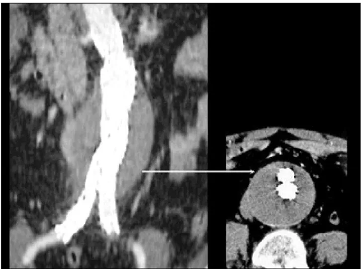
Fig. 1 - Abdominal CT scan with contrast showing the stent and an exclusion of the aneurysm.
It is worth noting that in this study no late ruptures oc-curred of the metallic structure of the endoprostheses nor did late migrations occur, and no cases were noted of ruptu-re-expansion of the aneurysm or late leakage of the endo-prostheses. The only late complication observed associa-ted with the endoprosthesis was an occlusion of the left branch after 1 month.
The 4 patients who continue under observation for leakage have not experienced, until now, dilation of the aneurysmatic balloon, suggesting that some degree of pro-tection is provided by the endoprosthesis.
The systemic complications in our study were few compared with those in to published investigations and considering the profile of our patients, who had an advan-ced mean age and thus were sufferers of co-morbidities.
We did not observe mortalities with elective surgical conversions, which suggests that we can be less restrictive in the indication of endovascular treatment because, when necessary, classical surgical treatment can be performed with the same traditional risks 12.
Arq Bras Cardiol 2003; 81: 522-5.
Palma et al Treatment of abdominal aortic aneurysm
5 2 5 sent with (a) an iliac-femoral system compatible with these
catheters and (b) a proximal neck adequate for the anchoring of the prosthesis.
Other complications observed were considered minor. Fever not related to infection is frequent and may respond to nonhormonal antiinflammatory drug therapy. This com-monly observed side effect may be an immune-mediated process, and the typical presentation is low-grade fever and mild leucocytosis. The use of antibiotics must be judi-ciously decided upon for this group of patients.
In conclusion, this successful initial experience in the treatment of severe cases with complications allows us to an-ticipate a promising future for this simple, less-invasive and low-risk approach. The follow-up of these cases shows it is possible to use the aortic stent-graft inserted in a catheter laboratory, with a low incidence of complications. However, a larger series, longer follow-up periods, and the development of prostheses and catheters (especially reducing the shaft size) that are more appropriate are required for this procedure to be more extensively used in the treatment of aortic disease.
1. Ashton HA, Buxton MJ, Day NE, et al. The Multicentre Aneurysm Screening Study (MASS) into the effect of abdominal aortic aneurysm screening on mortality in men: a randomized controlled trial. Lancet 2002; 360: 1531-9.
2. Brown PM, Zelt DT, Sobolev B. The risk of rupture in untreated aneurysms: the impact of size, gender, and expansion rate. Vasc Surg 2003; 37: 280-4. 3. Beebe HG, Kritpracha B. Imaging of abdominal aortic aneurysm: current status.
Ann Vasc Surg 2003: 24.
4. Parodi JC. Endovascular repair of abdominal aortic aneurysms and other arterial lesions. J Vasc Surg 1995; 21: 549-57.
5. Dake MD, Kato N, Michell RS. Endovascular stent-graft placement for the treatment of acute aortic dissection. N Engl J Med 1999; 340: 1546-52. 6. Nienaber CA, Fattori R, Lund G, et al. Nonsurgical reconstruction of thoracic
aortic dissection by stent-graft placement. N Engl J Med 1999; 340: 1539-45. 7. Palma JH, Souza JAM, Alves CR, et al. Self-expandable aortic stent-grafts for
treatment of descending aortic dissections. Ann Thorac Surg 2002; 73: 1138-45.
References
8. Palma JH, Almeida DR, Carvalho AC, et al. Surgical treatment of acute type B aortic dissection using an endoprosthesis (Elephant trunk). Ann Thorac Surg 1997; 63: 1081-4.
9. Dotter CT. Transluminally-placed coilspring endoarterial tube grafts: long-term patency in canine popliteal artery. Invest Radiol 1969; 4: 329-32.
10. Schermerhorn ML, Finlayson SR, Fillinger MF, et al. Life expectancy after endovascular versus open abdominal aortic aneurysm repair: results of a decision analysis model on the basis of data from EUROSTAR. J Vasc Surg 2002; 36: 1112-20.
11. Matsumura JS, Brewster DC, Makaroun MS, et al. A multicenter controlled clinical trial of open versus endovascular treatment of abdominal aortic aneurysm. J Vasc Surg 2003; 37: 262-71.