Arq Bras Cardiol 2003; 81: 479-82.
Duarte et al Transient left ventricular dilation assessed during myocardial Tc-99m sestamibi
4 7 9
Nuclear Medicine and Cardiology Unit, Fleury - Centro de Medicina Diagnóstica and Faculdade de Saúde Pública of USP
Mailing address: Paulo Schiavom Duarte - Seção de Medicina Nuclear Rua Cincinato Braga, 282 - Cep 01333-910 - São Paulo, SP, Brazil E-mail: [email protected]
Received 10/23/02 Accepted 1/14/03
English version by Stela Maris C. e Gandour
Arq Bras Cardiol, volume 81 (nº 5), 479-82, 2003
Paulo Schiavom Duarte, Paola Emanuela Smanio, Carlos Alberto Oliveira, Luiz Roberto Martins,
Luiz Eduardo Mastrocolla, Julio César Pereira
São Paulo, SP - Brazil
Clinical Significance of Transient Left Ventricular Dilation
Assessed during Myocardial Tc-99m Sestamibi Scintigraphy
Original Article
The balanced involvement of multiple arteries in coro-nary artery disease is a potential cause of false-negative results on myocardial perfusion scintigraphy. During exerci-se, that degree of coronary artery disease may cause perfu-sion defects homogeneously distributed throughout the myocardium, leading to uniform radiopharmaceutical upta-ke, and, consequently, to a false-negative examination 1.
Transient dilation of the left ventricle – also known as transient ischemic dilation (TID) – is considered present when the image of the left ventricular cavity seems to be sig-nificantly greater after stress as compared with that at rest. Transient ischemic dilation is a way to detect balanced co-ronary artery disease in patients with apparently normal myocardial perfusion scintigraphy.
Transient ischemic dilation has been classically descri-bed in myocardial perfusion examinations performed with the stress/redistribution thallium-201 protocol 1,2. In this
pro-tocol, the images following stress are obtained almost imme-diately after radiopharmaceutical injection, during the period when the myocardium is under the effects of physical stress. However, this signal has also been described in other myocardial perfusion scintigraphy protocols, such as the stress/rest sestamibi protocol 3, the tetrofosmin stress/rest
protocol 4, or the dual-isotope protocol (thallium
rest/ses-tamibi stress) 5. In these situations, the acquisition of images
representative of perfusion under stress is performed some time after the exercise test (30 to 60 minutes), when the myo-cardium has already had time to recover from the mechanical dysfunction caused by stress. Transient ischemic dilation has also been described in situations in which the myocar-dium has not undergone a real stress situation, such as in the examination performed with the pharmaceutical stimulus of dipyridamole or adenosine 6-9. Thus, some researchers
consider that the term dilation is imprecise and should represent, in most cases, an apparent dilation secondary to diffuse subendocardial ischemia. Although transient ischemic dilation has been frequently reported in the literature, scarce information exists about the real value of that parameter to assess coronary artery disease and about the values to be used to better separate the patients with
exten-Objective - To assess the clinical significance of tran-sient ischemic dilation of the left ventricle during myocar-dial perfusion scintigraphy with stress/rest sestamibi.
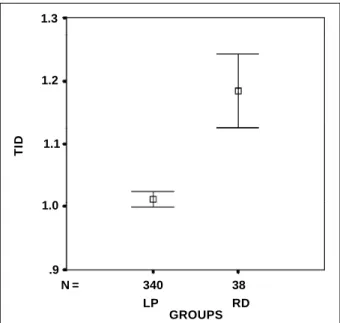
Methods - The study retrospectively analyzed 378 pa-tients who underwent myocardial perfusion scintigraphy with stress/rest sestamibi, 340 of whom had a low probabi-lity of having ischemia and 38 had significant transient defects. Transient ischemic dilation was automatically calculated using Autoquant software. Sensitivity, specifi-city, and the positive and negative predictive values were established for each value of transient ischemic dilation.
Results - The values of transient ischemic dilation for the groups of low probability and significant transient de-fects were, respectively, 1.01 ± 0.13 and 1.18 ± 0.17. The values of transient ischemic dilation for the group with significant transient defects were significantly greater than those obtained for the group with a low probability (P<0.001). The greatest positive predictive values, around 50%, were obtained for the values of transient ischemic dilation above 1.25.
Conclusion - The results suggest that transient ische-mic dilation assessed using the stress/rest sestamibi proto-col may be useful to separate patients with extensive myo-cardial ischemia from those without ischemia.
4 8 0
Duarte et al
Transient left ventricular dilation assessed during myocardial Tc-99m sestamibi
Arq Bras Cardiol 2003; 81: 479-82.
sive myocardial ischemia from those without ischemia, mainly, when the myocardial perfusion study is performed according to the 1-day protocol with stress/rest sestamibi.
Methods
This study aimed at assessing whether transient is-chemic dilation may be a useful index when observed in myocardial perfusion scintigraphy performed according to the 1-day protocol with stress/rest sestamibi, and at trying to establish values of transient ischemic dilation to separate patients with extensive myocardial ischemia from those with a low probability of having myocardial ischemia.
Three hundred and seventy-eight patients were re-trospectively studied. These patients had been referred to the nuclear medicine unit to undergo myocardial perfusion scintigraphy according to the 1-day protocol with stress/ rest sestamibi. Of the 378 patients, 340 (mean age, 52 years; 259 men, 81 women) had a low probability of ischemia (sum of the stress scores – SSS < 2, negative exercise test, no pre-vious history of infarction, myocardial revascularization, angioplasty, or typical angina) and 38 (mean age, 59 years; 32 men, 6 women) had extensive transient defects (sum of the differences of the scores – SDS > 9).
It is worth noting that the referred perfusion scores are analyses of perfusion in different myocardial segments and may be established through subjective visual assessment or automatically calculated with processing programs. In our study, the scores were automatically established with Autoquant software and adjusted by the nuclear physician, when necessary. The scores, as well as transient ischemic di-lation, are discussed in the I Guideline on Nuclear Cardiolo-gy published by the Brazilian Society of CardioloCardiolo-gy 10.
The 1-day protocol with sestamibi in the rest and stress phases was used for acquisition of tomographic ima-ges (SPECT) of myocardial perfusion. The imaima-ges at rest we-re acquiwe-red 30 minutes after injection of 370 MBq (10 mCi) of Tc-99m sestamibi. The stress phase was performed 4 hours after the rest phase. Cardiac stress was induced by program-med exercise on a treadmill according to one of the following protocols: Bruce, modified Bruce, or Ellestad protocol. During exercise peak or when the patient had limiting symptoms, 1.11 GBq (30 mCi) of sestamibi was intravenously injected. The images were acquired 45 to 60 minutes after injection. A scintillation camera with 2 detectors (Vertex plus MCD-AC) equipped with low energy and high resolution collimator was used. Forty-eight projections (25 s/projection) were obtained using a 64 x 64 matrix and a sweeping angle of 180°.
Data were processed with the Ultra-60 workstation (SUN Microsystems, Santa Clara, CA, USA). The reconstruc-tion was performed according to the technique of filtered retro-projection and Butterworth filter using Autospect plus software (Philips Medical Systems, Andover, MA, USA). Correction was made with the movement, when necessary. Transient ischemic dilation was automatically calculated using Autoquant software (Philips Medical Systems, Andover, MA, USA), which calculates the value of transient ischemic dilation by dividing the volume of the left
ven-tricular cavity after effort by the volume of the left venven-tricular cavity at rest (volume of the left ventricular cavity during stress/volume of the left ventricular cavity at rest).
The mean and standard deviation (SD) for that index were calculated for the 2 groups of patients. The nonpaired
t test was used to compare the means of the values of tran-sient ischemic dilation for the groups. Sensitivity, specifici-ty, and the positive and negative predictive values were es-tablished for each value of transient ischemic dilation, ran-ging from 0.7 to 1.6 to separate the group of patients with reversible defects from those with a low probability of ha-ving myocardial ischemia. The ROC curve was also plotted.
Results
The transient ischemic dilation values for the groups with a low probability and significant transient defects were 1.01 ± 0.13 and 1.18 ± 0.17 (mean ± SD), respectively. The dis-tribution of the values of transient ischemic dilation for the 2 groups of patients is shown in figure 1. The mean values of transient ischemic dilation for the group with significant transient defects was significantly greater than those obtai-ned for the group with a low probability (P<0.001) (fig. 2). Sensitivity, specificity, and the positive and negative pre-dictive values for the different transient ischemic dilation values are shown in figure 3. The ROC curve (fig. 4) showed an area below the curve of 0.79, with a 95% confidence inter-val ranging from 0.703 to 0.876, and a better relation bet-ween sensitivity and specificity (the most distant point from the diagonal line) at the transient ischemic dilation value of 1.05. At this point, the test had a sensitivity of 82% and a specificity of 69%.
Discussion
Transient ischemic dilation is a parameter obtained on
Fig. 1 - Box plot of the distribution of the transient ischemic dilation values for the 2 groups of patients. The transversal bar inside the box shows the median of the values. The box groups 50% of the values closer to the median, and the vertical bar groups the maximum and minimum values (I) except for the outlier (m) and extreme (n) values.
GROUPS
T
ID
1.8
.8
N = 340 38
1.6
1.4
1.2
1.0
Arq Bras Cardiol 2003; 81: 479-82.
Duarte et al Transient left ventricular dilation assessed during myocardial Tc-99m sestamibi
4 8 1
myocardial perfusion examination and may be automatically calculated with some types of software. This parameter may be useful to detect extensive and balanced coronary artery disease in patients with normal myocardialperfusion 1. It
was classically described using the protocol with stress/re-distribution thallium 1,2, but may be present when other
pro-tocols, such as the stress/rest sestamibi 3 or the
dual-isoto-pe protocols, are used 5. However, the transient ischemic
di-lation value in the 2 latter situations has been less analyzed. This study analyzed whether the transient ischemic di-lation value automatically calculated with Autoquant soft-ware is associated with moderate to extensive myocardial ischemia detected on the myocardial perfusion examinations performed using the 1-day protocol with rest/stress sesta-mibi. Because the cardiac catheterization results of patients were not available, a group of patients with balanced triple-vessel coronary disease could not be defined. For the pur-pose of this study, the patients with moderate to extensive reversible defects on myocardial perfusion examination were considered to have a transient ischemic dilation value similar to that of patients with balanced triple-vessel coro-nary disease.
The results obtained showed a significant difference between the mean values of transient ischemic dilation in the group of patients with reversible defects and in the group of patients with a low probability of having ischemia. Figure 3 shows that, although sensitivity and specificity reached values close to 75% for transient ischemic dilation values around 1.07, the positive predictive value was very low for this degree of transient ischemic dilation. The posi-tive predicposi-tive value progressively increased for transient ischemic dilation values ranging from around 0.90 to 1.25, where positive predictive values close to 50% were obtai-ned; beyond that point, the positive predictive value began to fluctuate. This fluctuation was due to the presence of a few patients with transient ischemic dilation values greater
than 1.25 in the 2 groups of patients. Therefore, any increa-se in the cut-off value can exclude a significant fraction of ischemic or nonischemic patients, which makes the positive predictive value vary a lot.
Figure 1 shows a subgroup of patients with outlier or extreme values in the low probability group. This subgroup, which comprises 9 patients (8 women and 1 man) with tran-sient ischemic dilation values greater than 1.30, accounts for the limited positive predictive value of transient ischemic dilation. Extending this analysis to the subgroup of patients with a low probability of having transient ischemic dilation > 1.25, this subgroup comprises 16 patients, 14 of whom are
Fig. 2 - Error bar of the means (o) for the 2 groups of patients with a 95% confidence interval (I).
GROUPS
N = 340
LP
38 RD .9
T
ID
1.3
1.2
1.1
1.0
Fig. 4 - ROC curve for the transient ischemic dilation values.
1.0
0.0
1 - Specificity
S
e
n
s
it
iv
it
y
1.23
1.05 1.02
0.96
1.12
ROC
.3 .5 .8 1.0
.8
.5
.3
0,0
Fig. 3 - Sensitivity (SENS), specificity (SP), positive predictive value (PPV), and negative predictive value (NPV) for the transient ischemic dilation values.
1.2
V
a
lu
e
s
SENS
TID .8
.4
.2
SPEC
PPV
NPV 0.0
.6 1.0
4 8 2
Duarte et al
Transient left ventricular dilation assessed during myocardial Tc-99m sestamibi
Arq Bras Cardiol 2003; 81: 479-82.
women. A possible reason for the predominance of women in this subgroup may be the difficulty of the processing software in defining the endocardial contour in patients with small ventricular cavities, mainly during the resting phase. Because the dose injected in this phase is approxi-mately 3 times smaller than that injected in the stress phase, blurring of the ventricular wall may occur. Therefore, in-correct delimitations of the ventricular cavity occur with a tendency towards an underestimation of the ventricular vo-lume measured at rest, with a consequent increase in tran-sient ischemic dilation values. Thus, trantran-sient ischemic dila-tion assessed on myocardial perfusion scintigraphy using the 1-day protocol with stress/rest sestamibi has limita-tions that may be associated with the definition of the ven-tricular cavity at rest in patients with a small ventricle.
The low positive predictive value of transient ische-mic dilation when the 1-day protocol with stress/rest sesta-mibi is used shows that transient ischemic dilation alone is not a good index to separate the 2 populations when used with no other information about the patient. Therefore, if we have a high transient ischemic dilation value in a patient with normal myocardial perfusion and without a high probability of disease assessed through clinical history and exercise testing, the transient ischemic dilation value should be ig-nored. On the other hand, in a patient with a high probability of having coronary artery disease established on exercise testing or clinical history, or both, and apparently normal myocardial perfusion study, the same transient ischemic di-lation value should be valued.
In a similar study, Kinoshita et al 4 reported that, for
1-day protocols using tetrofosmin stress/rest, a transient is-chemic dilation value of 1.012 had sensitivity, specificity, and accuracy of 91.4%, 76.9%, and 85%, respectively. These authors studied 75 patients (55 with coronary artery disease and 20 healthy controls) with a high prevalence of coronary artery disease; the use of low transient ischemic dilation values in this group of patients resulted in an ap-propriate positive predictive value. However, in daily clini-cal practice, the prevalence of extensive coronary artery di-sease in the population examined is not very high, which
1. Weiss AT, Berman DS, Lew AS, et al. Transient ischemic dilation of the left ventri-cle on stress thallium- 201 scintigraphy: a marker of severe and extensive coro-nary artery disease. J Am Coll Cardiol 1987; 9: 752-9.
2. Sugihara H, Katahira T, Shiga K, et al. Evaluation of transient dilation of the left ventricle on exercise thallium-201 scintigraphy. Kaku Igaku 1989; 26: 1549-53. 3. Marcassa C, Galli M, Baroffio C, Campini R, Giannuzzi P. Transient left ventricu-lar dilation at quantitative stress-rest sestamibi tomography: clinical, electrocar-diographic, and angiographic correlates. J Nucl Cardiol 1999; 6: 397-405. 4. Kinoshita N, Sugihara H, Adachi Y, et al. Assessment of transient left ventricular
dilatation on rest and exercise on Tc-99m tetrofosmin myocardial SPECT. Clin Nucl Med 2002; 27: 34-9.
5. Mazzanti M, Germano G, Kiat H, et al. Identification of severe and extensive coro-nary artery disease by automatic measurement of transient ischemic dilation of the left ventricle in dual-isotope myocardial perfusion SPECT. J Am Coll Cardiol 1996; 27: 1612-20.
References
6. Takeishi Y, Tono-oka I, Ikeda K, Komatani A, Tsuiki K, Yasui S. Dilatation of the left ventricular cavity on dipyridamole thallium-201 imaging: a new marker of triple-vessel disease. Am Heart J 1991; 121: 466-75.
7. Iskandrian AS, Heo J, Nguyen T, Lyons E, Paugh E. Left ventricular dilatation and pulmonary thallium uptake after single- photon emission computer tomogra-phy using thallium-201 during adenosine- induced coronary hyperemia. Am J Cardiol 1990; 66: 807-11.
8. Chouraqui P, Rodrigues EA, Berman DS, Maddahi J. Significance of dipyridamo-le-induced transient dilation of the left ventricle during thallium-201 scintigra-phy in suspected coronary artery disease. Am J Cardiol 1990; 66: 689-94. 9. Lette J, Lapointe J, Waters D, Cerino M, Picard M, Gagnon A. Transient left
ven-tricular cavitary dilation during dipyridamole- thallium imaging as an indicator of severe coronary artery disease. Am J Cardiol 1990; 66: 1163-70.
10. I Diretriz da Sociedade Brasileira de Cardiologia sobre Cardiologia Nuclear. Arq Bras Cardiol 2002; 78(Supl. III): 1-42.
may result in an inappropriate positive predictive value, if low transient ischemic dilation values are used to classify the patients as ischemic. Thus, we believe that transient is-chemic dilation values greater than those used by Kinoshita et al 4 should be adopted if we want to obtain reasonable
po-sitive predictive values when using the 1-day protocol with the stress/rest sestamibi. In addition, it is worth noting that the method used for estimating the index of left ventricular dilation may vary. Kinoshita et al 4 used a method other than
Autoquant software, and this may have been a reason for the difference in the transient ischemic dilation values ob-tained for classifying the patients. With Autoquant softwa-re, a transient ischemic dilation value similar to that reported (1.01) has a sensitivity of 85% and a specificity of 53%, which are lower than those obtained by those authors.
Marcassa et al 3 assessed the incidence and
signifi-cance of transient ventricular dilation on myocardial scinti-graphy using the 2-day protocol with stress/rest sestamibi in a population of 234 consecutive men. These authors ob-tained a normal reference value for transient ischemic dila-tion (mean ± SD) based on a group of 40 volunteers with a probability of having coronary artery disease lower than 5%, and they defined abnormal dilation of the endocardium as transient ischemic dilation values above 1.24. Although the method used by Marcassa et al 3 was not exactly the
same as that of Autoquant software, both were based on endocardial contour. It is worth noting that taking 1.24 as a cut-off value for classifying the patients as abnormal, these authors obtained a sensitivity of 37%, very similar to ours (39%) using the same cut-off value.