This work is licensed under the Creative Commons Attribution-NonCommercial-ShareAlike 4.0 International License.
Physical activity, sedentary time and bone tissue:
effects of an 8-months interdisciplinary program
with overweight/obese children
Atividade física, tempo sedentário e tecido ósseo: efeitos de um programa
interdisciplinar de oito meses com crianças com sobrepeso/obesidade
AUTHOR’SJúlio Brugnara Mello1
Luís Filipe Gomes Barbosa Pereira de Lemos2 Luísa Maria Aires3
Gustavo Silva3
Rafael Miranda Tassitano4 Jorge Augusto Mota5 Anelise Reis Gaya1
Clarice Maria de Lucena Martins6 1 Universidade Federal do Rio Grande do Sul, Porto Alegre, Rio Grande do Sul, Brasil. 2 Centro Universitário de João Pessoa, João Pessoa, Paraíba, Brasil.
3 Instituto Universitário de Maia, Maia, Portugal. 4 Universidade Federal Rural de Pernambuco, Recife, Pernambuco, Brasil.
5 Universidade do Porto, Porto, Portugal. 6 Universidade Federal da Paraíba, João Pessoa, Paraíba, Brasil.
CORRESPONDING Júlio Brugnara Mello
Escola de Educação Física, Fisioterapia e Dança da Universidade Federal do Rio Grande do Sul. Prédio LAPEX.
Rua Felizardo, 750, Jardim Botânico. Porto Alegre, Rio Grande do Sul, Brasil. CEP: 90690-200.
DOI
10.12820/rbafs.23e0053
ABSTRACT
Moderate to vigorous physical activity plays a recognized osteogenic effect on bone. Moreover, sed-entary time, and fat accumulation are unfavorable to bone health. Our study aimed (1) to examine changes in body composition, bone tissue, physical activity, and sedentary time; and (2) to explore whether changes in physical activity intensities and in sedentary time are associated with changes in bone outcomes after a school-based interdisciplinary intervention program. A total of 53 over-weight/obese students (10.6 ± 3.5 year-olds; 26 girls) participated in physical activity classes. Bone area, bone mass, and bone mineral density z-score, body composition (fat mass, fat lean mass), phys-ical activity, sedentary time and potential confounders (vitamin D and maturational status) were assessed at baseline, and 8 months later. General Linear Models were carried out and significance level was set at 5%. Changes in moderate to vigorous physical activity were positively correlated with changes in all bone mass indicators. We observed a significant overall effect of the intervention on bone mineral density z-score changes, however after adjustments for changes in sedentary time and moderate to vigorous physical activity, no effect was observed. Finally, variations in sedentary time and in moderate to vigorous physical activity play an important role in bone mass density in those participants of the interdisciplinary program.
Keywords: Motor activity; Sedentary lifestyle; Bones; Physical education. RESUMO
A atividade física moderada-vigorosa tem efeito osteogênico reconhecido no osso. Além disso, o tempo seden-tário e o acúmulo de gordura são desfavoráveis à saúde óssea. Este estudo objetivou: (1) examinar mudanças na composição corporal, tecido ósseo, atividade física e tempo sedentário; e (2) explorar se as mudanças nas intensidades de atividade física e no tempo sedentário estão associadas a mudanças nos resultados ósseos após um programa de intervenção interdisciplinar de base escolar. 53 estudantes com sobrepeso/obesidade (10,6 ± 3,5 anos; 26 meninas) participaram de aulas com atividade física. Área óssea, massa óssea e escore z de densidade mineral óssea, composição corporal (massa gorda, massa magra de gordura), atividade física, tempo sedentário e potenciais confundidores (vitamina D e status maturacional) foram avaliados no início e após 8 meses. Modelos Lineares Generalizados foram realizados e o nível de significância foi estabelecido em 5%. Alterações na atividade física moderada-vigorosa foram positivamente correlacionadas com as mu-danças em todos os indicadores de massa óssea. Observou-se um efeito geral significativo da intervenção nas alterações do escore z de densidade mineral óssea, entretanto, após ajustes para mudanças no tempo sedentário e atividade física moderada-vigorosa, nenhum efeito foi observado. Por fim, variações no tempo sedentário e na atividade física moderada a vigorosa desempenham importante papel na densidade da massa óssea nos participantes do programa interdisciplinar.
Palavras-chave: Atividade motora; Estilo de vida sedentário; Ossos; Educação física.
Introduction
Some behaviors, such as low levels of physical activity and increased sedentary time, are important public health problems1. In this sense, promoting physical
ac-tivity during childhood and adolescence is an insight-ful strategy for good health for young people.
Among the known benefits, a good bone health seems to be directly related to involvement with
vigor-ous physical activity in childhood2,3, once moderate to
vigorous physical activity (MVPA) plays a recognized osteogenic effect on bone4,5. However, the huge
in-crease in sedentary time among children6 especially in
Brazil7 leads to consider the effects of this behavior in
studies about PA and bone health.
A review study by Koedijk et al.8 suggest that
sed-entary behavior is associated with adolescent’s lower extremity bone outcomes, such as the bone mineral density of the femoral neck. Two high-quality articles include in this review indicated that objectively meas-ured total sedentary time was negatively associated with bone outcomes. The results showed that an additional hour of sedentary time was associated with 0.006 g/ cm2 lower femoral neck bone mineral density (BMD)
in 11–14-year-old boys and every additional hour of MVPA was associated with 0.02 g/cm2 increase in
fem-oral neck BMD. The authors further suggest that one hour less on sedentary time per day has the same effect on femoral neck BMD as 18 minutes of MVPA in boys.
This relationship clear when considering obese chil-dren. Despite of obesity is traditionally viewed as fa-vorable to bone health because of the positive effect of mechanical body weight´s loading on bone formation, epidemiological9 and animal10 studies strongly support
that fat accumulation is unfavorable to bone mass. Ad-ditionally, a recent review study11 showed that
longitu-dinal results remain inconclusive once only 50% of the included studies reported a positive effect of a structured intervention program on bone health. Results of the meta-analysis highlighted that structured interventions did not influence bone markers, even considering bene-ficial effects on general health of obese youth.
Though the benefits of physical activity on bone health are recognized in normal-weight children, the studies realized with obesity children should take into account potential confounders such as serum levels of vitamin D, or lean and fat mass. Thus, this was a twofold aim study: (1) to examine changes in body composition variables (fat mass; lean mass; body mass index (BMI)); bone tissue variables (BMD; bone area (BA) and bone mass (BM)) and physical activity intensities along with sedentary time, and (2) to explore whether changes in time spent in different physical activity intensities and in sedentary time are associated with changes in bone outcomes after eight months of a school-based intervention program.
Methods
The «ACORDA Project» (i.e. Obese Children and
Adolescent Involved in Physical Activity and Diet Program) is an interdisciplinary school-based inter-vention study, focused on overweight and obese youth, which, aimed to change behaviors by providing easy access to physical activity. The ACORDA´s purpose is to change obesity-related behaviors in youth by provi-ding easy access to supervised PA and associated nou-rishment counselling and clinical supervision.
The voluntary non-representative sample of this study comes from 6 public schools of Porto, Portugal. An invitation letter was sent to all parents, acknowl-edging the mission of the project and inviting them to participate in a meeting where they would be informed about the aims, contents and evaluation protocols. All volunteers were screened for eligibility criteria. Partic-ipants under regular medication or with diabetes mel-litus, endocrine disorders, inflammatory or infectious diseases don’t meet the inclusion criteria. Further de-tails may be seen elsewhere12.
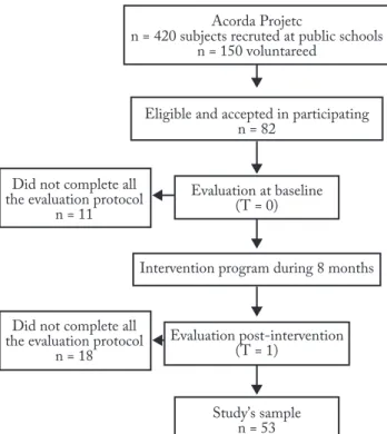
A total of 82 students (10 years old on average) of both genders recruited in Porto public schools were eligible and volunteered to participate in the study, al-though 29 did not complete all the evaluation protocol. Therefore, over a period of 8 months, 53 students were assigned, and: (1) did all the testing procedures at time point 0 (baseline – TP0) and time point 1 (post-inter-vention – TP1); (2) were not attending any other for-mal sports or physical activity program. All the partic-ipants were stimulated to modify their lifestyle habits and to engage in regular physical exercise classes.
The planning program converged in three hours of mandatory Physical Education formal classes and two hours of after-school sessions (1h each session), result-ing in a total of five hours per week, from October to June 2012/2013. The project´s coordinators previously planned the exercise sessions for each class and each after-school session. Two graduates in Sport Scienc-es, under the guidance of two researchers, taught the classes, ensuring that the type and variety of exercises would be performed according to previously planned, and equally applied to all schools. Physical exercise ses-sions included 15 minutes for warm-up with aerobic endurance and flexibility, 30 minutes of working cir-cuit for aerobics, strength training, coordination and balance (with balls, bows, strings and callisthenic exer-cises), 10 minutes of games to promote the enjoyment and five minutes of stretching. All activities were done in indoor schools sports facilities. Exercises and games were progressively intensified as individually tolerated,
according to their heart rate and subjective perception of effort. Training intensity and compliance between individuals were defined to induce heart rate (HR) higher than 70% of each child’s HRmax. To ensure this, 10 randomly selected children wore a portable HR monitor during sessions (Polar Team2 Pro, Polar, Finland). Attendance was in average of 85%.
The Regional Education Board approved the study protocol, and students, parents and schools agreed to participate. The nature, benefits, and risks of the study were explained to the volunteers, and a parent’s writ-ten informed consent was obtained before the study, consistent with the Helsinki Declaration. The exper-imental protocol was approved by the Review Com-mittee of the Scientific Board of the Faculty of Sport, University of Porto, as well as by the Foundation of Science and Technology under the process: PTDC/ DTP-DES/0393/2012. The project staff, according to the approved study protocol, did all the measurements.
Figure 1 – Study flow diagram of schools and participants through the 8-months intervention. Porto, Portugal, 2013 (n = 53).
This information was recorded in a cadastral record, along with the subject’s name, gender and age. Height and weight were measured with participants wearing only shorts and t-shirts. Height was measured using a Holtainstadiometer (Holtain Ltd., Crymmych, UK) and recorded in centimeters to the nearest millime-ter. Weight was measured to the nearest 0.1 kg with
the scale Tanita MC 180 MA. BMI was calculated by the ratio between weight and squared height (kg/m2).
Body mass index categories were set using internation-al recommended age-sex cut points13.
Maturational stage was determined on an individu-al basis during physicindividu-al examination. Each participant self-assessed his/her own stages of secondary sex char-acteristics: stage of breast development in females and pubic hair in males14. A previous study showed a high
correlation (r = 0.73) between ratings on two occasions (three days interval) in a sample of 50 selected sub-jects. Concordance between self-assessments of sexual maturity status and physician assessment ranged from 63% for girls and 89% for boys15. This information was
used as a co-variable in the statistical procedures. Whole body Dual-energy X-ray Absorptiometry (DXA) was performed using a Hologic Explorer con-figured with a software version 12.1 (Hologic, Bedford, MA). Measurements were analyzed using Hologic APEX 3.1 software (Hologic) according to standard pro-cedures set in the user’s guide for the DXA instrument, and BM, BA, and BMD z-score (primary outcome vari-ables). Body lean mass (BLM) and body fat mass (BFM) were also reported (secondary outcome variables). The in-vivo coefficient of variation (CV) was 0.5%.
In order to minimize DXA analysis error in a re-peated measurements approach, one operator analyzed all scans (baseline and follow-up) in a short period of time, so that consistency of procedures were optimized, and few changes were done from the default scan16.
After an overnight fast, blood was obtained by ven-ipuncture in EDTA containing tubes and processed within 2 hours of collection. Fasting serum samples were analyzed for 25 (OH) D (vitD) by chemilumines-cent assay. The same control and reagent lots were used for the analysis of the samples, and all of the samples, in duplicate, from a participant were run in the same assay. The inter-assay CVs for the low and high controls were 8% and 6%, respectively, and the intra-assay CVs for the low and high controls were 5% and 3%, respectively.
Physical activity was objectively assessed by accel-erometers (wGT3x, Actigraph, Florida) during sev-en consecutive days. Data was stored in raw mode in samplings of 30Hz. With a specific software (ActiLife, version 6.9, Actigraph, Florida), data was reduced into one-minute periods (epochs), organized into daily phys-ical activity and analyzed after data collection. Wear and nonwear time were determined according to established algorithms17. Time periods with at least 10 consecutive
minutes of zero counts recorded were excluded from analysis assuming that the monitor was not worn. A minimum recording of 8-hours/day (480-minutes/day) was the criteria to accept daily physical activity data as valid. Individual’s data were only accepted for analysis if at least three-week days and one weekend day were successfully assessed. The main outcomes of reduced accelerometer data were: total physical activity (total Physical Activity (counts/min/day)), time in sedentary behavior (sedentary time (min/day)), light physical ac-tivity (Low Physical Acac-tivity (min/day)), and moderate to vigorous physical activity (MVPA (min/day)).
International recommendations18,19 cut-points for
youths were used to determine time spent in PA of dif-ferent intensities. The following counts intervals were considered: 0-100 for sedentary time, 101-2295 for low physical activity, and ≥ 2296-4011 for MVPA18.
Descriptive data for continuous variables are pre-sented as mean, ± standard deviation for adjusted anal-yses. At baseline, preliminary Independent Student’s T-test was carried out to analyze differences between boys and girls in the entire sample, for all the stud-ied variables, and no statistically significant differences were seen (p < 0.05). Paired Student’s T-test was car-ried out to analyze differences between TP0 and TP1, and it was shown statistically significant differences between the two times for some variables.
For descriptive purposes, relative changes (%Δ) were calculated as ((TP1-TP0)/TP0)*100, and partial correlations between %Δ of the different bone variables and the other measured variables were analyzed, after
adjustments for sex, age at post-test (T1), maturational status, vitD at baseline, and %Δheight. The main re-sults for General Linear Models (GLM) - Repeated Measures Analysis of Covariance - were based in the analyses of BMD z-scores, according to the American Academy of Pediatrics Association guidance20.
GLM was carried out with adjustments for poten-tial confounders: sex, age, maturational status, %ΔvitD, %Δheight, %Δlean mass, and %Δfat mass (model 1). If this analysis was significant, it was considered to be indicative of a significant treatment (intervention) effect. Considering differences between the two time points, adjustments for sedentary time relative change (%ΔST) (model 2), and %ΔMVPA (model 3) were also analyzed. All analyses were performed using the SPSS 21.0 (SPSS Inc., Chicago, IL) for Mac OSX and sig-nificance level was set at 5%.
Results
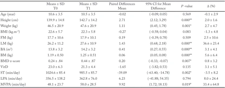
Table 1 shows descriptive data of the entire sample. At baseline, we ran t-test between gender and no statistically significant differences were found (data not showed). Pairwise comparisons indicated statistically significant increase between T0 and T1 for height lean mass, BA, BM (p < 0.01), as well as for BMD z-score, and MVPA (p < 0.05), while statistically significant decrease were found for weight, (p < 0.01) and sedentary time (p < 0.05).
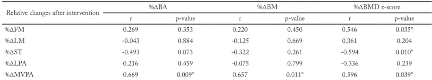
Partial correlations between relative changes in bone variables, body composition and physical activity lev-els are shown in Table 2. Correlations were adjusted for sex, age, maturational status, vitD at baseline, and
Table 1 - Descriptive analysis of the measured variables in overweight/obese children at the two time points. Porto, Portugal, 2013 (n = 53).
Means ± SD
T0 Means ± SDT1 Paired DifferencesMean 95% CI for Mean Difference P -value Δ (%) Age (year) 10.6 ± 3.5 10.5 ± 3.5 -0.02 (-0.09; 0.05) 0.569 -0.1 ± 2.9 Height (cm) 139.9 ± 14.8 142.7 ± 14.2 2.71 (2.12; 3.29) 0.000** 2.0 ± 1.6 Weight (kg) 46.5 ± 20.9 47.6 ± 20.9 1.11 (0.45; 1.78) 0.001* 2.7 ± 4.7 BMI (kg.m-2) 22.6 ± 5.7 22.3 ± 5.8 -0.27 (-0.58; 0.04) 0.083 -1.3 ± 4.8 FM (kg) 17.7 ± 10.6 17.9 ± 10.1 0.19 (-0.39; 0.78) 0.509 2.5 ± 10.6 LM (kg) 26.2 ± 11.2 27.6 ± 10.9 1.43 (0.68; 2.18) 0.000** 36.6 ± 23.4 BA (m2) 13.8 ± 3.2 14.2 ± 3.2 0.41 (0.27; 0.55) 0.000** 3.1 ± 4.1 BM (kg) 1.19 ± 0.50 1.25 ± 0.51 0.06 (0.05; 0.08) 0.000** 5.6 ± 6.4 BMD z-score 0.24 ± .84 0.44 ± .87 0.20 (-0.33; -0.07) 0.007* 0.8 ± 3.2 VitD 23.0 ± 6.3 21.3 ± 4.4 -1.65 (-3.82; 0.53) 0.135 3.1 ± 5.1 ST (min/day) 1024.6 ± 85.4 985.5 ± 85.7 -39.09 (-63.40; -14.78) 0.002* -3.5 ± 8.2 LPA (min/day) 356.5 ± 138.2 362.8 ± 76.0 6.23 (-41.88; 54.35) 0.794 8.0 ± 24.4 MVPA (min/day) 48.1 ± 23.7 58.0 ± 28.5 9.92 (1.72; 18.13) 0.019* 33.4 ± 64.8
BMI = body mass index; FM = fat mass; LM = lean mass; BA = bone area; BM = bone mass; BMD z-score = bone mineral density z-score; VitD = vitamin D; ST = sedentary time; LPA = low physical activity; MVPA = moderate to vigorous physical activity; Differences (* p < 0,05; ** p < 0,01) between the two time points (T0 and T1); SD = standard deviation.
%Δheight. Positive changes (increased levels) of MVPA were positive and significantly correlated with increased levels of BA, BM and BMD z-score. In addition, BMD z-score was inverse and significantly correlated with de-creased values of sedentary time and %ΔFM. Any other statistically significant differences were found.
The results for GLM (Table 3) showed a significant effect of the exposure in %ΔBMD z-score (F = 2.252; η2 = 0.132). After adjustments for relative changes in
sed-entary time and MVPA, no significant effect was observed.
Discussion
This study explored whether changes in physical acti-vity intensities as well as sedentary time are related to changes in bone outcomes in obese/overweight chil-dren and adolescents after 8 months of a school-based interdisciplinary intervention program. To the best of our knowledge, this is one of the few studies presenting associations between variables of bone mass with se-dentary time and physical activity intensities objectively measured in a group of overweight/obese youngsters.
Our study design aimed to assess the effect of a school-based physical activity program on a given popu-lation (overweight/obese youngsters), performing “pre-post” intervention measures. Nonetheless, it has the strength of temporality, which might suggest that the outcome is impacted by the intervention21. Noteworthy,
was the fact that our data showed a significant overall effect of the intervention on %ΔBMD z-score, that after adjustments for changes in sedentary time and MVPA, no significant effect was observed. These findings high-light that variations in sedentary time and in MVPA play an important role in BMD in those children and adolescents involved in the interdisciplinary program.
Indeed, our data agreed with previous studies showing that in Spanish boys, self-reported screen-based activities were associated with an increased risk for low whole body BM22. Other study has shown that
8 months of physical activity program lead towards a significant gain of bone mineral content in proximal femur and in the intertrochanteric region in children aged 8-10 year-olds23. In addition, and reinforcing
the role of physical activity, a randomized control tri-al study in obese children showed higher increase in bone mineral content in trained group compared to the diet-alone24. Further, increased bone mass during
childhood and adolescence (obese or non-obese) has been associated to the frequency, intensity and type of physical activity25, especially in Physical Education26.
The study by Ivuškāns et al.27 also investigated the
influence of the change in physical activity - evaluated by accelerometer - on bone variables in peripubertal boys (BMI average = 19.77). As in the present study, the main results described by authors were the ability of MVPA
Table 2 – Partial correlations between relative changesa of the measured variables in the overweight/obese children. Porto, Portugal, 2013. (n = 53).
Relative changes after intervention %ΔBA %ΔBM %ΔBMD z-score r p-value r p-value r p-value %ΔFM 0.269 0.353 0.220 0.450 0.546 0.035* %ΔLM -0.043 0.884 -0.125 0.669 0.361 0.204 %ΔST -0.493 0.073 -0.322 0.261 -0.594 0.010* %ΔLPA 0.216 0.459 -0.075 0.799 -0.336 0.239 %ΔMVPA 0.669 0.009* 0.657 0.011* 0.596 0.039*
a = Adjusted for sex, age, maturational status, vitD at baseline, %Δ weight and %Δ height; %ΔFM = relative change fat mass; %ΔLM = relative change lean mass; %ΔST = relative change sedentary; %ΔLPA = relative change low physical activity; %ΔMVPA = relative change moderate to vigorous physical activity; %ΔBA = relative change bone area; %ΔBM = relative change bone mass; %ΔBMD z-score = relative change bone mineral density z-score; * p < 0,05.
Table 3 – Generalized Linear Models analysis for the effect of the intervention in % Δ bone mineral density z-score in overweight/obese children. Porto, Portugal, 2013 (n = 53).
Relative changes in BMD TP0 TP1
Mean SE Mean SE Time effect F p-value Partial Eta² %Δ BMD z-score Model 1a 0.128 0.142 0.188 0.147 2.252 0.000** 0.132 %Δ BMD z-score Model 2 (%ΔST) 0.238 0.144 0.292 0.136 1.173 0.296 0.073 %Δ BMD z-score Model 3 (%ΔMVPA) 0.319 0.185 0.344 0.156 0.304 0.605 0.057
a = Adjusted for sex, age, maturational status, %Δ vitD, %Δ height, %Δ lean mass, and %Δ fat mass; %ΔBMD z-score = relative change bone mineral density z-score; %ΔST = relative change sedentary time; %ΔMVPA = relative change moderate to vigorous physical activity.** p < 0.01.
and sedentary time to influence the modification of bone mass in one year. Another common point is that light physical activity was not associated with any bone variable at cross-sectional and longitudinal analyzes.
Other studies also demonstrate light physical activ-ity as absent in relations with bone variables11,27.
How-ever, overweight/obese children and adolescents are more predisposed to light physical activity and seden-tary activities and these variables should be considered in studies with this population. Because light physical activity is an important variable of the children’s general physical activity behavior and sedentary time influences a reduction or stagnation of bone mass development.
The ability of physical activity to stimulate bone re-modeling is determined by the nature and magnitude of the load, the rate at which the load is applied, and the duration of the loading session. During childhood, bouts of high-impact activities augment bone mass accrual and enhance bone´s structural characteristics28. So, the
increased levels of MVPA levels achieved at T1 (post-in-tervention) may have positively influenced the better bone profile observed in our obese/overweight sample.
The osteogenic effect of high-level physical exer-cises, that is, the MVPA characterized by high vol-ume and intensity, has already been reported in other populations29. However, when thinking about
school-based interventions, the intensity and volume of activ-ities should be considered with caution. However, this is still one of the best intervention strategies for the young population in general. Besides having applica-bility, it demonstrates relevant results in bone health26.
Despite the intervention had last 8 months, and no follow-up measures has been taken, we do believe that variation in MVPA levels lead towards a better BM in these overweight/obese children. Indeed, a review suggested three possible mechanisms to explain the deleterious effect of obesity on bone metabolism, as such (1) obesity status, by increasing adipogenesis, may decrease osteoblastogenesis because adipocytes and osteoblasts are derived from a common multipotential stem-cell; (2) obesity is associated with chronic inflam-mation, and proinflammatory cytokines are the main mediators of osteoclast differentiation as well as bone reabsorption is increased in chronic inflammatory dis-orders, and, (3) a high fat diet may compromise in-testinal calcium absorption by insoluble calcium soaps produced from free fatty acids30. Though, even without
a significant decrease in FM, the positive increase ob-served in LM may have influenced the bone profile.
Our results refer to a population that presents high chances of development of several diseases throughout the life. Therefore, a strong point of this study is the rigor in the evaluation method of the variables (scien-tific point), mainly by the use of gold standard eval-uations to evaluate physical activity, sedentary time, blood and bone variables. And also, the proposition of an intervention applicable in the school environment (social point), whose effect is beneficial in an important area of the young people health.
Notwithstanding these points, some limitations must be considered. Our longitudinal results cannot be compared to a similar group of Portuguese children due to the absence of a control group. However, the authors used all available techniques to minimize this limitation.
In conclusion, the 8-months interdisciplinary in-tervention program showed positive correlations be-tween the variation in MVPA and the variations in all the measured bone outcomes. A significant overall ef-fect of the intervention on BMD z-score was observed. After adjustments for changes in ST and MVPA, no significant effect was observed. These findings high-light that in those children and adolescents involved in the program, variations in ST and in MVPA play an important role in BMD.
Conflict of interest
The authors declare no conflict of interest.
Authors’ contributions
MelloJB, collaborated with the analysis, interpretation and discussion
of the results, writing of the text and approval of the final version. Lemos LFGBP, collaborated with the data collection, execution of the intervention. Aires LM, collaborated with the design of the research project, data collection, execution of the intervention. Silva G, collabo-rated with the design of the research project, data collection, execution of the intervention. Tassitano RM, collaborated with the writing of the text. Mota JA, collaborated with the design of the research project, supervision of the writing of the text and approval of the final version. Gaya AR, collaborated with the analysis, interpretation and discussion of the results, wording of the text and approval of the final version. Martins CML, collaborated with the design of the research project, data collection, execution of the intervention, analysis of the data, su-pervision of the writing of the text and approval of the final version.
Funding
Mota JA was supported by grants FCT: SFRH/ BSAB/142983/2018 and UID/DTP/00617/2019 as well as Pro-grama de Bolsas Santander Universidades 2018.
Quote this article as:
Mello JB, Lemos LFGBP, Aires LM, Silva G, Tassitano RM, Mota JA, Gaya AR, Martins CML. Physical activity, sedentary time and bone tissue: ef-fects of an 8-months interdisciplinary program with overweight/obese children. Rev Bras Ati Fis Saúde. 2018;23:e0053. DOI: 10.12820/rbafs.23e0053
References
1. Trost SG, Blair SN, Khan KM. Physical inactivity remains
the greatest public health problem of the 21st century: Evidence, improved methods and solutions using the ‘7 investments that work’ as a framework. Br J Sports Med. 2014;48(3):169–70.
2. Janz KF, Thomas DQ, Ford MA, Williams SM. Top 10
research questions related to physical activity and bone health in children and adolescents. Res Q Exerc Sport. 2015;86(1):5-12.
3. Gómez-Bruton A, Matute-Llorente Á, González-Agüero A,
Casajús JA, Vicente-Rodríguez G. Plyometric exercise and bone health in children and adolescents: a systematic review. World J Pediatr. 2017;13(2):112-21.
4. Cadore EL, Brentano MA, Kruel LFM. Efeitos da atividade
física na densidade mineral óssea e na remodelação do tecido ósseo. Rev Bras Med do Esporte. 2005;11(6):373-79.
5. Bass SL, Naughton G, Saxon L, Iuliano-Burns S, Daly R,
Briganti EM, et al. Exercise and calcium combined results in a greater osteogenic effect than either factor alone: a blinded randomized placebo-controlled trial in boys. J Bone Miner Res. 2007;22(3):458-64.
6. Bucksch J, Sigmundova D, Hamrik Z, Troped PJ, Melkevik
O, Ahluwalia N, et al. International trends in adolescent screen-time behaviors from 2002 to 2010. J Adolescent Health. 2016;58(4):417-25.
7. Lopes AS, Silva KS, Barbosa Filho VC, Bezerra J, Oliveira
ES, Nahas MV. Trends in screen time on week and weekend days in a representative sample of Southern Brazil students. J Public Health. 2014;36(4):608-14.
8. Koedijk JB, van Rijswijk J, Oranje WA, van den Bergh JP,
Bours SP, Savelberg HH, et al. Sedentary behaviour and bone health in children, adolescents and young adults: a systematic review. Osteoporos Int. 2017;28(9):2507-19.
9. Weiler HA, Janzen L, Green K, Grabowski J, Seshia MM,
Yuen KC. Percent body fat and bone mass in healthy Canadian females 10 to 19 years of age. Bone. 2000;7(2):203-07.
10. Halade G V, El Jamali A, Williams PJ, Fajardo RJ, Fernandes
G. Obesity-mediated inflammatory microenvironment stimulates osteoclastogenesis and bone loss in mice. Exp Gerontol. 2011;46(1):43-52.
11. Chaplais E, Naughton G, Greene D, Dutheil F, Pereira B,
Thivel D, et al. Effects of interventions with a physical activity component on bone health in obese children and adolescents: a systematic review and meta-analysis. J Bone Miner Metab. 2018;36(1):12-30.
12. Aires L, Silva G, Martins C, Marques E, Lagoa MJ, Ribeiro
JC, et al. Exercise intervention and cardiovascular risk factors in obese children. Comparison between obese youngsters taking part in a physical activity school-based programme with and without individualized diet counselling: the ACORDA project. Ann Hum Biol. 2016;43(3):183-90.
13. Cole TJ. Establishing a standard definition for child
overweight and obesity worldwide: international survey. BMJ. 2000;320(7244):1240.
14. Tanner JM. Growth at Adolescence: With a General
Consideration of the Effects of Hereditary and Environmental Factors Upon Growth and Maturation from Birth to Maturity. 1962.
Duarte JA. Association of maturation, sex, and body fat in cardiorespiratory fitness. Am J Hum Biol. 2002;14(6):707-12.
16. Fulkerson JA, Himes JH, French SA, Jensen S, Petit MA,
Stewart C, et al. Bone outcomes and technical measurement issues of bone health among children and adolescents: considerations for nutrition and physical activity intervention trials. Osteoporos Int. 2004;15(12):929-41.
17. Choi L, Liu Z, Matthews CE, Buchowski MS. Validation
of accelerometer wear and nonwear time classification algorithm. Med Sci Sports Exerc. 2011;43(2):357-64.
18. Evenson KR, Catellier DJ, Gill K, Ondrak KS, McMurray
RG. Calibration of two objective measures of physical activity for children. J Sports Sci. 2008;26(14):1557-65.
19. Trost SG, Loprinzi PD, Moore R, Pfeiffer KA. Comparison
of accelerometer cut points for predicting activity intensity in youth. Med Sci Sports Exerc. 2011;43(7):1360-68.
20. Bachrach LK, Sills IN. Bone densitometry in children and
adolescents. Pediatrics. 2011;127(1):189-94.
21. Thiese MS. Observational and interventional study design
types; an overview. Biochem Medica. 2014;24(2):199-210.
22. Vicente-Rodríguez G, Ortega FB, Rey-López JP,
España-Romero V, Blay VA, Blay G, et al. Extracurricular physical activity participation modifies the association between high TV watching and low bone mass. Bone. 2009;45(5):925–30.
23. McKay HA, MacLean L, Petit M, MacKelvie-O’Brien K,
Janssen P, Beck T, et al. ‘Bounce at the Bell’: A novel program of short bouts of exercise improves proximal femur bone mass in early pubertal children. Br J Sports Med. 2005;39(8):521–26.
24. Yu CCW, Sung RYT, So RCH, Lui K-C, Lau W, Lam PKW,
et al. Effects of strength training on body composition and bone mineral content in children who are obese. J Strength Cond Res. 2005;19(3):667–72.
25. Gracia-Marco L, Vicente-Rodriguez G, Casajus JA, Molnar
D, Castillo MJ, Moreno LA. Effect of fitness and physical activity on bone mass in adolescents: the HELENA Study. Eur J Appl Physiol. 2011;111(11):2671–80.
26. Daly RM, Ducher G, Hill B, Telford RM, Eser P, Naughton
G, et al. Effects of a Specialist-Led, School Physical Education Program on Bone Mass, Structure, and Strength in Primary School Children: A 4-Year Cluster Randomized Controlled Trial. J Bone Miner Res. 2016;31(2):289–98.
27. Ivuškāns A, Mäestu J, Jürimäe T, Lätt E, Purge P, Saar M, et
al. Sedentary time has a negative influence on bone mineral parameters in peripubertal boys: a 1-year prospective study 2015 J Bone Miner Metab. 2015;33(1):85-92.
28. Macdonald HM, Ashe MC, McKay HA. The link between
physical activity and bone strength across the lifespan. Int J Clin Rheumtol. 2009;4(4):437–63.
29. Nasri R, Hassen Zrour S, Rebai H, Fadhel Najjar M, Neffeti
F, Bergaoui N, et al. Grip Strength is a Predictor of Bone Mineral Density Among Adolescent Combat Sport Athletes. J Clin Densitom. 2013;16(1):92–7.
30. Cao JJ. Effects of obesity on bone metabolism. J Orthop Surg
Res. 2011;6(1):30-6.
Received: 16/11/2018 Approved: 22/03/2019