Cop
yright
© ABE&M t
odos os dir
eit
os r
eser
vados
.
The effect of weight reduction
on antioxidant enzymes and
their association with dietary
intake of vitamins A, C and E
Efeito da redução de peso sobre as enzimas antioxidantes e associação destas com a ingestão de vitaminas A, C e E
Masoud Ramezanipour1, Mahmood Jalali1, Haleh Sadrzade-Yeganeh2, Seyed Ali
Keshavarz3, Mohammad Reza Eshraghian4, Minoo Bagheri2, Sara Seyed Emami5
ABSTRACT
Objective: Our goal was to assess the effects of weight loss on antioxidant enzymes of red blood cells and it’s relation with vitamins A, E and C intake in 30 obese women. Subjects and methods: General information, anthropometric measurements, 3-day food recall, and fasting blood samples were collected from 30 obese women at the beginning of the study and after 3 months intervention. Weight loss was set at about 10% of their weight before the intervention. Results: Glutathione reductase and catalase activities showed a signiicant increase (P < 0.01) after weight reduction, but no signiicant changes were seen in the superoxide dismutase and glutathione peroxidase activities. There was a positive linear correlation between daily vitamin C intake with superoxide dismutase enzyme after intervention (P = 0.004, r = 0.507). There was a negative linear correlation between vitamin E intake and glutathione peroxidase activity before intervention (P = 0.005, r = -0.5). A negative correlation was found between daily vitamin A intake and glutathione reductase enzyme before and after intervention (r = -0.385, r = -0.397, P < 0.05) respectively. No signiicant correlation was observed between vitamins A, C, E amounts and catalase activity. Conclusions: Ten percent weight reduction can have a signiicant role in in-creasing antioxidant enzymes activities, especially glutathione reductase, and catalase enzymes in obese women. However, it is important to take into consideration a balanced amount of cer-tain nutrients while administering a diet with limited energy. Arq Bras Endocrinol Metab. 2014;58(7):744-9
Keywords
Obesity; enzymic antioxidants; weight reduction
RESUMO
Objetivo: Nosso objetivo foi avaliar os efeitos da perda de peso sobre as enzimas antioxidantes de eritrócitos, e a relação destas com a ingestão das vitaminas A, E e C. Sujeitos e métodos: Foram coletadas informações gerais e medidas antropométricas, registro alimentar de três dias e amostras de sangue em jejum de 30 mulheres obesas no início do estudo e depois de três meses da intervenção. A perda de peso determinada antes da intervenção foi de 10% do peso. Resultados: As atividades da glutationa redutase e da catalase mostraram aumento signiicativo (P < 0,01) depois da perda de peso, mas não houve mudanças signiicativas nas atividades da superóxido dismutase e da glutationa peroxidase. Foi observada uma correlação linear positiva entre a ingestão diária de vitamina C e a enzima superóxido dismutase após a intervenção (P = 0,004, r = 0,507). Houve uma correlação linear negativa entre a ingestão de vitamina E e a atividade da glutationa peroxidase antes da intervenção (P = 0,005, r = -0,5). Foi observada uma correlação negativa entre a ingestão diária de vitamina A e a enzima glutationa redutase antes e depois da intervenção (r = -0,385, r = -0,397, P < 0,05), respectivamente. Não foram observadas correlações signiicativas entre as vitaminas A, C, E e os níveis e a atividade da catalase. Conclu-sões: Uma redução de 10% no peso pode ter um papel signiicativo no aumento da atividade das enzimas antioxidantes, especialmente na glutationa redutase e catalase em mulheres obesas. Entretanto, é importante levar em consideração uma ingestão equilibrada de certos nutrientes ao se recomendar uma dieta com níveis de energia restritos. Arq Bras Endocrinol Metab. 2014;58(7):744-9
Descritores
Obesidade; enzimas antioxidantes; redução de peso
1Department of Cellular and
Molecular Nutrition, School of Nutritional Sciences and Dietetic and School of Public Health, Tehran University of Medical Sciences, Tehran, Iran
2 Department of Community
Nutrition, School of Nutritional Sciences and Dietetic and School of Public Health, Tehran University of Medical Sciences, Tehran, Iran
3 Department of Clinical Nutrition,
School of Nutritional Sciences and Dietetic and School of Public Health, Tehran University of Medical Sciences, Tehran, Iran
4 Department of Epidemiology
and Biostatistics, School of Public Health, Tehran University of Medical Sciences, Tehran, Iran
5 Tehran University of Medical
Sciences, Tehran, Iran
Correspondence to: Haleh Sadrzadeh-Yeganeh Department of Community Nutrition, School of Nutritional Sciences and Dietetic and School of Public Health, Tehran University of Medical Sciences,
Poursina Avenue 14155-6446 – Tehran, Iran [email protected]
Received on Jan/2/2014 Accepted on July/6/2014
Cop
yright
© ABE&M t
odos os dir
eit
os r
eser
vados
.
INTRODUCTION
O
besity, which is deined as having a body mass index (BMI) of 30 kg/m2 and above, has beenon the increase in many countries in recent decades (1-3). It is a risk factor for many diseases including diabetes mellitus, hyperlipidemia, colon cancer, sudden death,
illnesses related to gall bladder, high blood pressure, ar-teriosclerosis, and cardiovascular diseases. Obesity pa-thogenesis is accompanied with the increase of oxygen derived free radicals (1-3). Recent studies show that obesity results in oxidative stress even in the absence of other risk factors including cardiovascular diseases (4) and therefore plays a role in development of the above--mentioned diseases (5-7). Likewise, adipose tissue was found to be an independent factor to produce oxidative stress (8).
The removal of free radicals by antioxidants happens through enzymic and non-enzymic reactions, but re-search on human has shown that an increase in weight can reduce the antioxidant capacity of plasma (5,9). Therefore, enzymic and non enzymic antioxidants can constitute one of the most important defense barrier of cells (9,10).
Obesity–associated oxidative stress could occur be-cause of increased oxygen utilization and consequent radical formation through mitochondrial respiration, more fat deposition and cell damage (12). Studies on human and animals show controversial results. Some studies have shown that antioxidant enzymes increase in obesity (13,14). Other studies have identiied no sig-niicant difference in antioxidant enzymes concentra-tions in obesity (1,15).
Dietary intake can play an important role in antioxi-dant defense system of the body by reducing the oxida-tive damage occurred in the presence of free radicals (16). Diets rich in fruits and vegetables contain anti-oxidant that can protect the body from oxidative dam-age (16,17). It was suggested that not taking enough antioxidant could increase oxidative damage (18,19). Most of the research done in this regard is related to the comparison of antioxidant enzymes activities in overweight people compared to people within a nor-mal BMI range. In this survey we studied the effects of the 10% weight loss (achieved through a limited energy diet) on antioxidant enzymes activities of red blood cells; glutathione peroxidase (GPX), superoxide dis-mutase (SOD), catalase (CAT), glutathione reductase (GR) and it’s relation with the intakes of vitamins A, E and C in obese women.
SUBJECTS AND METHODS
This study is a quasi experimental trail conducted on 30 obese women aged between 19 to 50 years, who visited a diet therapy clinic in the city of Tehran. The sample was chosen from women who frequented the diet therapy clinic and who met the requirements for participating in the study. The inclusion criteria were being women aged between 19 to 50 years and having a BMI of or above 30 kg/m2. Exclusion criteria were
being menopausal, smoking, having diseases related to the heart, kidneys, liver, cancer, diabetes mellitus, and
hyperlipidemia. Other exclusion criteria were having intestinal inlammatory diseases, taking supplements, taking medication, being pregnant or being breastfe-eding women. After attaining written consent, general information, anthropometric measurements and 3-day 24 hour food recall were completed by a nutritionist. Subjects went on a weight loss diet with an energy de-icit of 500-1,000 kcal/day through lower intake of macronutrients and higher intake of iber.For achieve-ment of the recommended calories by the nutritionist, the carbohydrate, fat and protein were set at 55%, 25% and 20% of the total amount of energy required for approximately 10% weight loss in 3 months, respective-ly. During the study participants were monitored every two weeks for weight changes and for compliance with the diet and the study protocol.
The participant’s weight was measured with digital scale (Seca GmbH & co.KG, Germany) with a preci-sion of 100 grams and height was measured with non-stretchable tape (SecaGmbH, Germany) with a preci-sion of 0.1 cm. BMI was obtained by dividing weight by the square of height. Triceps skin fold (TSF) was measured with a caliper with an accuracy of 0.1 ml. TSF, BMI and weight measurement were used to in-tensify that weight loss was achieved after the interven-tion.
Dietary intake
Three 24-hour dietary recalls for 2 weekdays and one weekend day were asked from all subjects both before and after the intervention and the collected data of be-fore and after the intervention was separately analyzed by Food Processor 2.
Cop
yright
© ABE&M t
odos os dir
eit
os r
eser
vados
.
a recommended diet, the information was gathered again. The amount of the recommended calories by the nutritionist was 500 to 1,000 kcal/day less than the calorie intake before intervention. In accordance, for achievement of the recommended calories by the nutritionist, the carbohydrate, fat and protein were set at 55%, 25% and 20% of the total amount of energy re-quired for approximately 10% weight loss in 3 months, respectively.
Biochemical measurements
In order to measure hemoglobin and antioxidants en-zymes activities per gram of hemoglobin, 10 mL fas-ting blood sample was drawn from the saphenous vein before and after intervention. The hemoglobin was measured according to Zistshimi kit method (Cat No: 10-532, Zistshimi company, Tehran, Iran).
SOD enzyme activity was measured according to Ransod Kit method (Cat No: SD 125, Randox-Ransod, UK) In this methodSOD activity was assessed by mea-suring the dismutation of superoxide radicals generated by xanthine oxidase and hypoxanthine (20). Catalase activity in erythrocytes was assessed by the method de-scribed by Hugo Aebi (21). In this method, activity of CAT was determined by following the decomposition of H2O2 in phosphate buffer pH 7.2 spectrophotomet-rically at 230 nm. GR activity was measured according to Sauberlich method (22) and GPX activity was mea-sured according to Paglia and Valentine method with modiications according to Lawrence and Burk (23).
The present study was conducted according to the guidelines laid down in the declaration of Helsinki and all procedures involving human subjects were approved by the Ethics Committee at Tehran University Medical Sciences. Written informed consent was obtained from all subjects.
Statistical analysis
The Food Processor II, nutrient analysis program sof-tware, was used to determine the calorie and nutrients intake of the participants. SPSS software (ver.11.5)was used for the analysis of the data. The paired t-test was used for the comparison of quantitative variables before and after intervention.
The percentage of energy from carbohydrates, fat and protein and the intake of iber before and after the intervention were examined. Furthermore, the associa-tion of vitamins A, C and E with antioxidant enzymes
was assessed since these vitamins were likely to inluen-ce the antioxidant enzymes activities. To explore the effect of vitamins A and E independent of dietary fat intake, stepwise regression analysis was performed.
In order to determine the amount of daily intake of A, E and C vitamins from food, participants were di-vided into two groups: the group that received 75% less than the recommended daily allowance (RDA) and the normal group who received equal or more than 75% of the RDA (24). The stepwise regression analysis was applied to assess the changes in quantitative variables before and after intervention on antioxidant enzymes activities. In order to measure the correlation between enzymes activities, the Pearson’s correlation coeficient was used.
RESULTS
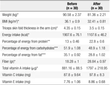
As it is shown in table 1, on the basis of the paired t-test, there was a signiicant difference between the weight, BMI, TSF, energy intake, dietary ibers, total vitamin A intake (P < 0.01), and the achieved energy percentage from the received carbohydrates and fat (P < 0.01) of the participants before and after intervention. Also the achievement of the energy percentage from the received protein of the participants showed a statistically signii-cant difference between before and after intervention (P < 0.05). No statistical signiicant difference was obser-ved between the intake amounts of vitamin E (tocophe-rol) and vitamin C before and after intervention.
Table 1. Mean and standard errors of independent variables before and after intervention
After (n = 30) Before
(n = 30)
81.36 ± 2.21 90.58 ± 2.37
Weight (kg)*
32.41 ± 0.81 36.1 ± 0.9
BMI (kg/m2)*
3.5 ± 0.15 4.35 ± 0.15
Triceps skin fold thickness in the arm (cm)*
1107.6 ± 46.2 1907.6 ± 76.1
Energy intake (kcal)*
22.8 ± 0.6 13 ± 0.46
Percentage of energy from protein**
48.9 ± 1.18 51.9 ± 1.08
Percentage of energy from carbohydrates***
28.8 ± 1.02 35.1 ± 0.92
Percentage of energy from fat***
28.84 ± 0.97 18.28 ± 1
Fiber (gr)*
1797 ± 218.95 881.16 ± 88.5
Total vitamin A intake (mg)*
97.8 ± 8.3 87.8 ± 9.64
Vitamin C intake (mg)
8.86 ± 0.68 7.76 ± 1.06
Vitamin E intake (mg)
Data are shown as mean and standard errors.
Cop
yright
© ABE&M t
odos os dir
eit
os r
eser
vados
.
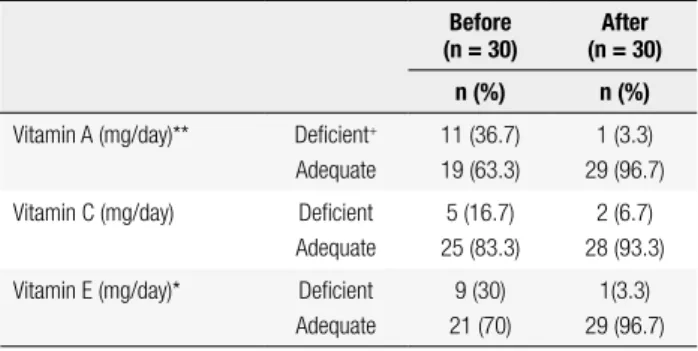
Table 2 shows the antioxidant vitamins intake status of the participants before and after intervention. Vita-min intake level less than 75% of RDA was classiied as deicient and intake level equal to more than 75%
of RDA were classiied as normal. By applying the Mc Nemar test, daily intake status of total amounts of the vitamins A and E showed a signiicant difference (P < 0.05) between before and after intervention. There was no signiicant difference between the vitamin C intake status of the participants before and after intervention.
GPX activity after intervention (P = 0.005, r = -0.5). A negative linear correlation was also found between vitamin A intake and GR activity both before and after the intervention (r = -0.397, r = -0.385, P < 0.05), res-pectively. There was no signiicant correlation between vitamins A, C and E intake with CAT activity.
DISCUSSION
This study, complying with a restricted energy diet which is low in fat and carbohydrate and rich in protein and vegetables and dairy products, resulted in changes in the nutrient intake of participants after intervention. The-refore, besides the effect of weight loss on antioxidant enzymes activities, the changes in vitamins A, C and E intake can affect the amount of antioxidants as well.
Crujeiras and cols. conducted a study to assess the effect of weight loss and diet modiication on antioxi-dant capacities of obese people. Total antioxiantioxi-dant ca-pacity improved in those who consumed more fruits and vegetables (25). A similar study showed that obesi-ty was an independent risk factor in draining protective enzymes in erythrocytes; individuals with normal BMI had higher SOD and GPX activities in comparison with obese people (1). Thus, weight loss can result in an increase in antioxidant enzymes activities. Melissas and cols. observed that weight loss and decrease in BMI obtained through placing a balloon in the stomach of extremely obese individuals for 6 months, increased the plasma antioxidant capacity (26). Bougoulia and cols. prescribed a calorie restricted diet to 36 obese women for 6 months. They showed that the reduction of BMI from 38.5 to 30.9 is accompanied with a signiicant in-crease in the GPX enzyme activity from 22.3 to 48.9 ng/mL (27).Contrary to present study, GPX activity had a signiicant increase after 20% weight reduction
In an interventional study conducted by Dworschak and cols., on obese individuals, in which they were given
Table 2. Daily vitamins A, C and E intake status of participants before and after intervention
After (n = 30) Before
(n = 30)
n (%) n (%)
1 (3.3) 29 (96.7) 11 (36.7)
19 (63.3) Deicient+
Adequate Vitamin A (mg/day)**
2 (6.7) 28 (93.3) 5 (16.7)
25 (83.3) Deicient
Adequate Vitamin C (mg/day)
1(3.3) 29 (96.7) 9 (30)
21 (70) Deicient
Adequate Vitamin E (mg/day)*
Mc Nemar test was used to assess before and after the intervention.
* P < 0.05 and ** P < 0.01 were considered statistically signiicant. Other variables showed no signiicant difference.
+ Deicient: < 75% of RDA & Adequate: ≥ 75% of RDA.
Table 3 shows the mean and standard error of the antioxidant enzymes activities before and after interven-tion using paired t-test. GR and CAT activities showed a signiicant increase after the intervention (P < 0.01).
Table 3. The antioxidant enzymes activities of participants before and after intervention
After (n = 30) Before
(n = 30)
431.8 ± 34.7 438.1 ± 32.4
Superoxide dismutase (U/grHb)
53.02 ± 2.44 50.6 ± 2.52
Glutathione peroxidase (U/grHb)
4.73 ± 0.51 2.47 ± 0.33
Glutathione reductase (U/grHb)*
231.4 ± 10.8 188.3 ± 8.95
Catalase (U/grHb)*
Data are shown as mean ± standard errors.
Paired t-test was used to assess before and after intervention.
* P < 0.01 was considered statistically signiicant. Other variables showed no signiicant difference.
In order to assess the effect of the weight reduction or nutrient intake on the antioxidant enzymes activities, stepwise regression analysis was conducted (Table 4). In the present study, a positive linear correlation was found between vitamin C intake and SOD activity before in-tervention (P = 0.004, r = 0.507). A negative linear correlation was identiied between vitamin E intake and
Table 4. Stepwise regression analysis of the GR activity changes and the alteration of the independent variables before and after intervention
T Standardized
coeficient Unstandardized
coeficient
Beta Std Error
Beta
-2.7
-1.94 -5.22
Constant
3.9 0.64
0.51 2.01
Difference in BMI*
-2.45 -0.4
0.08 -0.21
Difference in vitamin E**
Cop
yright
© ABE&M t
odos os dir
eit
os r
eser
vados
.
a restricted energy diet of for three months, SOD activity was reduced, whereas the GPX did not change (28). In the present study mean of SOD activity decreased some-what (but not signiicantly) after intervention. Con-trary to Bougoulia and cols.’s study, the GPX activity in Dworschak and cols. study did not change. In the pres-ent study there was a slight increase after intervpres-ention, although it was not signiicant. This could be because of the lower amount of weight reduction compared with Bougoulia and cols. study, where weight loss was 20%.
We found a positive, albeit statistically weak, cor-relation between vitamin C intake status and SOD activity after intervention (Table 2). Even though, no signiicant statistical difference was seen between mean vitamin C intake before and after intervention, most participants had normal intakes of vitamin C after in-tervention. Standard deviation for intake of vitamin C was high among participants. This could be because the power of the study was not enough to show a sig-niicant difference before and after intervention for this vitamin or to show a weak association between vitamin C status and SOD activity. Vitamin C as an antioxidant could be effective in maintaining and increasing SOD activity. This needs further investigation.
We observed a signiicant negative correlation be-tween vitamin E intake and GPX activity before inter-vention. Although mean intake of vitamin E did not
show any signiicant difference before and after the in-tervention, vitamin E status differed signiicantly after the intervention. This means the number of individuals with adequate intake of vitamin E increased after the intervention. This can be the reason for the decrease in GPX activity after intervention.
In the present study, GR activity increased signii-cantly after the intervention and a signiicant negative correlation was observed between vitamin A intake and GR activity before and after the intervention. As a re-sult of dietary intervention, the number of individuals with adequate intake of vitamin A increased after inter-vention. It is possible that if vitamin A intake had not increase after intervention, the increase of GR activity would have been higher.
The number of those that had adequate intake of vitamin A increased after the intervention. Also, a sig-niicant negative correlation between vitamin A intake and GR activity was observed before and after the in-tervention. An increase in intake of this vitamin can re-sult in a decrease of GR activity. In the present study, GR increased signiicantly after the intervention. It is
possible that if vitamin A intake did not increase after intervention, the increase of GR activity after interven-tion would be higher.
The results of the stepwise regression analysis showed that more weight loss causes an increase in GR activity and an increase in vitamin E intake causes a decrease in GR activity. However, the amount of in-crease in GR activity after intervention was almost twice as before the intervention. Perhaps if there had been more weigh loss among the participants, other enzymes would have been increased and the effects of weight loss would have been more apparent.
The irst limitation of our study was small sample size. Thus, when the intakes of vitamins A, E and C were analyzed in subgroups of the study, the power of the re-search might decrease. Also, the study group was limited to only women and therefore our indings can be gen-eralized only to women. We did not observe signiicant changes in activity of some enzymes after intervention. Besides the changes in antioxidant vitamin intake during intervention, one explanation could be that weigh re-duction was not enough in our study. We suggest further studies with more weigh reduction, longer duration of intervention, and different percentages of weight reduc-tion. Another reason that we did not observe signiicant correlation between intake of vitamins such as C could be that the sample size was not large enough for a high standard deviation for intake of this vitamin in our popu-lation. Furthermore, we did not assess the status of other oxidative stress factors. We recommend conducting studies which assess other oxidative stress factors.
In conclusion, a weight loss of about 10% of origi-nal weight can increase antioxidant enzymes especially CAT and GR activities. Also, activity of these enzymes is inluenced by certain nutrients in the diet. Because a correlation was observed between antioxidant vitamins amounts and antioxidant enzymes activities, daily in-take of these vitamins may induce signiicant changes in the antioxidant enzymes activities. The weight loss and BMI reduction might induce an increase of antioxidant enzymes activities in the obese individuals.
Cop
yright
© ABE&M t
odos os dir
eit
os r
eser
vados
.
Seyed Emami assisted in conducting the study. All authors have read and approved the inal manuscript.
Acknowledgements: the present study was supported by a grant from the Vice-chancellor for Research, Tehran University of Me-dical Sciences, Tehran, Iran. We are grateful to the cooperation of the participants.
Disclosure: no potential conlict of interest relevant to this article was reported.
REFERENCES
1. Olusi SO. Obesity is an independent risk factor for plasma lipid peroxidation and depletion of cytoprotectic enzymes in humans. Int J Obes Relat Metab Disord. 2002;26(9):1159-64.
2. Zwirska-Korczala K, Jochem J, Rybus-Kalinowska B, Polaniak R, Birkner E. Assessment of blood superoxide dismutase, gluta-thione peroxidase activity and malondialdehyde concentration as oxidation status parameters in obese women. Pol Arch Med Wewn. 2003;110(1):725-31.
3. DeBoer MD. Obesity, systemic inlammation, and increased risk for cardiovascular disease and diabetes among adolescents: a need for screening tools to target interventions. Nutrition. 2013;29(2):379-86.
4. Higdon JV, Frei B. Obesity and oxidative stress: a direct link to CVD? Arterioscler Thromb Vasc Biol. 2003;23(3)365-7.
5. Bełtowski J, Wójcicka G, Górny D, Marciniak A. The effect of dietary-induced obesity on lipid peroxidation, antioxidant enzy-mes and total plasma antioxidant capacity. J Physiol Pharmacol. 2000;51(4 Pt 2):883-96.
6. Erdeve O, Siklar Z, Kocaturk PA, Dallar Y, Kavas GO. Antioxidant superoxide dismutase activity in obese children. Biol Trace Elem Res. 2004;98(3):219-28.
7. Speakman JR, Mitchell SE. Caloric restriction. Mol Aspects Med. 2011;32(3):159-221.
8. Fernández-Sánchez A, Madrigal-Santillán E, Bautista M, Esquivel--Soto J, Morales-González A, Esquivel-Chirino C, et al. Inlamma-tion, oxidative stress, and obesity. Int J Mol Sci. 2011;12(5):3117-32. 9. Karaouzene N, Merzouk H, Aribi M, Merzouk SA, Berrouiguet AY,
Tessier C, et al. Effects of the association of aging and obesity on lipids, lipoproteins and oxidative stress biomarkers: a com-parison of older with young men. Nutr Metab Cardiovasc Dis. 2011;21(10):792-9.
10. Aaseth J, Norsth T. Copper. In: Friberg L, Nordberg GF, Vouk VB. Handbook on the toxicology of metals. Vol. II. New York: Elsevier Publishing; 1996. p. 233-49.
11. Block G, Jensen CD, Morrow JD, Holland N, Norkus EP, Milne GL, et al. The effect of vitamins C and E on biomarkers of
oxi-dative stress depends on baseline level. Free Radic Biol Med. 2008;45(4):377-84.
12. Vincent HK, Powers SK, Dirks AJ, Scarpace PJ. Mechanism for obesity-induced increase in myocardial lipid peroxidation. Int J Obes Relat Metab Disord. 2001;25(3):378-88.
13. Koboyasi R, Akamine EH, Davel AP, Rodrigues MA, Cavalho CR, Rossoni LV. Oxidative stress and inlammatory mediators contri-bute to endothelial dysfunction in high-fat diet-induced obesity in mice. J Hypertens. 2010;28(10):2111-9.
14. Nakao C, Ookawara T, Sato Y, Kizaki T, Imazeki N, Matsubara O, et al. Extracellular superoxide dismutase in tissues from obese (ob/ ob) mice. Free Radic Res. 2000;33(3):229-41.
15. Brown LA, Kerr CJ, Whiting P, Finer N, McEneny J, Ashton T. Oxi-dant stress in healthy normal-weight, overweight and obese indi-viduals. Obesity (Silver Spring). 2009;17(3):460-6.
16. Bisbal C, Lambert K, Avignon A. Antioxidants and glucose meta-bolism disorders. Curr Opin Clin Nutr Metab Care. 2010;13(4):439-46.
17. Peairs AD, Abbey EL. Antioxidants and inlammation in obesity. In: Walson R, Preedy V (eds.). Bioactive Food as Dietary Interven-tions for Diabetes. Boston: Academic Press; 2013. p. 413-34. 18. Fang YZ, Yang S, Wu G. Free radicals, antioxidants, and nutrition.
Nutrition. 2002;18(10):872-9.
19. Ulrich-Merzenich G, Zeitler H, Vetter H, Kraft K. Synergy research: vitamins and secondary plant components in the maintenance of the redox-homeostasis and in cell signaling. Phytomedicine. 2009;16(1):2-16.
20. Lohe L, Otting F. Superoxid dismutase assays. Methods in Enzy-mology. 1984;105:93-104.
21. Aebi H. Catalase in vitro. Methods Enzymol. 1984;105:121-6. 22. Sauberlich HE. Laboratory tests for the assessment of nutritional
status. Boca Raton: CRC press; 1999.
23. Lawrence RA, Burk RF. Glutathione peroxidase activity in se-lenium-deicient rat liver. Biochem Biophys Res Commun. 1976;71:952-8.
24. Lee R.D, Neiman DC. Nutritional assessment. 3rd ed. Boston: Mc Graw Hill; 2003. p. 19-20.
25. Crujeiras AB, Parra MD, Rodríguez MC, Martínez de Morentin BE, Martínez JA. A role for fruit content in energy-restricted diets in improving antioxidant status in obese women during weight loss. Nutrition. 2006;22(6):593-9.
26. Melissas J, Malliaraki N, Papadakis JA, Talampas P, Kampa M, Castanas E. Plasma antioxidant capacity in morbidly obese pa-tients before and after weight loss. Obes Surg. 2006;16(3):314-20. 27. Bougoulia M, Triantos A, Koliakos G. Plasma Interleukin-6 levels,
glutathione peroxidase and isoprostane in obese women before and after weight loss. Association with cardiovascular risk fac-tors. Hormones (Athens). 2006;5(3):192-9.