Impact of radiotherapy on the orofacial region and
management of related conditions
*
Repercussões da radioterapia na região orofacial e seu tratamento
Ana Emília Holanda Rolim1, Lino João da Costa2, Luciana Maria Pedreira Ramalho3
Head and neck radiotherapy causes countless sequelae in irradiated patients, affecting the stomatognathic system, with significant systemic implications. Sequelae of exposure to ionizing radiation may be extensive and sometimes permanent, particularly in the salivary glands and bone tissue. It is of utmost importance that the surgeon dentist be aware of adverse reactions and appropriate forms of treatment to alleviate discomfort and improve the quality of life of the irradiated patient. Therefore, awareness and motivation of the patient, with promotion of oral health through the adaptation of the oral environment and guidance on preventive measures are essential to get a better prognosis.
Keywords: Head and neck tumors; Radiotherapy; Acute effects; Late effects.
A radioterapia em região de cabeça e pescoço provoca inúmeras sequelas ao paciente irradiado, afetando o sistema estomatognático e com repercussões sistêmicas importantes. As sequelas da radiação ionizante podem ser extensas e, algumas vezes, permanentes, em especial nas glândulas salivares e no tecido ósseo. É relevante que o cirurgião dentista tenha conhecimento das reações adversas e das formas adequadas de prevenção e tratamento para ameni-zar o desconforto e melhorar a condição de vida do paciente irradiado. Portanto, a conscientização e motivação deste paciente, com a promoção de saúde oral através da adequação do meio bucal e orientações sobre ações preventivas, são essenciais para se obter o melhor prognóstico.
Unitermos: Tumores de cabeça e pescoço; Radioterapia; Efeitos agudos; Efeitos tardios. Abstract
Resumo
* Study developed at Universidade Federal da Bahia (UFBA), Salvador, BA, Brazil.
1. Surgeon Dentist, Fellow PhD degree – Organs/Systems Interactive Processes, Program of Post-graduation, Universidade Federal da Bahia (UFBA), Salvador, BA, Brazil.
2. PhD of Diagnosis of Oral Cavity, Full Professor, Department of Clinical Practice and Social Odontology – Universidade Fede-ral da Paraíba (UFPB), João Pessoa, PB, Brazil.
3. PhD of Odontology, Associate Professor, Department of Diagnosis and Therapeutics – Universidade Federal da Bahia (UFBA), Salvador, BA, Brazil.
Mailing Address: Dra. Ana Emília Holanda Rolim. Rua Poetisa Cora Coralina, 229, Edifício Solar do Vale, ap. 204, Santa Te-resa. Salvador, BA, Brazil, 40265-070. E-mail: anaemilia.rolim@ gmail.com
Received May 11, 2011. Accepted after revision September 22, 2011.
Rolim AEH, Costa LJ, Ramalho LMP. Impact of radiotherapy on the orofacial region and management of related conditions. Radiol Bras. 2011 Nov/Dez;44(6):388–395.
of the cases and are characterized by the presence of malaise, nausea, occasional vomiting, anorexia and fatigue(2).
The aim of this present updated liteture review is to describe the action of ra-diotherapy on neoplastic oral lesions, its in-dications, describing biological mecha-nisms, side effects, current treatment pro-tocols, and to promote a better clinical ap-proach by the dental surgeon in the assis-tance to such irradiated patients.
MATERIALS AND METHODS
Updated literature review was under-taken covering articles approaching the theme at the following data bases: Bireme, Medline, Cancerlit, Scirus, Portal Capes, SciELO, Medscape, PubMed. The follow-ing descriptors were utilized in the search for publications: oral mucositis, radio-therapy, stomatitis, neoplasm, xerostomia, osteoradionecrosis, candidiasis, cancer, dentistry, laser therapy, LLLT, head and neck tumors, sequelae. Searches with the lective, and also affects healthy cells, which
makes it toxic for the organism(2).
The radiation dose is measured in gray (Gy) and patients with head and neck car-cinoma generally receive between 50 and 70 Gy (1 Gy = 1 J/kg = 100 rads) as cura-tive dose. Such a dose is generally fraction-ated over a period of five to seven weeks, once a day, five days a week with a daily dose to the tumor of approximately 2 Gy(3,4). With doses as low as 10 Gy, side
effects are already noticeable (cutaneous radiation syndrome, mucositis and glandu-lar changes)(4).
High radiation doses may cause hy-poxia, decrease in blood supply, necrosis and susceptibility to infection. The irradia-tion field skin may develop the following sequelae: erythema, desquamation, devel-opment of blisters, necrosis, and also pain and burning. In the buccal mucosa, besides histophysiological changes, structural and functional changes may also occur in the support tissues(5). The most frequent
sys-temic complications occur in 65% to 100%
INTRODUCTION
Radiotherapy is a treatment modality that relies on the utilization of electromag-netic or corpuscular ionizing radiation ca-pable of interacting with tissues for treat-ment of malignant neoplasias. The elec-trons move through the tissues, ionizing the medium and causing chemical and biologi-cal effects, such as damage to DNA, im-pairing the replication of neoplastic cells(1).
se-same descriptors in Portuguese were also undertaken.
One hundred scientific papers pub-lished in the period from 1985 to 2011 were retrieved for analysis. After exploratory reading and critical analysis of all the ar-ticles, 59 were excluded, either for present-ing methodological limitations or for bepresent-ing published before 2000, without a high number of citations. Thus, 41 studies were selected for analysis, including books, re-views, case studies, multicentric clinical trials and publications by Instituto Nacio-nal de Câncer and by the Cancer Preven-tion Research Institute of Texas. Most of these references corresponded to the period from 2000 to 2011, in the English and Por-tuguese languages, published in national and international journals. Exceptionally, five articles published in the English lan-guage by highly regarded journals before 2000, which presented a number of cita-tions above three, were included. Besides two reference books, seven articles were written in the Portuguese language and published in Brazilian journals.
The collected data were organized on a Microsoft Office Excel worksheet, version 2003, including: the reference, year of pub-lication, number of citations; type of study, main interventions for prevention and treat-ment of sequelae of radiotherapy accord-ing to the findaccord-ings in the literature review; and presentation of the interventions most recommended by the authors.
LITERATURE REVIEW
Radiotherapy causes chemical, physi-cal and biologiphysi-cal changes at cellular level, by direct action and obliteration of local microcirculation. The positive effect of such a therapy depends on the repair, repopulation, redistribution and reoxy-genation capacity of the cells, tissues and organs. Thus, the side effects vary accord-ing to each individual’s capability of bio-logical response and depend on the irradi-ated area extent, radiation dose delivered, type and radio-sensitivity of the healthy tissue involved by the radiation, dose fractioning, age and systemic condition of the patient. Additionally, other modifying factors must be considered, such as
alco-holism and smoking, and mainly the situ-ations that might compromise the oral mu-cosa integrity, such as a poorly adapted prosthesis, dental caries and pre-existing periodontal disease, deficient hygiene hab-its, status of previous restorations and en-dodontic treatments, patient awareness and cooperation during radiotherapy treat-ment(2,6).
Deleterious effects of radiotherapy may occur immediately during treatment and/ or months or even years after its conclu-sion. The most frequent effects and acute symptoms of head and neck radiotherapy are the following: dysphagia, odyn-ophagia, mucositis, bleeding, presence of opportunistic infections such as candidi-asis, xerostomia, dysgeusia, periodonto-pathy, weight loss, hoarseness and skin changes(7,8).
The following late effects of treatment with ionizing radiations are highlighted: ra-diation caries, subcutaneous tissue fibrosis, trismus, skin and/or mucosa ulcerations, infections, cartilage necrosis, fistulas, hear-ing changes, ocular changes, hormonal changes (hypothyroidism), face and neck edema, pain, hair loss, upper limbs numb-ness and/or tingling, cervical osteomyeli-tis, osteoradionecrosis(6,9).
PREVENTION AND MANAGEMENT OF RADIOTHERAPY SEQUELAE
The necessity of maintaining the oral health of oncologic patients submitted to radiotherapy is aimed at providing such patients with better quality of life(10). The
periodontium must be maintained in healthy conditions by means of routine procedures, during and after irradiation. The guidance on oral hygiene techniques, the patient’s motivation and cooperation capacity are essential to achieve the best prognosis(11–13).
Bruins et al.(14) have proposed a method
to assess the need for dental extractions be-fore radiotherapy. Among the criteria for such evaluation, the following may be mentioned: dental status (periodontal and endodontic conditions, presence of im-pacted teeth), location and role of the tooth in the oral cavity, radiation dose and peri-odontal disease degree(15).
Patients with previous history of peri-odontal disease with considerable bone loss may face severe complications such as osteoradionecrosis in cases of extractions. Dental extraction is indicated before radio-therapy treatment in cases where periodon-tal pockets are > 6 mm or > 4 mm with grade I tooth mobility(11).
The decreased blood perfusion in the ir-radiated tissues leads to a progressive de-crease in tissue oxygenation, as well as in the immune response from the host and in the quantity and quality of saliva(8). The
saliva loses its antimicrobial, buffering and remineralization potential. Thus, the devel-opment of some microorganisms is en-hanced in the oral environment, among those the obligate anaerobe Porphyro-monas gingivalis, which are present in ir-radiated patients six months after treat-ment. Such bacteria take part in the devel-opment of lateral periodontitis, endodon-tic infections, acute abscesses, anginas and facial cellulitis. Porphyromonas gingivalis induce the release of cytokines that may contribute to an ineffective local and sys-temic response by the host, the inflamma-tory condition.
Dental plaque accumulation increases the severity of mucosal infections, besides predisposing to gingivitis. If the patient present low platelet count, spontaneous bleeding may occur. Additionally, septic episodes in neutropenic patients are related to the oral microbiota(16). Thus, the
man-agement of oral cavity infections is of ut-most importance, as it allows the continu-ation of the radiotherapy itself, since the presence of uncontrolled infections is a limiting factor for the success of such therapy, and the use of antibacterial drugs is problematic for such patients. Therefore, guidance regarding appropriate oral hy-giene is indispensable for such patients.
Still, regarding the oral environment, amalgam restorations pose a risk as such material emits secondary radiation and li-chenoid reactions may develop due to the contact between amalgam and the oral mucosa(16). As an alternative, 5 mm-thick
For such patients, therefore, treatment must be planned with a multidisciplinary and personalized approach, taking the patient’s needs into consideration. Some examples of therapeutic approaches found in the lit-erature are described below.
MUCOSITIS
Mucositis is characterized by oral mu-cosal desquamation, erythema, develop-ment of pseudomembrane and ulceration. Typically, its onset occurs seven days after the beginning of the therapy, as the radia-tion dose achieves 10 to 30 Gy, and it may disappear between two and four weeks af-ter conclusion of the treatment(18).
Symp-toms such as pain and burning occur par-ticularly upon ingestion of spicy and rough-textured foods, making food swallowing and oral hygiene more difficult. The in-creased incidence of mucositis has been related to the increase in alcohol and to-bacco consumption, and is aggravated by local traumatic factors such as poorly adapted prostheses(2,19,20). The World
Health Organization has established scores for grading mucositis, with grade 0 repre-senting absence of mucositis; grade 1, erythema; grade II, presence of erythema, edema, painful ulcer in patients who are still able to eat solid foods; grade III, severe cases with oral ulcerations in patients who can only swallow liquid foods; and grade IV, as the patient is unable eat through the mouth, requiring enteral or parenteral nu-tritional support(1,20).
Treatment
Laser therapy is a recommended alter-native for the management of mucositis, as the laser light stimulates cellular activity and enhances the release of macrophage growth factors, the proliferation of keratinocytes, increases the population and degranulation of mast cells and enhances angiogenesis. Such effects accelerate the injury healing process, in part because of the decrease in the acute inflammation duration. Additionally, the daily laser ap-plication reduces the intensity, severity and duration of mucositis, besides reducing the pain and the need for morphine administra-tion(21–24). Before such a therapy, both the
patient and the laser therapy parameters must be carefully evaluated, and laser ap-plications must be avoided in tumor areas. In order to minimize the radiation effects, in the management of mucositis and xeros-tomia, the application of low level laser has been suggested, with a wave length of 685 nm, at a power level of 35 mW and 1 to 4 J/point, on the following sites: three points on each parotid gland; one right and one left submandibular points; two points on the jugal mucosa at left and right; one point on each side of the floor of the mouth; two points on the tongue; one point on each tonsillar pillars; and 1 point on the uvula(23). Laser therapy delivered to similar points of application (parameters: 830 nm, 15 mW, 12 J/cm2 and 2,4 J), applied on a daily ba-sis from the first to the last day of radio-therapy, before each session during five consecutive days, has demonstrated more satisfactory results as compared with the administration of 310 mg/5 ml aluminum hydroxide, four times a day for the duration of radiotherapy(25). However, in another study with laser application in 75 oncologic patients (parameters: 660 nm, 10 mW, 2,5 J/cm2 and 0,1 J), satisfactory results in the reduction of grades III and IV mucositis have not been observed(26).
Repair or elimination of all the poten-tial irritation sources, such as sharp and fractured cusps, broken dental pins, poorly adapted prosthesis or orthodontic bands are important before radiotherapy(4).
It is also important to instruct the patient to avoid hard, hot, acid and spicy foods and to always maintain proper hydration level. The use of cocoa butter lip balm is recom-mended(5).
In some cases, it is necessary to pre-scribe topical anesthetics (triamcinolone acetonide; topical benzocaine or solution; 2% lidocaine hydrochloride suspension; topical lidocaine hydrochloride and prilo-caine), or systemic analgesics in cases of more severe painful symptoms(27).
In cases of secondary opportunistic in-fections, the utilization of corticoids asso-ciated with antibiotics may be necessary, such as 40 to 80 mg prednisone oral solu-tion daily for a week; 5 ml dexamethasone, every 8 hours, for seven days, alternating with 250 mg/5 ml erythromycin
mouth-washes every 8 hours for seven days(27). In
cases of mucositis associated with candidi-asis, the utilization of antifungal drugs is recommended as follows: nystatin suspen-sion with the swish-and-swallow tech-nique, four times a day; or 10 mg clotri-mazole tablets five times a day; and in cases where Candida is located under a prosthe-sis or in the labial commissure, nystatin cream 100,000 units/g (15 g or 30 g tubes) should be applied three times a day. In cases of children, nystatin suspension, 1/2 to 3/4 table spoon for each ice cube tray is recommended to be used as popsicles or ice cubes(27).
The use of 0.12% or 0.2% clorexidine solution twice a day, or 100 ml povidone/ iodine solution (1:8) may be instituted as treatment. A solution comprising diphen-hydramine hydrochloride (aqueous solu-tion at 0.25%), lidocaine hydrochloride at 2%, nystatin (100,000 u/ml) and an anti-acid compound (200 mg of magnesium hydroxide, 200 mg of aluminum hydroxide and 20 mg of dimethicone) at a rate of 30 ml of each component, four to six times a day, helps alleviate mucositis symp-toms(2,27).
Bicarbonate water (a tea spoon of so-dium bicarbonate in two cups of water) every two hours, or saline water (a tea spoon of salt for 1/4 cup of water) helps reduce the mucosal irritation, increases moisture in the mouth and removes secre-tions and debris, being also recommended in the treatment of leukemic gingivitis(5). Such a solution may be associated with benzidamine hydrochloride for pain reduc-tion.
The utilization of cytoprotective agents is reported in literature. For example, ami-nophostine, 15 to 30 minutes before each radiotherapy session, for six to 7.5 weeks, stimulates the proliferation of basal cells and tissue repair(28). Satisfactory results are
also noticeable with the use of silver ni-trate(28), steroids, E vitamin and oral
sup-plementation with glutamine(29–32).
HYPOGEUSIA AND DYSGEUSIA
sensa-tion(4). Such changes start around the first
or second week of irradiation, sometimes with progression up the end of the treat-ment. Such changes in taste sensation are affected by the decrease in salivary flow and in mucositis(33).
Hypogeusia is defined as decrease or substantial loss of the four tastes, resulting from the compromising of the taste buds and also as a consequence of stomatitis and xerostomia. For most patients, the taste sensation returns in four months, however in some cases the sequelae are permanent. Dysgeusia occurs before mucositis symptoms(4). The taste buds undergo
atro-phy with doses around 10 Gy. The percep-tion of the sour and bitter tastes is most commonly affected in early phases of the radiotherapy, with later changes in sweet and salty tastes perception. Such changes are transitory and reversible, with recovery of taste perception occurring between two and four months after radiotherapy(1,3,4).
Treatment
Because of all such changes, the patient presents weakness, malaise, dehydration, loss of appetite with a negative impact on the patient’s general condition. In such cases, where the oral and systemic health are compromised, frequent patient’s weight monitoring and follow-up by a nutritionist are essential(5).
Preventive supplementation with zinc and copper during the whole radiotherapy period and for some time after the conclu-sion of the treatment may reduce dysgeu-sia(31,34).
OPPORTUNISTIC INFECTIONS
Mucositis may be aggravated by fungal (Candida albicans), viral and bacterial (Gram-positive and Gram-negative bacilli) infections. The following factors should be evaluated in patients with oral infections: presence of endocrine dysfunctions, le-sions in the mucosa, poor oral hygiene, prolonged treatment with antibiotics and corticosteroids. Irradiated patients are im-munosuppressed, namely, their immune system is debilitated and cannot fight mi-croorganisms in their own oral microbiota and other opportunistic microorganisms.
Decreased salivary flow reduction and poor quality of the saliva are other predisposing factors in irradiated patients(18).
The local infection origin, either in the mouth or systemic, is many times caused by pre-existing periodontal or endodontic infection.
In neutropenic patients, buccal candidi-asis may cause fungal septicemia, with 60% of death cases being associated with pre-existing infections. Presence of ulcer-ative lesions of the mucosa or compromis-ing of the gastrointestinal tract may be re-lated to systemic fungal infection(16).
Viral lesions caused by herpes infec-tions are also commonly observed in onco-logic patients undergoing treatment and may compromise not only the keratinized mucosa, but any area of the buccal mu-cosa(16).
Treatment
Fungal infections must be treated with hydrogen peroxide salt or topical anti-fun-gal agents such as nystatin, preferably in powder or suspension forms, with no sugar in its composition, clotrimazole, ketocona-zole and chlorhexidine. More severe infec-tions require prescription of systemic anti-fungal agents such as 200 mg ketoconazole once a day for up to one week after the signs and symptoms disappear, or fluco-nazole 100 mg per day for 7 to 14 days, or amphotericin B(27).
Chlorhexidine at 0.12% may also be prescribed. Mouthwash is recommended at least 30 minutes before or after the utiliza-tion of any topical antifungal agent and of teeth brushing. It must be used twice a day for a maximum of seven days, as the pro-longed use may cause changes in the oral flora, retards healing, and the alcohol may dehydrate the oral mucosa. Additionally, chlorhexidine may cause teeth and restora-tions staining, reduce toothpaste and nys-tatin effectiveness, enhance bacterial growth (Pseudomonas), present unpleasant taste and affect taste sensation(2,5).
In cases of bacterial infections, the use of topical or systemic antibiotics is recom-mended. The utilization of hydrogen per-oxide, in the form of diluted solutions (1:4) for short periods of one to two days is rec-ommended in periodontal bacterial
infec-tions. The adverse effects of its prolonged use are the following: delayed wound heal-ing, enamel demineralization, induction of emesis, dry mouth, thirst and discomfort, enhancement of fungi growth, and dis-agreeable taste(5).
In cases of viral infections, particularly by herpes, in immunosuppressed adults, the use of 400 mg acyclovir, in the form of two 200 mg tablets every six hours, during five days is recommended. In children, the acyclovir dose is 30 mg/kg per day, every 8 hours, or the option for topical use every four hours, during seven days(16). Low level
laser is also effective in cases of herpes at the lesion healing phase(12).
XEROSTOMIA
Radiation in doses between 40 and 65 Gy promotes a degenerative inflammatory reaction, particularly in the salivary glands serous acinar cells. Additionally, anxiety and depression, many times present in such patients, favor the occurrence of xerosto-mia(5).
In patients irradiated in the regions of head and neck, the salivary flow may de-crease by up to 90% or to as low as 0.3 ml/ min, with the saliva becoming thick, with more organic contents, decreased transpar-ency and yellowish. In most severe cases, the patients present the mucosa without any moisture, with impaired chewing and preparation of alimentary bolus, and con-sequently impaired swallowing and even impaired speech. There are changes in tast-ing, causing dysphagia, nutritional changes with appetite and weight loss, affecting the patient’s quality of life(3).
injury; incapacity of wearing dental pros-thesis(5).
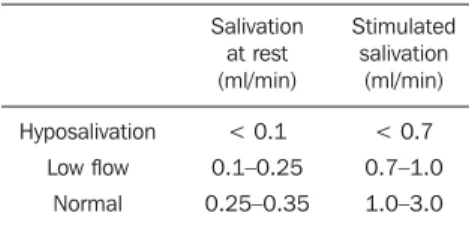
Permanent xerostomia is one the most prevalent late effects(1). It is necessary to
evaluate the salivary flow, by means of rest-ing and stimulated sialometry (Table 1).
topical use of pilocarpine spray or gel should be avoided as the effects are infe-rior to those in systemic administration(5,13).
Urecholine is an acetylcholine analogue, and a dose of 25 mg three times a day, for two to three weeks, may be prescribed. However its use should be interrupted for one week in case another sialogogue is being used. Anetholtrithione and cevi-meline, 30 mg every four hours, are also systemic salivary stimulants(13).
Saline solution at 0.9% (0.9 mg NaCl in100 ml of water) or a bicarbonate solu-tion mouthwashing for 12 to 30 seconds every 6 hours or at 15-30-minute intervals, depending on the patient’s need, may also be recommended(5).
Acupuncture in the salivary glands for 2 to 20 minutes per week, over 6 weeks has also demonstrated acceptable results(13).
Laser therapy with 685 nm wave length in a continuous form, with a power level of 35 mW, applied on three points in each parotid gland, one right and one left sub-mandibular point, but far from tumor areas, brings positive results for patients present-ing with xerostomia(23).
The surgical displacement of the mandibular gland to a nonirradiated sub-mental area is a conservative and effective approach(13).
Topical and/or systemic anesthetics, anti-inflammatory and antihistaminic drugs must be prescribed after evaluation by the oncologist(5).
In some cases, it is important that the pa-tient is referred to non-pharmacological therapies for pain management. Physical activities, massotherapy, trans-cutaneous electrical nerve stimulation, acupuncture, cognitive-behavioral strategies, relaxation techniques, hypnosis, group therapy, psy-chosocial interventions are therapeutic al-ternatives that may bring improvement of the quality of life for oncologic patients(5).
RADIATION CARIES
From three weeks to one year after ra-diotherapy, caries lesions may develop, usually around the cervical margins. The absence of the salivary buffering action that regulates the oral cavity pH leads to alter-ation of organic and inorganic components
of the teeth so as they may become more susceptible to decalcification(5).
Treatment
It is absolutely necessary to instruct the patient about oral hygiene, with advice about appropriate hydration and a balanced diet, with less sugar-containing foods(31).
Artificial saliva containing fluoride and sialogogues, such as oral pilocarpine 5 mg, three or four times a day starting one day before the beginning of the treatment and maintained for the duration of the treat-ment, may be necessary to re-establish the balance of the oral microflora(5).
The surgical displacement of the mandibular glands to a nonirradiated sub-mental area preserves their function and prevents the development of xerostomia and caries(36).
During radiotherapy, dental follow-up and fluoride prophylaxis must be frequent, at every 6 to 8 weeks. Restoration of incipi-ent caries must be done after radiotherapy. The follow-up must proceed for at least 12 months or longer after radiotherapy, in cases where xerostomia persists. The daily utilization of neutral 1% sodium fluoride gel with an individual mold for 5 to 10 minutes is recommended. Acidulated gels are not recommended. Fluoride solutions at 1.0 to 1.1% once a week for one minute, or 0.05% fluoride solution twice a day, for one minute may also be utilized. Brushing with 0.4% stannous fluoride gel is also ef-fective in cases of radiation caries, but its utilization should be avoided because of low fluoride concentration and acidic pH. Fluoride varnish (22,600 ppm of fluoride), two to three times a week may be an alter-native, particularly in pediatric patients(5).
Prescription of chlorhexidine gel at 1% or at 0.2%, twice a day, or chlorhexidine 0.12% solution for one minute twice a day, is also recommended to reduce the cari-ogenic bacterial flora(5).
In such patients, preferably, one should opt for dual-curing resins or glass ionomer, avoiding amalgam restorations(5).
OSTEORADIONECROSIS
Osteoradionecrosis is one of the most severe complications of radiotherapy, with Table 1 Sialometry.
Hyposalivation
Low flow
Normal
Salivation at rest (ml/min)
< 0.1
0.1–0.25
0.25–0.35
Stimulated salivation
(ml/min)
< 0.7
0.7–1.0
1.0–3.0
Treatment
The following measures are recom-mended to irradiated patients with xeros-tomia: correct water intake (8 to 12 glasses per day), sugarless drinks, sugarless chew-ing gum and candies, foods rich in ascor-bic acid, malic acid or citric acid, avoid-ing coffee, soft drinks, teas and salty foods, avoiding smoking and alcoholic drinks(31,32,34).
Salivary flow tests must be performed not only previously to the radiotherapy, but also periodically to evaluate possible func-tional damages to the salivary glands. In ir-radiated patients, the control of oral micro-organisms is necessary, and for such a pur-pose chlorhexidine gel 2%, for 5 minutes per day over 14 days promotes satisfactory results. However, in some cases, it is nec-essary to repeat the use every three or four months, until the salivary flow becomes normal(5,35).
Artificial saliva or mouth moisturizers represent alternatives in cases of hypo-salivation. They will keep the mouth pH between 6.0 and 7.0, and their composition can comprise important components for dental remineralization as follows: car-boxymethylcellulose, xylitol, fluoride, amino acids enzymes, glycerol, calcium and phosphate ions(5).
higher incidence in the elderly (10% to 37%), and occurs seven times more fre-quently in the mandible than in the maxilla, because of its higher bone density e smaller vascularization. According to Ben-David et al.(17), osteoradionecrosis may occur up to two years after the conclusion of radio-therapy.
According to Thorn et al.(37), 74% of the cases occur within the first three years following radiotherapy, with higher fre-quency in patients who received doses above 60 Gy.
The ionizing radiation results in nar-rowing of the vascular channels (endarteri-tis obliterans), which decreases the blood flow, producing an area with low resis-tance to trauma and with difficult regen-eration capability, as there is also a de-crease in viable osteocytes and osteocytes in the affected bone. The presence of pe-riodontal or endodontic disease prior to radiation favors the access of buccal cav-ity microorganisms to systemic areas and bone necrosis(38).
In cases of osteoradionecrosis, bone cells and vascularization may be irrevers-ibly injured. Additionally, the risk for os-teoradionecrosis remains indefinitely after irradiation(8). In chronic osteonecrosis,
mixed anaerobic infections may occur, with prevalence of some types of bacteria such as Aggregatibacter actinomycetemco-mitans, Fusobacterium, Parvimonas and Staphylococcus, originated from endodon-tic and periodontal infections(1,8).
In general, extractions must be per-formed at least two weeks before the ra-diotherapy and at least one year after the conclusion of the treatment. There is a greater risk for osteoradionecrosis during radiotherapy in cases where the involved tooth is in the path of the radiation beam(17).
The risk for post-extraction osteoradi-onecrosis is higher as the tumor and the ex-traction are located in the mandible, with doses higher than 5,000 cGy, particularly in patients with periodontal disease, poor buccal hygiene and in the elderly(2). In cases
with dubious or poor prognosis, patients must have antibiotic coverage from the day before surgery up to the cicatrization is completed(15).
Patients who utilize bisphosphonates, particularly zoledronic acid, present a greater risk for osteoradionecrosis(5). For
that reason, traumatic dental procedures such as extractions, scaling and prosthesis installation must be carefully planned. Post-radiotherapy extractions must be carefully performed, with minimum muco-periosteal detachment and alveoloplasty, without filling the alveolus and with ap-propriate suture(39). It is important to assess
the risk for odontogenic infection in onco-logic patients. For such reason, the patient’s complete blood count must be evaluated. In case the leucocytes count is > 3,500 cells/mm3, and platelets count is > 100,000 cells/mm3, clinical procedures will be safer. However, in case of leuko-penia or thrombocytoleuko-penia, it is necessary to evaluate the need for dental intervention in hospital environment, with platelet transfusion5 as the number of platelets is < 50,000 cells/mm3.
Treatment
Hyperbaric oxygenation is a treatment modality that improves healing of affected areas, enhancing angiogenesis and reduc-ing free radicals. In some cases, 25 sessions with 90 minutes each are recommended(2). It is recommended to contact the on-cologist for the interruption in the use of biphosphonates in those patients requiring extractions(5).
In the treatment of osteoradionecrosis, the antibiotic association of amoxicillin, clavulanate and metronidazole is pre-scribed, for 10 days. Additionally, daily local irrigation with 0.2% chlorhexidine is necessary. Debridement and complete re-section of the necrotic area and reconstruc-tion with new antibiotic therapy should be carefully considered(5,17).
PERIODONTAL DISEASE
The periodontium, like all other tissues, is also sensitive to the effects of high radia-tion doses(40). The blood vessels, not only
in the periodontium but also in the perios-teum, are affected. Radiographically, changes in the alveolus such as periodon-tal ligament space widening and trabecu-lar bone destruction are observed. Such
changes increase the risk for periodontal disease, as there is a decrease in the capac-ity of bone repair and remodeling(4).
Addi-tionally, xerostomia and immune suppres-sion lead to changes in the overall bacte-rial flora of the oral cavity, enhancing the growth of gram-negative bacteria (Strepto-coccus mutans, Lactobacilos and Actino-myces naeslundi, P. gingivalis)(8)
. Patients who present thrombocytopenia (< 30,000/mm3) cannot perform effective brushing and flossing, because of the risk for hemorrhage. In such cases, the utiliza-tion of cotton swabs or gauze with saline solution and mouth rinsing with antiseptic solutions are recommended for the removal of bacterial plaque.
Dental and periodontal assessment as a pre-radiotherapy step is critical for the patient’s prognosis. The literature is con-troversial about the appropriate period for teeth extraction. In teeth with periodontal pockets > 4 mm and/or grade I mobility(16),
or with periodontal pockets > 6 mm and furcation involvement, with the tooth close to the irradiation field, high radiation doses, poor oral hygiene and poor patient coop-eration(14), are indicative factors for
pre-ra-diotherapy extraction in order to avoid os-teoradionecrosis.
Treatment
Appropriate guidance on oral hygiene before and after radiotherapy is of utmost importance, particularly for patients with periodontal disease before the radiotherapy treatment. Scaling and root planning pro-cedures should be instituted preferably ei-ther before or after radioei-therapy(11).
The removal of factors causing plaque accumulation, such as poorly adapted pros-thesis, restorations excess, root fragments, fractured teeth, cavitated caries lesions, as well as extraction of teeth with major peri-odontal compromising, must be performed before radiotherapy. The reconstructive treatment of periodontal disease by means of the utilization of bone grafts may be performed before radiotherapy(38), but it
should be carefully planned to avoid osteo-radionecrosis.
TRISMUS
Trismus is a sequel whose onset gener-ally occurs between the third and sixth weeks after conclusion of radiotherapy treatment and that limits buccal opening, impairing feeding, speech, the assessment of the oral cavity, dental treatment, oral hygiene and causes intense discom-fort(1,2,4,33). Patients with pharynx tumors located in retromolar regions and posterior regions of the palate are most commonly affected. Still, in cases where the mastica-tory muscles are in the irradiation field, secondary effects such as edema, cellular destruction and muscle fibrosis will be observed(4,6).
Treatment
Physiotherapy exercising the involved masticatory muscles, with the utilization of dynamic bite openers to elongate such muscles is effective in increasing buccal opening(27).
Non-steroid anti-inflammatory drugs and muscle relaxants (cyclobenzaprine, 10 mg, 8/8 hours for seven days) are recom-mended to reduce painful symptoms. Pentoxifylline is effective in cases of tris-mus, considering its immunomodulatory action, which regulates certain cytokines that mediate the post-irradiation fibrogenic reaction(27).
DEVELOPMENTAL CHANGES
The following developmental changes can be mentioned as the most common changes in pediatric patients: abnormalities in craniofacial skeletal development (man-dibular micrognathia, or maxillary retro-gnathism), changes in odontogenesis, enamel hypoplasia, interruption in the de-velopment of dental organs, agenesias, microdontia and changes in rhizogenesis such as interruption, thinning and widen-ing of the pulp chamber(16).
CONCLUSIONS
A protocol intended to minimize the sequelae of radiotherapy must be instituted and monitored before, during and after the conclusion of the treatment. The integral
multidisciplinary clinical approach allows the prevention, diagnosis and management of radiotherapy side effects. The psycho-logical aspects are relevant, and patient’s self-esteem may favor the adherence to the proposed treatment, personal and dental care.
REFERENCES
1. Langendijk JA. New developments in radio-therapy of head and neck cancer: higher precision with less patient discomfort? Radiother Oncol. 2007;85:1–6.
2. Salazar M, Victorino FR, Paranhos LR, et al. Efei-tos e tratamento da radioterapia de cabeça e pes-coço de interesse ao cirurgião dentista: revisão da literatura. Odonto (São Bernardo do Campo). 2008;16:62–8.
3. Huber MA, Terezhalmy GT. The head and neck radiation oncology patient. Quintessence Int. 2003;34:693–717.
4. Vissink A, Jansma J, Spijkervet FK, et al. Oral sequelae of head and neck radiotherapy. Crit Rev Oral Biol Med. 2003;14:199–212.
5. Rankin KV, Jones DL, Redding SW. Oral health in cancer therapy. A guide for health care profes-sionals. 3rd ed. [Internet]. 2008. [cited 2010 Dec 14]. Available from: http://www.doep.org/images/ OHCT_III_FINAL.pdf
6. Silverman S Jr. Oral cancer: complications of therapy. Oral Surg Oral Med Oral Pathol Oral Radiol Endod. 1999;88:122–6.
7. Dib LL, Gonçalves RCC, Kowalski LP, et al. Abordagem multidisciplinar das complicações orais da radioterapia. Rev Assoc Paul Cir Dent. 2000;54:391–6.
8. Geraldes AM, Jardim Júnior EG, Campos MJA, et al. Ocorrência de Porphyromonas gingivalis na microbiota bucal de pacientes submetidos à ra-dioterapia para tratamento de lesões malignas de cabeça e pescoço. Rev Odontol Araçatuba. 2009; 30(Supl 1):41.
9. Niehoff P, Springer IN, Açil Y, et al. HDR brachytherapy irradiation of the jaw – as a new experimental model of radiogenic bone damage. J Craniomaxillofac Surg. 2008;36:203–9. 10. Epstein JB, Robertson M, Emerton S, et al.
Qual-ity of life and oral function in patients treated with radiation therapy for head and neck cancer. Head Neck. 2001;23:389–98.
11. Faloni APS, Lorenzon AP, Margonar R, et al. Importância dos procedimentos periodontais pré-vios à radioterapia em região de cabeça e pes-coço. Rev Int Periodontia Clin. 2005;2:93–9. 12. Wright WE, Haller JM, Harlow SA, et al. An oral
disease prevention program for patients receiving radiation and chemotherapy. J Am Dent Assoc. 1985;110:43–7.
13. Shiboski CH, Hodgson TA, Ship JA, et al. Man-agement of salivary hypofunction during and af-ter radiotherapy. Oral Surg Oral Med Oral Pathol Oral Radiol Endod. 2007;103 Suppl:S66.e1–19. 14. Bruins HH, Jolly DE, Koole R. Preradiation den-tal extraction decisions in patients with head and neck cancer. Oral Surg Oral Med Oral Pathol Oral Radiol Endod. 1999;88:406–12.
15. Santos MG, Silva LCF, Lins CA, et al. Fatores de
risco em radioterapia de cabeça e pescoço. RGO – Rev Gaúcha Odontol (Porto Alegre). 2010;58: 191–6.
16. Albuquerque RA, Morais VLL, Sobral APV. Pro-tocolo de atendimento odontológico a pacientes oncológicos pediátricos – revisão da literatura. Rev Odontol UNESP. 2007;36:275–80. 17. Ben-David MA, Diamante M, Radawski JD, et al.
Lack of osteoradionecrosis of the mandible after intensity-modulated radiotherapy for head and neck cancer: likely contributions of both dental care and improved dose distributions. Int J Radiat Oncol Biol Phys. 2007;68:396–402.
18. Neville BW, Damm DD, Allen CM, et al. Patolo-gia oral e maxilofacial. 2ª ed. Rio de Janeiro, RJ: Guanabara Koogan; 2004.
19. Karbach J, Callaway A, Kwon YD, et al. Compari-son of five parameters as risk factors for peri-mucositis. Int J Oral Maxillofac Implants. 2009; 24:491–6.
20. Raber-Durlacher JE, Elad S, Barasch A. Oral mucositis. Oral Oncol. 2010;46:452–6. 21. Bensadoun RJ, Franquin JC, Ciais G, et al.
Low-energy He/Ne laser in the prevention of radiation-induced mucositis. A multicenter phase III ran-domized study in patients with head and neck cancer. Support Care Cancer. 1999;7:244–52. 22. Kelner N, Castro JFL. Laser de baixa intensidade
no tratamento da mucosite oral induzida pela ra-dioterapia: relato de casos clínicos. Rev Bras Cancerol. 2007;53:29–33.
23. Campos L, Simões A, Sá PHRN, et al. Improve-ment in quality of life of an oncological patient by laser phototherapy. Photomed Laser Surg. 2009;27:371–4.
24. Simões A, de Campos L, de Souza DN, et al. Laser phototherapy as topical prophylaxis against radia-tion-induced xerostomia. Photomed Laser Surg. 2010;28:357–63.
25. Lima AG, Antequera R, Peres MP, et al. Efficacy of low-level laser therapy and aluminum hydrox-ide in patients with chemotherapy and radio-therapy-induced oral mucositis. Braz Dent J. 2010;21:186–92.
26. Gouvêa de Lima A, Villar RC, de Castro G Jr, et al. Oral mucositis prevention by low-level laser therapy in head-and-neck cancer patients under-going concurrent chemoradiotherapy: a phase III randomized study. Int J Radiat Oncol Biol Phys. 2010 Dec 14. [Epub ahead of print].
27. Andrade ED. Terapêutica medicamentosa em odontologia. São Paulo, SP: Artes Médicas; 1998. 28. Antonadou D, Pepelassi M, Synodinou M, et al. Prophylactic use of amifostine to prevent radio-chemotherapy-induced mucositis and xerostomia in head-and-neck cancer. Int J Radiat Oncol Biol Phys. 2002;52:739–47.
29. Rosenthal C, Karthaus M. Current approaches in prevention and therapy of chemo- and radio-therapy-induced oral mucositis. Wien Med Wochenschr. 2001;151:53–65.
30. Noé JE. L-glutamine use in the treatment and prevention of mucositis and cachexia: a naturo-pathic perspective. Integr Cancer Ther. 2009;8: 409–15.
32. Brasil. Ministério da Saúde. Instituto Nacional de Câncer. Consenso nacional de nutrição oncoló-gica. [Internet]. 2009. [acessado em 6 de setem-bro de 2011]. Disponível em: http://www.inca. g o v . b r / i n c a / A r q u i v o s / p u b l i c a c o e s / Consenso_Nutricao_internet.pdf
33. Harrison JS, Dale RA, Haveman CW, et al. Oral complications in radiation therapy. Gen Dent. 2003;51:552–60.
34. Moss RW. Should patients undergoing chemo-therapy and radiochemo-therapy be prescribed antioxi-dants? Integr Cancer Ther. 2006;5:63–82. 35. Lerman MA, Laudenbach J, Marty FM, et al.
Management of oral infections in cancer patients. Dent Clin North Am. 2008;52:129–53.
36. Jha N, Seikaly H, McGaw T, et al. Submandibu-lar salivary gland transfer prevents radiation-in-duced xerostomia. Int J Radiat Oncol Biol Phys. 2000;46:7–11.
37. Thorn JJ, Hansen HS, Specht L, et al. Osteoradi-onecrosis of the jaws: clinical characteristics and relation to the field of irradiation. J. Oral Maxillofac. Surg. 2000;58:1088–93.
38. Jegoux F, Malard O, Goyenvalle E, et al. Radia-tion effects on bone healing and reconstrucRadia-tion: interpretation of the literature. Oral Surg Oral Med Oral Pathol Oral Radiol Endod. 2010;109: 173–84.
39. Agbaje JO, Jacobs R, Michiels K, et al. Bone healing after dental extractions in irradiated
pa-tients: a pilot study on a novel technique for vol-ume assessment of healing tooth sockets. Clin Oral Investig. 2009;13:257–61.
40. Epstein JB, Lunn R, Le N, et al. Periodontal at-tachment loss in patients after head and neck ra-diation therapy. Oral Surg Oral Med Oral Pathol Oral Radiol Endod.1998;86:673–7.