Arq Neuropsiquiatr 2009;67(2-B):496-498
496
Clinical / Scientiic note
Cerebellopontine angle lipomaS
Magnetic resonance imaging indings in two cases
Rafael S. Borges
1, Cecília Castelo Branco Brito
2, Gustavo A. Carvalho
3,
Romeu C. Domingues
4, Emerson L. Gasparetto
5lipomaS do ângulo ponto-Cerebelar: aChadoS de reSSonânCia magnétiCa em doiS CaSoS
Clínica de Diagnóstico por Imagem, Multi-Imagem and Department of Radiology of the University of Rio de Janeiro, Rio de Janeiro RJ, Brazil: 1Medical
Student, Universidade Federal do Rio de Janeiro School of Medicine, Rio de Janeiro RJ, Brazil; 2Medical Resident in Diagnostic Radiology, Hospital
Uni-versitário Clementino Fraga Filho, Universidade Federal do Rio de Janeiro School of Medicine, Rio de Janeiro RJ, Brazil; 3Neurosurgeon, Neurosurgical
Department of the Clínica Bambina and Hospital Silvestre, Rio de Janeiro RJ, Brazil; 4Medical Director and Radiologist, Clínicas CDPI and Multi-Imagem,
Rio de Janeiro RJ, Brazil; 5Radiologist, Clínicas CDPI and Multi-Imagem, and Associated Professor, Universidade Federal do Rio de Janeiro School of
Medicine, Rio de Janeiro RJ, Brazil.
Received 21 October 2008, received in inal form 5 January 2009. Accepted 31 March 2009.
Dr. Emerson L. Gasparetto – Hospital Universitário Clementindo Fraga Filho da UFRJ - Rua Professor Rodolpho Paulo Rocco 255 - 21941-913 Rio de Janeiro RJ - Brasil. E-mail: [email protected]
Vestibular schwannomas and meningiomas are the most common lesions of the cerebellopontine angle (CPA), accounting for approximately 85–90% of the
tu-mors seen in this location1. Lipomas are rare at this
to-pography, representing about 0.15% of the CPA lesions2,3.
These tumors are maldevelopmental masses that arise
from abnormal differentiation of the meninx primitiva1,4,5.
Clinically, CPA lipomas can cause slowly progressive neu-rological symptoms and signs affecting cranial nerves or
brain stem3,6-8. Because these lesions usually are strongly
attached to the surrounding structures, any surgical at-tempts of complete resection can result in neural or vas-cular damage, reinforcing the importance of the
pre-op-erative imaging diagnosis1-3,7,9,10. Although the CT indings of
CPA lipomas can be typical, the magnetic resonance (MR) imaging, especially the fat suppression sequences, had im-proved the identiication of these lesions.
We aimed to report two patients with a CPA lipoma, emphasizing the MR imaging indings.
CaSe
Case 1
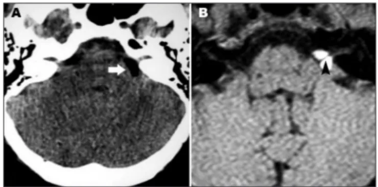
A 13-year-old female patient was evaluated due to a 1-year history of headache and hearing loss. The physical examination was unremarkable. The audiometric evaluation demonstrated a discrete sensorineural hearing loss on the right side. The CT scan revealed a markedly hypodense non-enhancing mass in the right CPA. The MR imaging showed a lesion measuring 2.1 × 2.0
× 1.7 cm in the right CPA cistern. The mass was hyperintense on T1-weighted images and isointense with hypointense halo (chemical-shift) on T2-weighted images, with very low signal on T1-weighted images with fat suppression (Figs 1 and 2). The VII and VIII cranial nerves were seen as linear images with low sig-nal inside the CPA mass. The diagnosis of CPA lipoma was
Arq Neuropsiquiatr 2009;67(2-B)
497
Cerebellopontine angle lipomas: MRI Borges et al.
gested and the surgical treatment was chosen once the patient was young and the chance of lesion growing and future com-plications was considerable. A craniotomy with posterior fos-sa approach was performed, the lesion was partially removed, and the histological examination conirmed the diagnosis of li-poma. Six months after the surgery the patient remains asymp-tomatic. The parent signed the informed consent agreeing with the study.
Case 2
A 35-year-old woman presented with a six-month history of vertigo, without signiicant abnormalities on physical examina-tion. A CT scan revealed a left-sided hypodense non-enhancing CPA mass. The MR imaging showed a left CPA cistern hyperin-tense lesion on T1-wheighted images and isoinhyperin-tense with hypoin-tense halo (chemical-shift) on T2-weighted images, measuring 1.4
× 1.3 cm and showing no enhancement after contrast administra-tion (Fig 3). The diagnosis of CPA lipoma was suggested and the patient was managed conservatively. The symptoms were con-trolled with medical therapy. The follow-up MR imaging per-formed one year later showed no signiicant modiications. The patient signed the informed consent agreeing with the study.
diSCuSSion
Intracranial lipomas are rare lesions, corresponding to
less than 0.1% of all intracranial tumors3,10,11. Some authors
have suggested that lipomas are congenital malformations because their lack of cellular atypia, dysplasia and other evidences of malignancy, as well as due to the fact that
they are usually associated to other malformations4,7,11.
They may originate from persistence of the meninx prim-itive, a precursor of pia mater and arachnoid, which
de-velop into fat4. Most of the intracranial lipomas are
per-icallosal asymptomatic lesions found incidentally during
neuroimaging studies6. On the other hand, the most
com-mon extra-axial site of lipomas in the posterior fossa is
the CPA4,7. These tumors can cause symptoms related to
the VIII nerve involvement, such as hearing loss, tinnitus,
vertigo and nausea. However, trigeminal symptoms such as neuralgia, paresthesia or headache, can also occur in patients with CPA lipomas extending to the trigeminal
nerve3,5,7,8. Our patients presented with headache,
verti-go and sensorineural hearing loss.
Due to their peculiar imaging indings, the diagnosis of intracranial lipomas is highly suggestive on the basis
of imaging studies3,5. The CT scan demonstrates a marked
hypodense nonenhancing lesion in the CPA, with atten-uation characteristics similar to adipose tissue (–40 to
–100 HU)1,5,9,10. Regarding the MR imaging indings,
lipo-mas have signal characteristics similar to the subcutane-ous tissue, with high-signal intensity on T1-weighted ages, and iso- to hypointense signal on T2-weighted
im-ages, usually without contrast enhancement1. The use of
the MR imaging with fat suppression is extremely
help-ful to clearly demonstrate the lipomas12. The
disappear-ance of a CPA mass with fat suppression techniques, such as short-time inversion recovery (STIR), and T1-weighted images with fat suppression, is highly suggestive of lipo-mas. In addition, the chemical-shift artifact, usually seen on T2-weighted images, also corroborates the diagnosis of lipoma. This artifact produces a ring of low signal intensi-ty around the tumor and is virtually diagnostic of a fatintensi-ty
lesion3. The MR imaging artifact is a result of the
differ-ence of the resonance frequency between lipid and
wa-ter protons12. The high-density structures seen inside the
lesion on CT scan, which are hypointense on T1-weighted MR imaging, most likely represent cranial nerves. The dif-ferential diagnosis of CPA lipomas should include vestib-ular schwannoma and meningiomas, as well as other
fat-ty tumors, such as epidermoids and dermoids cysts5,8,11,13.
In our cases the CT scan showed a CPA hypodense lesion. Furthermore, the MR imaging studies reveal a mass with signal intensity similar to the subcutaneous fat on T1 and T2-weighted images, with chemical-shift artifact around the lesion and no signal of fat suppression sequences.
Unlike vestibular schwannomas, complete surgical re-Fig 2. Patient 1. [A Coronal STIR MR image demonstrates the lipoma
with very low signal intensity due to the fat suppression. [B] Coro-nal contrast-enhanced T1-weighted image with fat suppression also shows the signal suppression of the lesion.
Arq Neuropsiquiatr 2009;67(2-B)
498
Cerebellopontine angle lipomas: MRI Borges et al.
section of CPA lipomas is dificult to achieve and not fre-quently indicated. These tumors are indolent, but inil-trate along cranial nerves, making complete removal dif-icult due to the high risk of postoperative cranial nerves deicit. Subtotal resection of CPA lipomas is only indicat-ed in patients with brain stem compression or signiicant cranial nerve deicit, such as intractable headache, trigem-inal neuralgia, facial spasm, vertigo and nauseas that are
resistant to clinical treatment5,7,8,13. Because surgical
inter-vention in patients with CPA lipoma is usually avoided,
correct imaging diagnosis is essential3,5. Regarding our
cas-es, the patient 1 underwent a partial resection of the CPA lipoma, relieving her headache. The surgical management was chosen once the patient was young and the
possibili-ty of lesion growing was considered3. Further studies with
longer follow-up of these cases are needed to exclude
this potential growth3. The patient remains
asymptomat-ic six months after surgery. However, the conservative ap-proach was adopted in case 2 because the symptoms were controlled with medical therapy. A one year later follow-up MR imaging showed no considerable alterations.
In conclusion, CPA lipomas are very rare tumors, which can be accurately diagnosed with CT scan and/or MR im-aging. Regarding the MR imaging sequences, T1-weighted images with and without fat suppression are fundamen-tal for the diagnosis. In addition, because CPA lipomas are slowly progressive tumors, imaging follow-up is suggest-ed, especially in asymptomatic patients. Finally, due to it’s benign potential and slow growth, a conservative follow-up should be preferred to surgical resection.
reFerenCeS
1. Bonneville F, Sarrazin JL, Marsot-Dupuch K, et al. Unusual le-sions of the cerebellopontine angle: a segmental approach. Ra-diographics 2001;21:419-438.
2. Krainik A, Cyna-Gorse F, Bouccara D, et al. MRI of unusual
lesions in the internal auditory canal. Neuroradiology 2001; 43:52-57.
3. Bigelow DC, Eisen MD, Smith PG, et al. Lipomas of the inter-nal auditory cainter-nal and cerebellopontine angle. Laryngoscope 1998;108:1459-1469.
4. Truwit CL, Barkovich AJ. Pathogenesis of intracranial lipoma: an MR study in 42 patients. AJR Am J Roentgenol 1990;155: 855-864.
5. Tankere F, Vitte E, Martin-Duverneuil N, Soudant J. Cerebello-pontine angle lipomas: report of four cases and review of the literature. Neurosurgery 2002;50:626-631.
6. Bonneville F, Savatovsky J, Chiras J. Imaging of cerebellopon-tine angle lesions: an update. Part 2: intra-axial lesions, skull base lesions that may invade the CPA region, and non-enhanc-ing extra-axial lesions. Eur Radiol 2007;17:2908-2920. 7. Zimmermann M, Kellermann S, Gerlach R, Seifert V.
Cerebel-lopontine angle lipoma: case report and review of the litera-ture. Acta Neurochir (Wien) 1999;141:1347-1351.
8. Fagundes-Pereyra WJ, Marques JA, Carvalho GT, Sousa AA. Lipoma of the cerebellopontine angle: case report Arq Neu-ropsiquiatr 2000;58:952-957.
9. Schuhmann MU, Ludemann WO, Schreiber H, Samii M. Cere-bellopontine angle lipoma: a rare differential diagnosis. Skull Base Surg 1997;7:199-205.
10. Yildiz H, Hakyemez B, Koroglu M, Yesildag A, Baykal B. In-tracranial lipomas: importance of localization. Neuroradiology 2006;48:1-7.
11. Budka H. Intracranial lipomatous hamartomas (intracranial b lipomas Q). A study of 13 cases including combinations with medulloblastoma, colloid and epidermoid cysts, angiomatosis and other malformations. Acta Neuropathol 1974;28:205-222. 12. Delfaut EM, Beltran J, Johnson G, Rousseau J, Marchandise X,
Cotten A. Fat suppression in MR imaging: techniques and pit-falls. Radiographics 1999;19:373-382.
![Fig 1. Patient 1. Coronal [A] and axial [B] T1-weighted MR image show a hyperintense mass in the right CPA involving the VII and VIII cranial nerves](https://thumb-eu.123doks.com/thumbv2/123dok_br/15432405.594948/1.955.75.839.710.905/patient-coronal-axial-weighted-hyperintense-involving-cranial-nerves.webp)