RBCCV 44205-1632 DOI 10.5935/1678-9741.20150026
Factors associated with moderate or severe left
atrioventricular valve regurgitation within 30 days
of repair of incomplete atrioventricular septal defect
Fatores associados à insuiciência moderada ou grave da valva atrioventricular esquerda nos primeiros
30 dias de correção de defeito de septo atrioventricular incompleto
Marcelo Felipe Kozak
1, MD; Ana Carolina Leiroz Ferreira Botelho Maisano Kozak
1, MD; Carlos
Henrique De Marchi
1, MD; Moacyr Fernandes de Godoy
2, MD, PhD; Ulisses Alexandre Croti
1,
MD, PhD; Airton Camacho Moscardini
1, MD
1Department of Pediatrics and Pediatric Surgery, Hospital de Base, São José
do Rio Preto Medical School, São José do Rio Preto, SP, Brazil.
2Department of Cardiology, Hospital de Base, São José do Rio Preto
Medi-cal School, São José do Rio Preto, SP, Brazil.
Work carried out at Faculdade de Medicina de São José do Rio Preto (FAMERP), São José do Rio Preto, SP, Brazil and Hospital de Base (HB), São José do Rio Preto, SP, Brazil.
No inancial support.
Correspondence address: Marcelo Felipe Kozak
Faculdade de Medicina de São José do Rio Preto (FAMERP)
Av. Brigadeiro Faria Lima, 5416 – Vila São Pedro- São José do Rio Preto, SP, Brazil - Zip code: 15090-000
E-mail: [email protected]
Article received on November 26th, 2014
Article accepted on April 6th, 2015
Abstract
Introduction: Left atrioventricular valve regurgitation is the most concerning residual lesion after surgical correction of atrioventricular septal defect.
Objective: To determine factors associated with moderate or greater left atrioventricular valve regurgitation within 30 days of surgical repair of incomplete atrioventricular septal defect.
Methods: We assessed the results of 51 consecutive patients 14 years-old and younger presenting with incomplete atrioven-tricular septal defect that were operated on at our practice be-tween 2002 and 2010. The following variables were considered: age, weight, absence of Down syndrome, grade of preoperative left atrioventricular valve regurgitation, abnormalities on the left atrioventricular valve and the use of annuloplasty. The me-dian age was 4.1 years; the meme-dian weight was 13.4 Kg; 37.2% had Down syndrome. At the time of preoperative evaluation, there were 23 cases with moderate or greater left atrioven-tricular valve regurgitation (45.1%). Abnormalities on the left atrioventricular valve were found in 17.6%; annuloplasty was performed in 21.6%.
Results: At the time of postoperative evaluation, there were
12 cases with moderate or greater left atrioventricular valve regurgitation (23.5%). The variation between pre- and post-operative grades of left atrioventricular valve regurgitation of patients with atrioventricular valve malformation did not reach
signiicance (P=0.26), unlike patients without such
abnormali-ties (P=0.016). During univariate analysis, only absence of Down
syndrome was statistically signiicant (P=0.02). However, after a
multivariate analysis, none of the factors reached signiicance.
Conclusion: None of the factors studied was determinant of a moderate or greater left atrioventricular valve regurgitation
within the irst 30 days of repair of incomplete atrioventricular
septal defect in the sample. Patients without abnormalities on
the left atrioventricular valve beneit more of the operation.
Descriptors: Endocardial Cushion Defects. Mitral Valve
In-suficiency. Heart Defects, Congenital. Postoperative Period.
Resumo
INTRODUCTION
A non negligible number of patients who undergo sur-gical repair of incomplete atrioventricular septal defect (AVSD) are discharged from hospital with residual left atrioventricular valve regurgitation (LAVVR)[1,2]. Reop-eration rates for LAVVR are still relatively high, varying between 7% and 22%[1,3-6]. Many factors associated to reop-eration have been proposed[1,2,4,6], but there is a lack of data reporting factors associated with immediate postoperative LAVVR, although the reports of valve replacement and of in-hospital deaths due to signiicant residual LAVVR within 30 days of operation[1,5,7].
To improve these statistics, a clear outline of the predis-posing factors leading to a worse immediate surgical out-come is essential. The goal of this study was to assess wheth-er some of the previous risk factors published in the litwheth-erature would be associated to an at least moderate LAVVR within 30 days of surgical repair at our practice, in Brazil.
METHODS
This study was approved by the ethics committee of our institution (protocol CEP 3802/2010), a tertiary-care hospi-tal with a division of pediatric cardiology and cardiovascular surgery in Brazil, which waived the need for patient consent. The medical records of all patients 14 years old and younger who had undergone repair of incomplete (partial or transi-tional) AVSD at our practice between March 2002 and April 2010 were reviewed. Patients with any right ventricle
ob-Abbreviations, acronyms & symbols
AV Atrioventricular
AVSD Atrioventricular septal defect LAVVR Left atrioventricular valve regurgitation
struction, and those who had a previous pulmonary banding were excluded.
The reports of the transthoracic echocardiograms per-formed before and after operation were reviewed. These exams were performed by one of two physicians using commercially available machines, HDI 5000CV (ATL Ultra-sound), Envisor-C and HD11 (Philips Ultrasound, Bothell, WA, USA), with 3 to 8 MHz probes. For further analysis, there were considered the exam before surgery and the exam closer to the 30th postoperative day, while still being within 1 month of the repair.
These were the factors assessed: preoperative (age, weight, absence of Down syndrome and grade of LAVVR), intraoper-ative (abnormalities on the AV valve morphology and the need for use of annuloplasty). Abnormalities on the AV valve mor-phology were subjectively described by the surgeon. Trans-esophageal echocardiography was not available during the period in which the patients were operated on.
Pre- and post-surgical LAVVR was graded based on the appearance of the color Doppler jets in relation to the left atrium (8): I=absent or trivial; II=mild; III=moderate; IV=se-vere. The categorizations were based just on oficial written summaries of the exams. Images stored on tapes or in digital media were not assessed.
Statistical Analysis
Continuous variables were expressed as mean or median, and comparisons were made using the two-sided Mann-Whit-ney test. Categorical variables were expressed using frequen-cy distribution and percentages, and comparisons were made Objetivo: Determinar fatores associados à insuiciência da
valva atrioventricular esquerda de grau moderado ou impor-tante nos primeiros 30 dias após correção de defeito de defeito de septo atrioventricular.
Métodos: Avaliamos os resultados em 51 pacientes consecu-tivos menores de 14 anos com defeito de septo atrioventricular incompleto, operados em nosso serviço entre 2002 e 2010.
Ava-liamos as seguintes variáveis: idade, peso, ausência de síndrome
de Down, grau de insuiciência da valva atrioventricular esquer
-da antes -da correção, anormali-dades na valva atrioventricular e uso de anuloplastia. A mediana da idade foi de 4,1 anos e a do
peso de 13,4 Kg; 37,2% tinham síndrome de Down; antes da
operação, 23 apresentavam insuiciência da valva atrioventricu
-lar esquerda pelo menos moderada (45,1%); anormalidades na
valva atrioventricular foram encontradas em 17,6% dos casos; anuloplastia foi realizada em 21,6% dos pacientes.
Resultados: Após a correção cirúrgica, 12 casos
apresenta-ram insuiciência da valva atrioventricular esquerda pelo me
-nos moderada (23,5%). A variância entre os graus de insuici
-ência da valva atrioventricular esquerda pré e pós-operatória
nos pacientes com anormalidades na valva atrioventricular não
teve signiicância estatística (P=0,26), ao contrário daqueles sem
tais anormalidades (P=0,016). Pela análise univariada, apenas
a ausência de síndrome de Down teve signiicância estatística
(P=0,02). Porém, após análise multivariada, nenhum dos fatores
teve signiicância.
Conclusão: Nenhum dos fatores estudados foi determinante
de insuiciência da valva atrioventricular esquerda de grau mo
-derado ou importante nos primeiros 30 dias após a correção de defeito de septo atrioventricular incompleto na população ava-liada. Pacientes sem anormalidades na valva atrioventricular se
beneiciam mais da operação.
Descritores: Coxins Endocárdicos. Insuiciência da Valva
using the Fisher exact test. For the analyses of variances, the Kruskal-Wallis test was used. Univariate odds ratios and their 95% conidence intervals (95% CI) were estimated for variables found to have a statistically signiicant (P≤0.05) or borderline signiicant (P≤0.2) relationship with moderate or greater post-operative LAVVR. These variables were includ-ed in the multivariate analysis, which was completinclud-ed using logistic regression. A P-value of 0.05 or less was considered signiicant. All statistical analyses were conducted using the software StatsDirect, version 2.7.2. 2008 (Cheshire, UK).
Patient Population
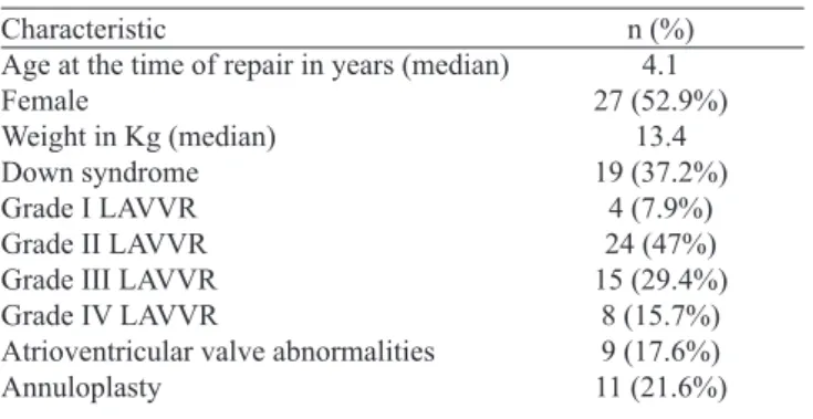
We included 51 patients (27 females and 24 males): 35 with partial AVSD and 16 with transitional AVSD, 32 without Down syndrome (62.7%). Age at the time of repair ranged from 4.2 months to 12.8 years (median=4.1 years). Weight varied between 3.8 and 44.3 Kg (median=13.4 Kg). At the time of preoperative evaluation, there were 4 cases with grade I LAVVR (7.9%), 24 with grade II (47%), 15 with grade III (29.4%), and 8 with grade IV (15.7%) (Table 1). Less than 50% of the patients presented minor associated heart defects (Table 2). Patients with Down syndrome had lower grades of preoperative LAVVR than those without Down syndrome, but it did not reach statistical signiicance (P=0.17).
Operative management
The atrial septal defect was closed with a preserved bovine pericardium patch in 50 of the 51 cases, and with direct suture in 1 case. The ventricular septal defect was not found in 8 of the 16 patients with transitional AVSD. Among the 8 other cas-es with ventricular septal defect, this defect was closed with di-rect suture in 3, with a preserved bovine pericardium patch in 2, with the same patch used for the closure of the atrial septal defect in 2, and it was closed using implantation of the superior bridging left on the superior rim of the defect in another one.
The cleft was completely closed with interrupted 6-0 polypropilene sutures in 49 patients (96.1%). For those 11 patients (21.6%) who presented annular dilation based on the surgeon’s judgment, both cleft closure and posterior an-nuloplasty were used. There were no statistically signiicant differences in the preoperative grade of LAVVR of these 11 patients when comparing to those who had not undergone annuloplasty (P=0.94).
Among the nine patients in whom abnormalities on the AV valve were diagnosed, the cleft was left untouched in one patient with a dysplastic valve. In one of the cases with an accessory cleft, the attempt of closing it caused worsening of regurgitation, thus this cleft was left intact. Another patient presented a big atrial septal defect and a severe LAVVR; the choice for this case was to close de cleft and to leave a small residual atrial septal defect. Annuloplasty was performed in four out of these nine patients.
RESULTS
The cardiopulmonary bypass time was longer in patients with AV valve malformation: mean=83±24.13 (median 94.5 min) vs. 64.7±20.2 (median 70 min); P=0.03. The postop-erative time on a mechanical ventilator ranged from 2.6 to 44.7 hours (mean=9.4±7.1 hours, median=7.2 hours) and the time of inotropic support varied between 10 and 157 hours (mean=49.9±26 hours, median=46 hours). The length of hos-pital stay ranged from 1 to 22 days (mean=7.9±4.6 days, me-dian=7 days). There were two deaths (3.9%), one within the irst 24 hours due to a 3rd degree AV block not properly treat-ed and another due to low cardiac output syndrome on the 2nd postoperative day. The postoperative LAVVR grades on these patients were II and I, respectively, and none of them presented AV valve malformation. Before treatment, both of these patients had a grade I LAVVR.
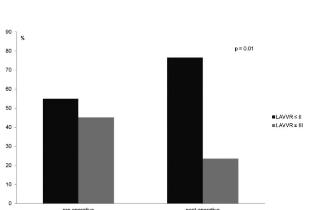
Postoperative echocardiographic exams were performed between zero and 30 days after surgery (mean=12.6±9.4 days). There were 8 cases with grade I LAVVR (15.7%), 31 with grade II (60.8%), 11 with grade III (21.5%), and 1 with grade IV (2%) (Figure 1). The mechanisms of valve regur-gitation were not available on the medical reports. The vari-ance between pre- and postoperative grades of valvar regur-gitation was statistically signiicant (P=0.01).
Table 1. Pre- and intraoperative characteristics of the patients enrolled in the study.
Characteristic
Age at the time of repair in years (median) Female
Weight in Kg (median) Down syndrome Grade I LAVVR Grade II LAVVR Grade III LAVVR Grade IV LAVVR
Atrioventricular valve abnormalities Annuloplasty
n (%) 4.1 27 (52.9%)
13.4 19 (37.2%)
4 (7.9%) 24 (47%) 15 (29.4%)
8 (15.7%) 9 (17.6%) 11 (21.6%)
LAVVR=left atrioventricular valve regurgitation.
Table 2. Associated minor heart defects* noted before surgery.
Defect
Ostium secundum atrial septal defect Common atrium
Additional ventricular septal defect Patent ductus arteriosus
Left superior caval vein Left atrium isomerism Coarctation of the aorta Subaortic stenosis
N 15 3 2 1 1 1 1 1
*Defects not mutually exclusive.
% 29.4
Nine cases (17.6%) of abnormalities on the AV valve mor-phology were found and they were described according to the surgeon’s report: dysplastic valve lealets (n=5), accesso-ry cleft (n=2), tendinous cordae rupture (n=1), and small left AV valve oriice (n=1). Six of these 9 patients presented with moderate or severe LAVVR before operation (66.7%), a rate greater than that seen in patients without such abnormalities (40.5%) (P=0.27). After repair, 4 of these 9 patients presented the same grade of preoperative LAVVR; there was worsening in 1, and improvement in 4. The variance between pre- and postoperative grades of LAVVR in this group of patients with AV valve malformation did not reach signiicance (P=0.26), unlike patients without such abnormalities (P=0.016). Among the 19 patients with Down syndrome, 2 (10.5%) presented AV valve abnormalities; whereas among the 32 without Down syndrome, 7 (21.9%) presented it (P=0.7).
In 17 patients, the trivial or mild LAVVR found before operation was maintained after the procedure. In 15 of the 23 (65.2%) patients with moderate or severe pre-operative LAVVR, the postoperative LAVVR was found to be trivial or mild. A one-grade worsening on the LAVVR was found in 8 patients (15.7%). Of the 2 cases in which the cleft was
left open, the grade of LAVVR didn’t change. Regarding the improvement on the grade of LAVVR, there was no statisti-cal signiicance comparing those patients who had undergone annuloplasty, with those who had not undergone it (P=0.84).
Other indings on postoperative echocardiograms: 12 cases (23.5%) of insigniicant residual ventricular septal defect, which is deined as a ventricular septal defect < 3 mm (8 of them were not seen during surgery, conirming that they were too small); four cases (7.8%) of insigniicant residual atrial septal defect, which is deined as an atrial septal defect < 3 mm; two (3.9%) cases of mild left AV valve stenosis; and one (2%) case of a istula from the left ventricle to the right atrium.
Univariate analysis revealed that absence of Down syn-drome was associated with moderate or severe post-operative LAVVR (P=0.02; OR=9.43). Severe preoperative LAVVR and AV valve abnormalities were only marginally signiicant (P=0.07 and 0.19, respectively) (Table 3). However, none of the factors were found to be associated with moderate or severe postoperative under multivariate analysis. Absence of Down syndrome reached only a borderline signiicance (P=0.06; OR=8.4) (Table 4).
DISCUSSION
Despite different ages, different weight at repair, and dif-ferent physiology, patients with both complete and incomplete AVSD present a similar risk of reoperation for LAVVR[1,3-6,9-14]. What both variations of the same disease have in common are the typical anatomical landmarks of AVSD (common AV junc-tion, a common 5-lealet AV valve, distinct papillary muscle displacement and a narrow and elongated left ventricle outlow tract)[15,16], as well as a high prevalence of individuals with Down syndrome[17]. Therefore, the clue to understanding this frequent complication may be related more to these two aspects than to another factor such as age, weight, or AV valve malformation.
In the study by Kanani et al.[18], in which the anatomy of the subvalvar apparatus of normal hearts was compared to those of hearts with complete and incomplete AVSD with an intact left AV valve, the structural and geometric disarray of the tendinous cords of those hearts with AVSD defect was clearly visible, along with its possible rule on the mechanisms of valve regurgitation. Moreover, in the work by Bharucha et al.[19], with the use of three-dimensional echocardiography, it was found that a more acute angle of the components of the common AV valve against the plane of the common AV junc-tion would be a predictor of postoperative valve funcjunc-tion.
Performing an intraoperative transesophageal exam is routine in many centers[5,10,20]; though, there is often too much discrepancy between its indings and those obtained through transthoracic exams in the days or weeks after repair[12,21]. In our
Table 3. Univariate relations between variables and moderate or severe postoperative left atrioventricular valve regurgitation (LAVVR).
Age in months (median) Weight in Kg (median) Abscence of Down syndrome Grade IV pré-operative LAVVR AV valve abnormality
Annuloplasty
LAVVR ≤ II n=39
53.3 13.1 21 (53.8%)
4 (10.2%) 5 (12.8%) 8 (20.5%)
LAVVR=left atrioventricular valve regurgitation; AV=atrioventricular; CI=conidence interval; OR=odds ratio. LAVVR ≥ III
n=12 45.3 15.4 11 (91.7%)
4 (33.3%) 4 (33.3%) 3 (25%)
Univariate 95% CI
1.12-427.9 0.64-28.31 0.53-19.7 OR
9.43 4.4 3.4
P
0.97 0.87 0.02 0.07 0.19 0.7
Table 4. Multivariate relations between variables and moderate or severe postoperative left atrioventricular valve regurgitation (LAVVR).
Abscence of Down syndrome Grade IV Preoperative LAVVR AV valve abnormality
LAVVR ≤ II n=39 21 (53.8%)
4 (10.2%) 5 (12.8%)
LAVVR=left atrioventricular valve regurgitation; AV=atrioventricular; CI=conidence interval; OR=odds ratio. LAVVR ≥ III
n=12 11 (91.7%)
4 (33.3%) 4 (33.3%)
Multivariate 95% CI 0.9-79.5 OR
8.4
P
0.06 0.25 0.32
study, which was performed without the use of intraoperative transesophageal echocardiography, a decrease of the number of patients with moderate or greater LAVVR from 45.1% to 23.5% was similar to that reported by Kaza et al.[22] in a study evalu-ating data from seven centers in North America. These results show that all techniques that were used, including cleft closure and annuloplasty, had an impact, but not enough of an impact to completely avoid early post-operative LAVVR in some patients. These patients, if previously identiied, might receive an alterna-tive approach to minimize the grade of LAVVR.
Some studies found the AV valve malformation to be asso-ciated with reoperation or valve replacement[9,14]. In the present study, the results could not prove any relationship between the presence of malformation and moderate or severe postoper-ative LAVVR. However, it was found that the operation for patients with such abnormalities did not produce the same ben-eits as it was seen for patients without them. The longer car-diopulmonary bypass time and the impossibility of closing the cleft in two out of nine patients demonstrate how dificult these cases can be. The diagnosis of these valvar abnormalities can be dificult. Ando et al.[21] found a weak correlation between preoperative echocardiographic indings and the surgeon’s judgment in regard to the diagnosis of these malformations. Moreover, one should know that little consistency between the indings of two- and three-dimensional Echocardiography, considering the presence of AV valve malformation, has been shown by some authors. In the study by Takahashi et al.[23], for instance, the correlation between the indings of both methods was lower than 46% in the evaluation of the mural lealet and in the evaluation of the commissural abnormalities of the left AV valve lealets. The three-dimensional echocardiogram was more accurate and more reliable.
In conclusion, for this sample we were not able to identi-fy any risk factor for early moderate or severe postoperative LAVVR at our center, but we could observe that patients with AV valve abnormalities may be a challenge for surgeons.
Study Limitations
It was a retrospective study, and was therefore subject to limitations in terms of how correctly the information in the medical records was iled. The small number of patients may, in some ways, account for the lack of statistical signiicance among the factors studied. There were no pre- and postoper-ative echocardiographic information about the mechanisms of valve regurgitation (residual cleft, mobility of the lealets, etc), precluding its impact on results.
Authors’ roles & responsibilities
MFK Analysis and/or interpretation of data; statistical analysis;
inal approval of the manuscript; study design; writing of the
manuscript or critical review of its content
ACLFBMK Analysis and/or interpretation of data; inal approval of the
manuscript; writing of the manuscript or critical review of its content
CHM Analysis and/or interpretation of data; inal approval of the
manuscript; writing of the manuscript or critical review of its content
MFG Statistical analysis
UAC Analysis and/or interpretation of data; inal approval of the
manuscript; operations and/or experiments conduct; writing of the manuscript or critical review of its content
ACM Analysis and/or interpretation of data; inal approval of the
manuscript; study design; writing of the manuscript or criti-cal review of its content
REFERENCES
1. Aubert S, Henaine R, Raisky O, Chavanis N, Robin J, Ecochard R, et al. Atypical forms of isolated partial atrioventricular septal defect increase the risk of initial valve replacement and reoperation. Eur J Cardiothorac Surg. 2005;28(2):223-8.
2. Minich LL, Atz AM, Colan SD, Sleeper LA, Mital S, Jaggers J, et al.; Pediatric Heart Network Investigators. Partial and transitional atrioventricular septal defect outcomes. Ann Thorac Surg. 2010;89(2):530-6.
3. Al-Hay AA, Lincoln CR, Shore DF, Shinebourne EA. The left atrioventricular valve in partial atrioventricular septal defect: management strategy and surgical outcome. Eur J Cardiothorac Surg. 2004;26(4):754-61.
4. Murashita T, Kubota T, Oba J, Aoki T, Matano J, Yasuda K. Left Atrioventricular valve regurgitation after repair of incomplete atrioventricular septal defect. Ann Thorac Surg. 2004;77(6):2157-62.
5. Chowdhury UK, Airan B, Malhotra A, Bisoi AK, Kalaivani M,
Govindappa RM, et al. Speciic issues after surgical repair of
partial atrioventricular septal defect: actuarial survival, freedom from reoperation, fate of the left atrioventricular valve, prevalence
of left ventricular outlow tract obstruction, and other events. J
Thorac Cardiovasc Surg. 2009;137(3):548-55.
6. Welke KF, Morris CD, King E, Komanapalli C, Reller MD, Ungerleider RM. Population-based perspective of long-term outcomes after surgical repair of partial atrioventricular septal defect. Ann Thorac Surg. 2007;84(2):624-9.
7. Stulak JM, Burkhart HM, Dearani JA, Cetta F, Barnes RD, Connolly HM, et al. Reoperations after repair of partial atrioventricular septal defect: a 45-year single-center experience. Ann Thorac Surg. 2010;89(5):1352-9.
8. Zoghbi WA, Enriquez-Sarano M, Foster E, Grayburn PA, Kraft CD, Levine RA, et al.; American Society of Echocardiography. Recommendations for evaluation of the severity of native valvular regurgitation with two-dimensional and Doppler echocardiography. J Am Soc Echocardiogr. 2003;16(7):777-802.
9. Al-Hay AA, MacNeill SJ, Yacoub M, Shore DF, Shinebourne EA. Complete atrioventricular septal defect, down syndrome, and surgical outcome: risk factors. Ann Thorac Surg. 2003;75(2):412-21.
10. Crawford FA. Atrioventricular canal: single-patch technique. Semin Thorac Cardiovasc Surg Pediatr Card Surg Annu. 2007;11-20.
12. Dragulescu A, Fouilloux V, Ghez O, Fraisse A, Kreitmann B, Metras D. Complete atrioventricular canal repair under 1 year: Rastelli one-patch procedure yields excellent long-term results. Ann Thorac Surg. 2008;86(5):1599-606.
13. Lange R, Guenther T, Busch R, Hess J, Schreiber C. The presence of Down syndrome is not a risk factor in complete atrioventricular septal defect repair. J Thorac Cardiovasc Surg. 2007;134(2):304-10.
14. Suzuki T, Bove EL, Devaney EJ, Ishizaka T, Goldberg CS, Hirsch
JC, et al. Results of deinitive repair of complete atrioventricular
septal defect in neonates and infants. Ann Thorac Surg. 2008;86(2):596-603.
15. Ho SY, Rigby ML, Anderson RH. Echocardiography in congenital heart disease made simple. London: Imperial College Press; 2005.
16. Mahle WT, Shirali GS, Anderson RH. Echo-morphological correlates in patients with atrioventricular septal defect and common atrioventricular junction. Cardiol Young. 2006;16 Suppl 3:43-51.
17. Torfs CP, Christianson RE. Anomalies in Down syndrome individuals in a large population-based registry. Am J Med Gen. 1998;77(5):431-8.
18. Kanani M, Elliott M, Cook A, Juraszek A, Devine W, Anderson
RH. Late incompetence of the left atrioventricular valve after repair of atrioventricular septal defects: the morphologic perspective. J Thorac Cardiovasc Surg. 2006;132(3):640-6.
19. Bharucha T, Sivaprakasam MC, Haw MP, Anderson RH, Vettukattil JJ. The angle of the components of the common atrioventricular valve predicts the outcome of surgical correction in patients with atrioventricular septal defect and common atrioventricular junction. J Am Soc Echocardiogr. 2008;21(10):1099-104.
20. Cope JT, Fraser GD, Kouretas PC, Kron IL. Complete versus partial atrioventricular canal: equal risks of repair in the modern era. Ann Surg. 2002;236(4):514-21.
21. Ando M, Takahashi Y. Variations of atrioventricular septal defects predisposing to regurgitation and stenosis. Ann Thorac Surg. 2010;90(2):614-21.
22. Kaza AK, Colan SD, Jaggers J, Lu M, Atz AM, Sleeper LA, et al.; Pediatric Heart Network Investigators. Surgical interventions for atrioventricular septal defect subtypes: the pediatric heart network experience. Ann Thorac Surg. 2011;92(4):1568-75.
23. Takahashi K, Guerra V, Roman KS, Nii M, Redington A, Smallhorn JF. Three-dimensional echocardiography improves the understanding of the mechanisms and site of left atrioventricular valve regurgitation in atrioventricular septal defect. J Am Soc