F
ACULDADE DE
M
OTRICIDADE
H
UMANA
B
ODY
C
OMPOSITION IN
A
THLETES
:
FROM METHODOLOGY TO APPLICATION
Dissertação elaborada com vista à obtenção do grau de Doutor emMotricidade Humana na especialidade de Atividade Física e Saúde
Orientador: Doutora Analiza Mónica Lopes de Almeida Silva Co-orientador: Doutora Cláudia Sofia Ferreira Correia Minderico
JÚRI:
Presidente: Reitor da Universidade Técnica de Lisboa Vogais:
Doutor Manuel João Coelho e Silva, Professor Associado com Agregação da Faculdade de Desporto e Educação Física da Universidade de Coimbra
Doutora Maria Isabel Caldas Januário Fragoso, Professora Associada com Agregação da Faculdade de Motricidade Humana da Universidade Técnica de Lisboa
Doutor André Filipe Teixeira e Seabra, Professor Auxiliar da Faculdade de Desporto da Universidade do Porto
Doutora Cláudia Sofia Ferreira Correia Minderico, Professora Auxiliar da Universidade Lusófona de Humanidades e Tecnologias
Doutora Maria Helena Santa-Clara Pombo Rodrigues, Professora Auxiliar da Faculdade de Motricidade Humana da Universidade Técnica de Lisboa
Doutora Analiza Mónica Lopes de Almeida Silva, Professora Auxiliar da Faculdade de Motricidade Humana da Universidade Técnica de Lisboa
Apesar do carácter individual de uma tese o trabalho desenvolvido ao longo destes últimos quatro anos não teria sido possível sem a enorme colaboração de várias pessoas que estiveram comigo durante o meu processo de formação.
Por tudo isto gostaria de agradecer em primeiro lugar à minha orientadora, Professora Doutora Analiza Mónica Silva pois esteve sempre comigo em todos os momentos que precisei. O seu perfil de cientista contribuiu sem dúvida para que eu quisesse ser cada vez mais e melhor.
Também um agradecimento muito especial para a minha coorientadora, Professora Doutora Cláudia Sofia Minderico, desde o meu ano de estágio que tive o privilégio de conhecer alguém com tanto conhecimento e ao mesmo tempo tanta disponibilidade e simpatia. A sua experiência com atletas foi sem dúvida uma mais valia para o desenvolvimento do meu trabalho.
O desenvolvimento deste trabalho não teria sido possível sem o Professor Doutor Luís Bettencourt Sardinha, a sua trajetória cientifica permite que hoje tenhamos à nossa disposição um dos melhores laboratórios de composição corporal do mundo. Sem o seu trabalho e dedicação à ciência não seria possível ter acesso a métodos estado de arte para desenvolver este projeto.
Agradeço também ao Professor Doutor Paulo Rocha por toda a ajuda na avaliação dos atletas mas também por todo o conhecimento que me transmitiu.
A todos os Professores e colegas do Exercício Saúde, que sempre me acolheram neste núcleo e me transmitiramo gosto pela investigação e acima de tudo pelo trabalho
Um agradecimento muito especial à minha colega e amiga Catarina Matias, ela partilhou comigo todas as madrugadas em que tínhamos de vir recolher dados, todos os
deadlines para congressos, submissões de artigos e também todos os momentos em que
apetecia atirar a toalha ao chão. Ela ajudou-me a que ficasse erguida e lutasse sempre. À Inês Quintas, por toda a ajuda e amizade durante os anos de recolha de dados. A todos os meus colegas do “spot 8”, Gobbo, Ezequiel, Margarida, Pedro Júdice, Xavier, Mafalda, Nuno… e claro, ao João, que apesar de não estar instalado no nosso spot, está lá em coração e ajudou-me sempre em todos os momentos mais difíceis.
O trabalho aqui desenvolvido não teria sido possível claro, sem a colaboração de todos os atletas, que disponibilizaram horas e horas do seu tempo para estarem connosco.
Um agradecimento muito especial para a Ana Vasco, já lá vão uns anos… acompanhou-me desde o primeiro ano de licenciatura e ainda hoje sei que posso contar com a sua ajuda e amizade.
Também para a Joaninha vão algumas palavras de agradecimento, a sua amizade tem sido uma constante em todos os momentos importantes da minha vida.
Ao meu cunhado Frederico por toda a amizade e ajuda (especialmente na estatística) que foram um incentivo para o desenvolvimento do meu trabalho.
À minha mãe, ao meu pai, e aos meus manos, Inês e Henrique. Todo o incentivo que me deram durante esta fase é indescritível mas claro, um agradecimento por desde
chegar com sucesso ao fim desta etapa.
Ao Valter, namorado, marido, amigo e companheiro. Sem ti não teria conseguido passar estes quatro anos difíceis. Todo o apoio e paciência nesta fase têm sido um enorme apoio.
Por último um agradecimento muito especial ao meu bebé Miguel, que está comigo apenas nesta última fase deste processo mas que me permite ser a pessoa mais feliz do mundo e ter vontade de continuar sempre a lutar.
The work presented in this dissertation was supported by the Portuguese Foundation for Science and Technology (Grant: SFRH/BD/46503/2008).
Acknowledgements/Agradecimentos ... I Abbreviations ... XV Abstract ... XVII Resumo ... XIX
CHAPTER 1
1. Introduction to the dissertation ... 21
1.1. Dissertation structure ... 23
1.2. List of articles and conference abstracts as first author ... 24
CHAPTER 2 2. Literature Review ... 27
2.1. Overview ... 29
2.2. Body composition rules ... 30
2.3. Body composition methodology ... 38
2.4. Body composition alterations ... 54
2.5. Body composition in athletes ... 58
2.6. The aim of the investigation ... 76
2.7. References ... 77
CHAPTER 3 3. Methodology ... 93
3.5. Statistical analysis ... 108
3.6. References ... 109
CHAPTER 4 4. Body composition in taller individuals using DXA: a validation study for athletic and non-athletic populations ... 111
4.1. Abstract ... 113 4.2. Introduction ... 114 4.3. Methods ... 115 4.4. Results ... 118 4.5. Discussion ... 123 4.6. Conclusions ... 126 4.7. References ... 126 CHAPTER 5 5. Validity of a combined heart rate and motion sensor for the measurement of free-living energy expenditure in very active individuals ... 129
5.1. Abstract ... 131 5.2. Introduction ... 132 5.3. Methods ... 133 5.4. Results ... 137 5.5. Discussion ... 141 5.6. Conclusions ... 144 5.7. References ... 145
players ... 149 6.1. Abstract ... 151 6.2. Introduction ... 152 6.3. Methods ... 153 6.4. Results ... 160 6.5. Discussion ... 164 6.6. Conclusions ... 170 6.7. References ... 170 CHAPTER 7 7. Dual Energy X-Ray Absorptiometry and Anthropometry Reference Values for Athletes ... 175 7.1. Abstract ... 177 7.2. Introduction ... 178 7.3. Methods ... 179 7.4. Results ... 185 7.5. Discussion ... 191 7.6. Conclusions ... 194 7.7. References ... 194 CHAPTER 8 8. General Discussion ... 199 8.1. Overview ... 201
APPENDICES
Tables
CHAPTER 2
Table 2.1. Assumed constants of composition and density (at 36ºC) of fat, fat-free mass, and body mass [17] ... 33 Table 2.2. Examples of body composition molecular models to estimate fat mass (kg) ... 41 Table 2.3. Fat mass (%) in samples of male athletes in several sports [adapted from [126]] ... 60 Table 2.4. Fat mass (%) in samples of female athletes in several sports [adapted from [126]] ... 61 Table 2.5. Investigations that characterized body composition with 4-component models in athletes. ... 63 Table 2.6. Investigations that presented information related to the sum of seven skinfolds (7SKF). ... 67 Table 2.7. Summary of total and resting energy expenditure, physical activity level, and energy intakes in different sports determined by the doubly labeled water method, including energy intake if available. ... 72
CHAPTER 3
Table 3.1 Basic characteristics of each study: sampling and design ... 95 Table 3.2 Intra and inter coefficient of variations (CV) for circumferences measured by the two anthropometrists ... 96 Table 3.3 Intra and inter coefficient of variations (CV) for skinfolds measured by the two anthropometrists ... 98
CHAPTER 4
Table 4.1. Descriptive characteristics (mean ± SD) of athletes, non-athletes, and whole sample ... 119 Table 4.2. Validation of whole-body and subtotal bone mineral content, fat mass, and fat-free mass (n=96) ... 121 CHAPTER 5
Table 5.1. Descriptive statistics and data from the combined HR and motion sensor monitor and from doubly-labelled water (results are expressed as mean ± SD). ... 138 Table 5.2. Validity of the energy expenditure models from the combined heart rate and motion sensor monitor. ... 139 CHAPTER 6
Table 6.1. Body composition at the pre-season training period, competitive training period, and respective changes (results are expressed as mean ± SD). ... 160 Table 6.2. Associations between body composition and performance at the pre-season training period, competitive training period, and respective changes. ... 162 CHAPTER 7
Table 7.1. Number of participants and respective age by sport and sex. ... 180 Table 7.2. Intra and inter coefficient of variations (CV) for skinfolds measured by the two anthropometrists ... 182 Table 7.3. Coefficients of variation in our laboratory for Dual-energy X-ray Absorptiometry measurements ... 183 Table 7.4. List of percentiles derived from dual energy X-ray absorptiometry (DXA) and anthropometry measures. ... 185 Table 7.5. Body composition for the main anthropometry outputs. ... 186
Figures
CHAPTER 2
Figure 2.1. The study of human body composition: three research areas [4] ... 29 Figure 2.2. The five-levels of human body composition. ECS and ECF, extracellular solids and fluids, respectively [4] ... 31 Figure 2.3. Molecular level components. ... 35 Figure 2.4. Classification of in vivo body composition methods [18] ... 39 Figure 2.5. Fundamental principle of dual-energy X-ray absorptiometry (DXA): the DXA measures the transmission of rays through the body at high and low energies. The X-ray beam energy is attenuated with the passage through tissue. The DXA body composition approach assumes that humans consist of three components that are distinguishable by their X-ray attenuation properties: bone mineral, fat tissue, and lean soft tissue (LST). [73] ... 47 Figure 2.6. Doubly labelled water technique [134] ... 57
CHAPTER 4
Figure 4.1. Participants’ position and delimitation marks in DXA scan area, for the reference (a), head (b) and trunk and limbs (c) scans. ... 117 Figure 4.2. Linear regression (left panel) for whole-body bone mineral content, fat mass, and lean soft tissue estimation using the reference method and bone mineral content, fat mass, and lean soft tissue using the sum of head plus trunk and limbs scan (Panel A) and the respective residual plots (Panel B). ... 122
total and activity energy expenditure using branched equation models, using the individual HR calibration (ACC+HRstep) or using the group HR calibration (ACC+HRgroup). ... 140 CHAPTER 6
Figure 6.1. Differences in whole body, molecular, cellular, and tissue-system body composition levels of analysis. ... 163 Figure 6.2. Fat-free mass density and the contribution of water, protein, and mineral components at the beginning of the pre-season and at the end of the competitive period... 164 CHAPTER 8
Figure 8.1. Interconnection between the three body composition research areas and the studies from the present dissertation... 202
∑7SKF Sum of seven skinfolds (triceps, subscapular, biceps, suprailiac, abdominal, thigh, and medial calf)
∑appSKF sum of appendicular skinfolds (triceps, biceps, thigh, and medial calf) ∑armSKF sum of arm skinfolds (triceps and biceps)
∑legSKF sum of leg skinfolds (thigh and medial calf)
∑trunkSKF sum of trunk skinfolds (subscapular, suprailiac, and abdominal) ADP air displacement plethysmography
AEE activity energy expenditure ALST appendicular lean soft tissue BCM body cell mass
BIA bioimpedance analysis
BIS bioelectrical impedance spectroscopy
BM body mass
BMD bone mineral density
BV body volume
CT computed tomography Db body density
DIT diet-induced thermogenesis DLW doubly labelled water
DXA Dual Energy X-ray Absorptiometry ECF extracellular fluid
ECS extracellular solids ECW extracellular water EE energy expenditure
FFMD fat-free mass density
FM fat mass
HR heart rate
ICF intracellular fluid ICW intracellular water LST lean soft tissue
Mc muscle circumefrences Mo bone mineral
MRI magnetic resonance imaging Ms soft-tissue mineral
PAL physical activity level REE resting energy expenditure SM skeletal muscle mass TBK total body potassium TBW total body water
TEE total energy expenditure UWW underwater weighting
It is recognized that an accurate body composition assessment is relevant for prescribing adequate training and nutritional regimens in highly trained athletes. The present dissertation presents four research studies conducted under the scope of the body composition methodological and alteration research areas. In the methodological area, an alternative solution to evaluate participants taller than the DXA scan area was valid and simple to be used in athletes engaged in sports recognised for including very tall competitors. Another study was performed to test the validity of a combined motion sensor (accelerometer and heart rate monitor) in assessing total and activity energy expenditure of highly trained athletes. Using doubly labelled water as the reference method, the combined sensor was accurate for estimating energy expenditure at a group level but was of limited validity for assessing energy requirements in athletes. Under the scope of the research area on body composition alterations, a very detailed characterization of body composition changes at the molecular, cellular, tissue, and whole-body level of analysis in elite junior basketball players during the course of a season was studied. The season was associated with an improved body composition profile, particularly in males. Considering the relevance of an accurate body composition and energy balance regulation over the season, a last study was conducted to provide reference percentiles (5th, 25th, 50th, 75th, and 95th) for several anthropometric and DXA body composition variables, stratified by sex and sport. These reference percentiles should be a helpful tool for coaches and nutritionists, in both laboratory and field settings, to prescribe exercise training and dietary intake regimens that assure an adequate energy requirement regulation in athletic populations over the season.
Key-words: body composition; athletes; season; dual-energy X-ray absorptiometry;
dilution techniques; multi-component models; reference values; energy expenditure; doubly labelled water; physical activity monitors
É reconhecida a relevância de uma avaliação válida da composição corporal para a prescrição adequada de programas de treino e dieta alimentar em atletas de alta competição. Esta dissertação apresenta quatro trabalhos de investigação conduzidos no âmbito de duas grandes áreas de investigação da composição corporal, metodologia e alterações. No âmbito da área metodológica foi avaliada uma alternativa para determinar a composição corporal em atletas cuja estatura excede a área de scan da DXA. Esta solução mostrou-se válida e simples para avaliar atletas muito altos, normalmente envolvidos em desportos onde esta característica apresenta uma vantagem competitiva. Foi realizado um outro estudo para testar a validade de um sensor de movimento que combina acelerometria e cardiofrequencímetro na avaliação do dispêndio energético total e em atividade física de atletas de alta competição. Tendo como referência a técnica da água duplamente marcada, o sensor combinado apresentou-se como um método válido na estimação do dispêndio energético num grupo de atletas, embora na avaliação das necessidades energéticas individuais este equipamento tenha apresentado uma validade muito limitada. No âmbito da área de investigação das alterações da composição corporal foi conduzido um estudo que caracterizou de forma muito detalhada as alterações da composição corporal ao nível molecular, celular, tecidular e de corpo inteiro em jogadores de basquetebol ao longo de uma época desportiva. Foi observada uma associação entre a época desportiva e a melhoria do perfil de composição corporal, de forma mais notória nos basquetebolistas do sexo masculino. Dada a relevância de uma avaliação válida da composição corporal assim como da regulação do balanço energético ao longo de uma época desportiva, foi conduzido um último estudo que estabelece percentis de referência (percentis 5, 25, 50, 75 e 95) para diversas variáveis obtidas através de técnicas antropométricas e pela DXA, em função do género e desporto. Estes percentis de referência podem ser instrumentos muito úteis para treinadores e nutricionistas, quer a nível laboratorial como não laboratorial, de forma a prescrever um regime de treino e dieta alimentar que garanta o equilíbrio das necessidades energéticas da população atlética ao longo de uma época desportiva.
Palavras chave: composição corporal; atletas; época desportiva; densitometria
radiológica de dupla energia; técnicas de diluição; modelos multi-compartimentais; valores de referencia; dispêndio energético; água duplamente marcada; equipamentos de
CHAPTER 1
1.1. Dissertation structure
The study of body composition in the athletic field has played an important role in monitoring athletic performance, training regimens, and also the athletic health status. The present dissertation, entitled “Body Composition in Athletes: from methodology to application” aimed to review some methodological issues relevant to the athletic field and to provide sports professionals a direction to use and apply body composition methodologies but also to understand and compare the several body components with proposed sex and sports specific references.
The present dissertation incorporates a compilation of four research articles already published, in press, or submitted for publication in peer-review journals with an established ISI Impact Factor. To clarify the framework of these studies this dissertation is organized as follows:
Chapter 2 includes a literature review of the topic, highlighting how the study of body composition is organized, particularly by looking in detail to the three body composition research areas (rules, methodology, and alterations). In addition, based on this organization, we reviewed the current literature regarding body composition along with the main gaps that currently exist regarding the study of body composition in the athletic field. This section finishes by highlighting the main research goals of the dissertation.
A detailed review of the methodology used in the present dissertation is showed in Chapter 3. Apart from the fact that in the four studies we included a methods section, we found relevant the inclusion of a methodology chapter. In this chapter we will provide a more detailed explanation of the methods used through the studies, specifically if a general description was provided.
Chapters 4 to 7 correspond to the four studies that were conducted to answer the research goals that were stated in chapter 2.
The Chapter 8 corresponds to a general discussion that provides a summary and integrated discussion of the main findings obtained within the four studies of this dissertation. This section was organized taking into account the three research areas that
were explained in Chapter 2 (literature review). Practical applications, taking in consideration the main findings, were also pointed out in the end of this section.
The bibliographic references were presented by the end of each section adopting a number format.
In the end the appendices section includes material that is mentioned across the dissertation that is essential to the integrity of the work presented.
1.2. List of articles and conference abstracts as first author
The investigation carried out as part of the present doctoral research program resulted in the following publications, and communications (oral/poster) as first author:
1.2.1.
P
EER-
REVIEWED ARTICLES PUBLISHED OR IN PRESS THAT ARERELATED TO THE DISSERTATION
:
Santos DA, Silva AM, Matias CN, Magalhães JP, Minderico CM, Ekelund U, Sardinha LB (in press) Validity of a combined heart rate and motion sensor for the measurement of free-living energy expenditure in very active individuals.
Journal of Science and Medicine in Sports.
Santos DA, Silva AM, Matias CM, Rocha PM, Alison DB, Sardinha LB (in press). Association of an entire season with body composition in elite junior basketball players. The Journal of Sports Medicine and Physical Fitness.
Santos DA, Gobbo LA, Matias CM, Petroski EL, Gonçalves EM, Cyrino ES, Minderico CS, Sardinha, LB, Silva AM (2013). Body composition in taller individuals using DXA: A validation study for athletic and non-athletic populations. Journal
of Sports Sciences. 31(4): 405-13. DOI: 10.1080/02640414.2012.734918.
Santos DA, Matias CN, Monteiro CP, Silva AM, Rocha PM, Minderico CS, Sardinha LB, Laires MJ (2011). Magnesium intake is associated with strength performance in elite basketball, handball and volleyball players. Magnesium
Santos DA, Silva AM, Matias CN, Fields DS, Heymsfield SB, Sardinha LB (2010). Accuracy of DXA in estimating body composition changes in elite athletes using a four compartment model as the reference method. Nutrition & Metabolism. 7: 22. DOI: 10.1186/1743-7075-7-22.
1.2.2.
O
THER PEER-
REVIEWED ARTICLES PUBLISHED AS FIRST AUTHOR:
Santos DA, Silva AM, Baptista F, Santos R, Mota J Sardinha LB (2012). Sedentarybehavior and physical activity are independently related to functional fitness in older adults. Experimental Gerontology. 47(12): 908-12. DOI: 10.1016/j.exger.2012.07.011.
Santos DA, Silva AM, Baptista F, Gobbo LA, Mota J, Sardinha LB (2012). Are cardiorespiratory fitness and moderate-to-vigorous physical activity independently associated to overweight, obesity, and abdominal obesity in elderly? American Journal of Human Biology. 24(1): 28-34. DOI: 10.1002/ajhb.21231.
1.2.3.
A
BSTRACTS THAT ARE RELATED TO THE DISSERTATION:
Santos DA, Silva AM, Matias CN, Sardinha LB (2011) Accuracy of a combined heart rate and motion sensor for the measurement of energy expenditure in elite junior basketball players. In Book of Abstracts of the 2nd International conference on Recent Advances and Controversies in Measuring Energy Metabolism,
Maastricht, The Netherlands, 2nd to 4th November 2011. p. 55
Santos DA, Silva AM, Matias CN, Rocha PM, Sardinha LB (2011). Effects Total body Water and Body Fluid Distribution Changes on Strength in Elite Basketball Players. In International Journal of Obesity, 35(Supp 2): S10-S27
Santos DA, Silva AM, Matias CN, Rocha PM, Sardinha LB (2011). Changes in Fat-Free Mass Composition and Density in Elite Basketball Players over an Entire Season. In International Journal of Obesity, 35(Supp 2): S10-S27
CHAPTER 2
2.1. Overview
Body composition refers to ‘‘the chemical or physical components that collectively make up an organism’s mass, defined in a systematic way’’ [1]. Conjecture on human body composition dates back to antiquity, about 440 B.C. with Hippocrates. By this time the Greeks believed that humans were made of the same basic elements that make up the cosmos: fire, water, air, and earth. Ingested food consisted of these elements, and digestion was thought to convert them to the four body juices, or humors: blood, phlegm, black bile, and yellow bile. Health was attributed to a balance of these four constituents of the body [2, 3]. Recently, human body composition research has become known as a distinct area of scientific investigation that studies various body components and their quantitative steady-state relations or rules. However the study of human body composition remounts more than 100 years and it is still an active area of basic science and clinical research. Almost every aspect of clinical nutrition, selected areas within many medical specialties and components of exercise science are touched on by the study of body composition [4]. Likewise, body composition plays an important role in the athletic field as it is associated with both sports performance [5-8] and health [9] of the athletic population.
The study of body composition is organized into three separate but interconnected research areas: body composition rules, body composition methodology, and body composition alterations (Figure 2.1) [4, 10].
Figure 2.1. The study of human body composition: three research areas [4]
First area: Body Composition RULES Third area: Body Composition ALTERATIONS Second area: Body Composition METHODOLOGY
The first area relates to body composition rules and studies the proportions of various components and their steady-state associations among five distinct levels: atomic, molecular, cellular, tissue-system, and whole body levels. The second area is body composition methodology and focus on in vivo methods of measuring various body components. Finally, the third area is the alteration in body composition caused by various influencing factors like growth, aging, nutrition, physical activity, race, sex, and several diseases [4, 10].
These three interacting areas of body composition research will be the basis of this chapter; first we will examine the rules behind body composition and its applications on the athletic population. After, we will describe the most commonly used methods for body composition assessment. Also we will look over body composition alterations with a particular focus on physical activity and energy expenditure as this is a major influencing factor in this research area. Finally we will include a section for body composition in athletes where we will review the investigations regarding the body composition rules, methodology, and alterations in athletes.
2.2. Body composition rules
With the purpose of organizing and systematizing the study of human body composition Wang et al. [4] as proposed a five-level model. In this comprehensive model body mass (BM) can be viewed as five distinct and separate but integrated levels of increasing complexity. The five levels are I, atomic; II, molecular; III, cellular; IV; tissue-system; and V, whole body (Figure 2.2).
Each of these levels is distinct, they do not overlap and the sum of all the components at each level of analysis is equivalent to body mass. An important concept when considering this five-level model is that components at higher body composition levels are composed of lower-level components. For example, adipose tissue is a tissue-system level component, includes adipocytes at the cellular level, lipids at the molecular level, and carbon at the atomic level. [11].
Another important concept when looking at the five-level model is the existence of a body composition steady-state in which quantitative associations exist over a specified time interval between components at the same or different levels. A
steady-state or dynamic homeostasis exists during a specified time period if body mass and the mass of various components on the different levels are maintained relatively constant. The important implication of a steady-state is that there are stable proportions among the different components on the same or different levels. The steady state of body composition indicates that although there are more than 30 major components at the five levels of body composition, differing from each other, they are well organized according to determinable quantitative relations [4].
The existence of a steady state within this first research area, the rules, allowed investigators to establish various characteristics of body components at each level of analysis and their quantitative relationships to one another within or between levels. Several commonly applied rules are that 16% of protein is nitrogen [12], 77% of fat mass (FM) is carbon [13], total body potassium/body cell mass = 109.1 mmol/kg [14] or that the ration of total body water (TBW) to fat-free mass (FFM) is 0.732 [15].
In the next sections we will review the main rules that are applied in each level of the proposed model.
Abbreviations: ECS, extracellular solids; ECF, extracellular fluids
Figure 2.2. The five-levels of human body composition. ECS and ECF, extracellular solids and fluids, respectively [4]
2.2.1.
A
TOMICL
EVEL OFB
ODYC
OMPOSITIONThe atomic level represents the foundation of body composition analysis and it is the starting point for the five-level approach [4].
Atoms or elements are the fundamental building blocks of the human body. About 50 of the 106 elements are found in the human body and their distributions in the various tissues and organs are well documented [16]. At this level 11 major elements are considered (equation 1) and six of these elements (oxygen, carbon, hydrogen, nitrogen, calcium, and phosphorus) account for > 98% of body mass. Oxygen alone constitutes more than 60% of total body mass in the Reference Man [16].
The equation for body mass, as defined at the atomic level of body composition is:
BM = O + C + H + N + Ca + P + S + K + Na + Cl + Mg + Residual (1) Where BM is body mass, O is oxygen, C is carbon, H is Hydrogen, N is nitrogen, Ca is calcium, P is phosphorous, S is sulphur, K is potassium, Na is sodium, Cl is chlorine, and Mg is magnesium.
Elements maintain relatively stable associations with other elements and with components at higher levels. The most common accepted rules in this level are: Sulphur/Nitrogen = 0.062 kg/kg, Nitrogen/protein = 0.16 kg/kg; Carbon/triacyglycerol = 0.77 kg/kg; or Hydrogen/body mass = 0.10 kg/kg [11]
2.2.2.
M
OLECULARL
EVEL OFB
ODYC
OMPOSITIONThe eleven principal elements described at the atomic level are incorporated into molecules that form >100,000 chemical compounds that can be found in the human body. Regardless it is neither useful nor possible to assess all of these chemical compounds individually in living humans, instead researchers consider chemical compounds in categories of closely related molecular species [4]
The molecular level of body composition analysis consists of five major components: water, protein, carbohydrates (glycogen), minerals (bone and soft tissue minerals), and lipid.
Table 2.1. Assumed constants of composition and density (at 36ºC) of fat, fat-free mass, and body mass [17]
Body component Density (g/cm3) Fat-free mass (%) Reference body (%)
Water 0.9937 73.8 62.4 Protein 1.34 19.4 16.4 Mineral Osseous Nonosseous 3.038 2.982 3.317 6.8 5.6 1.2 5.9 4.8 1.1 Fat-free mass 1.100 100 84.7 Fat 0.9007 15.3 Reference body 1.064 100 Water.
The most abundant chemical compound in the human body is water, which comprises about 60% of body mass in the Reference Man [16]. Water is distributed into the intracellular compartment (34% of BM) and the extracellular compartment (26% of BM) the last including five sub compartments: interstitial, plasma, connective tissue, bone, and gastrointestinal tract [18, 19]. Water is the largest component of fat-free mass (FFM), accounting for 73.8% and its density at 36 ºC is 0.9937 g/cm3 [17] (Table 2.1).
Protein.
There are many different families of proteins but the term protein in body-composition research usually includes almost all compounds containing nitrogen, ranging from simple amino acids to complex nucleoproteins. The most widely used representative stoichiometry for protein is C100H159N26O32S0.7 [4]. The density used for
total body protein is 1.34 g/cm3 and comprises 19.4% of FFM, however, specific proteins differ in density [17].
Glycogen.
The primary storage form of carbohydrate is glycogen which is found in the cytoplasm of most cells. There is less than 1 kg of glycogen in healthy adults; the principal distribution is within skeletal muscle (~2% wet weight) and liver (~1% wet weight). The stoichiometry of glycogen is (C6H10O5)x with an average density of 1.52
Mineral.
The term mineral describes a category of inorganic compounds containing an abundance of metal elements (e.g. calcium, sodium, and potassium) and non-metal elements (e.g. oxygen, phosphorus, and chlorine). The term ash is an important concept and represents the residue of a biological sample heated for a prolonged period to more than 500ºC, consisting of the non-volatile portion of mineral compounds. Total body ash is slightly lower in mass than mineral mass because of the loss of carbon dioxide from some carbonate groups and the release of tightly bound water during the heating period [4, 16, 20]. Minerals comprise 6.8% of body mass and are distributed in two main compartments: bone mineral (Mo) and soft-tissue mineral (Ms) (non-osseous) [17]. The bone mineral contains > 99% of total body calcium and about 86% of total body phosphorus [16]. Soft-tissue minerals include potassium, sodium, and chlorine [4]. The density of bone mineral is 2.982 g/cm3 at 36ºC. The densities of soft-tissue minerals range from 3.07 g/cm3 for potassium bicarbonate to 4.99 g/cm3 for magnesium chlorine, these densities were then multiplied by their relative contributions yielding an overall density of 3.317 g/cm3 for soft-tissue minerals [17].
Lipid.
Lipids are defined as a group of chemical compounds that are insoluble in water and very soluble in organic solvents like diethyl ether, benzene, and chloroform [21, 22]. From the molecular components described above, lipids are the most confusing since the terms lipid and fat are used interchangeably.
Lipids can be divided into fat and non-fat according to distribution, function, and solubility characteristics. In an adult about 90% of total body lipids are fat. Fat consists almost entirely of triglycerides that are simple lipids. The term fat is therefore used as a synonymous of triglycerides [21, 22]. The non-fat lipids include phospholipids, sphingolipids, and steroids.
Lipids can also be classified physiologically into two groups: essential (Le) and nonessential (Ln) lipids. The essential lipids like sphingomyelin and phospholipids, serve important functions such as forming cell membranes. The nonessential lipids, largely in the form of triglycerides, provide thermal insulation and a storage depot of
mobilizable fuel. The essential lipids comprise about 10% of total body lipids while 90% are nonessential [16].
It is assumed that the density of fat at 36ºC is 0.9007 g/cm3 and this value is stable between subjects [17].
In Figure 2.3 are illustrated the several molecular components that were described above.
Figure 2.3. Molecular level components.
2.2.3.
C
ELLULARL
EVEL OFB
ODYC
OMPOSITIONThe coordinated functions and interactions between cells are central to the study of human physiology. The cellular level is therefore an important area of body composition research. The cellular level of body composition is the first that includes living cells, which are the base of human physiology in health and disease but also for athletic performance. Despite its importance in the study of human body composition, little research has been conducted at this level, possibly because of the difficulty in quantifying some of the components [4].
The traditional cellular level model consists of three components: cell mass, extracellular fluid (ECF), and extracellular solids (ECS) [2, 4, 11] (equation 2).
There are many relatively stable cellular-level relationships that are used in body composition research, and some of the most important are described as follows: potassium/intracellular water = 152 mmol of potassium/kg of water; calcium/ECS = 0.177 kg/kg; and potassium/body cell mass = 109.1 mmol/kg [11, 23].
Body cell mass.
Body cell mass (BCM) can be defined as the total mass of “oxygen-exchanging, potassium-rich, glucose-oxidizing, work-performing” cells of the body [24] and corresponds to 20 to 55% of body mass in healthy adults [24, 25]. From a physiological or clinical perspective, the concept of BCM has more importance than that of the FFM. The BCM corresponds to 50 to 60% of FFM and is most likely to show the earliest effects of disease progression, medications, changes in nutrition, or physical activity level than FFM by itself [19, 26]. The BCM is therefore a valuable “core” reference standard for energy exchange and work performance [27].
Body cell mass includes water (intracellular), protein, and minerals in all cell types, and water is the largest chemical component of body cell mass [24, 28]. The ‘‘typical’’ mammalian cell contains 70% water, 18% protein, 5% phospholipids, 1% inorganic ions (e.g., K+, Na+, Mg2+, Cl-), 1.35% RNA and DNA, 2% polysaccharides, and 3% miscellaneous small metabolites [29]. Therefore BCM hydration (intracellular fluids) is assumed to be a mean value of 0.70 [2].
Extracellular fluid.
Extracellular fluid is a nonmetabolizing component that surrounds cells and provides an intermediate for gas exchange, transfer of nutrients, and excretion of metabolic end products. Extracellular fluid is distributed into two main compartments, with about one-sixth as plasma in the intravascular space and the remaining five-sixths as interstitial fluid in the extravascular space [2, 25]. Extracellular fluid consists of water, protein, and minerals [30, 31] and its hydration is assumed to be about 0.98 (i.e., a proportional mix of plasma and interstitial fluid) [2].
Extracellular solids.
The extracellular solids are a non-metabolizing portion of the human body that consists of organic and inorganic chemical compounds. From a clinical perspective they
are not of much interest, as they consist mainly of bone minerals (calcium and phosphorus) and collagen, reticular, and elastic fibres. Other inorganic components are also present in extracellular solids, including bicarbonate, citrate, magnesium, and sodium [4].
2.2.4.
T
ISSUE-S
YSTEML
EVEL OFB
ODYC
OMPOSITIONThe human body can be organized into tissues, organs and systems, this organization corresponds to the fourth level of body composition – the tissue-system [4]. Tissues contain cells that are similar in appearance, function, and embryonic origin [32]. The main tissue-system level components are adipose-tissue, skeletal muscle, bone, visceral organs, and brain [11]. Altogether the adipose, muscular, and bone tissues comprise approximately 75% of the Reference Man’s body mass [16].
Adipose tissue.
Adipose tissue is a type of connective tissue made up of adipocytes with collagenous and elastic fibers, fibroblasts, and capillaries. Adipose tissue can be divided into four types according to its distribution: subcutaneous, visceral (i.e. loosely surrounds organs and viscera), interstitial (i.e. intimately interspersed among the cells of organs), and yellow marrow [16]. Additionally it is now recognized a distinction between white and brown adipose tissue. The traditional role attributed to white adipose tissue is energy storage, with fatty acids being released when fuel is required [33]. Brown adipose tissue is a specialized tissue for thermogenesis in mammals, and it has been considered as a heating system in the body for burning excess calories. The function of brown adipose tissue is to dissipate large amounts of chemical/food energy as heat, thus maintaining the energy balance of the whole body [34].
Muscular tissue.
Muscle tissue can be subdivided into striated skeletal, smooth, and cardiac tissues. The skeletal muscle is also known as voluntary or striated, representing about 30% to 40% of body mass [16]. The majority of the skeletal muscle mass is found in the legs, with lesser amounts in the head, trunk, and arms [35].
Bone tissue.
Bone is a specialized form of connective tissue that consists of bone cells surrounded by a matrix of fibbers and ground substance. The distinguishing feature of bone is that the ground substance is calcified and accounts for nearly 65% of dry bone mass [16]. The calcified ground substance is mainly hydroxyapatite and a small amount of calcium carbonate [36].
2.2.5.
W
HOLEB
ODYL
EVEL OFB
ODYC
OMPOSITIONThe fifth level of body composition is the whole body level and it concerns body size, shape, and physical characteristics. Both humans and some primates have similar body compositions at the atomic, molecular, cellular, and tissue-system levels. The complex characteristics that distinguish humans from all other primates are found at the whole body level of body composition. There are ≥ 10 suggested dimensions at the whole body level. Examples of commonly used measures at this level of analysis are height, body mass, body mass index, segment lengths, body breaths, circumferences, skinfold thickness, body surface area, body volume, or body density. Changes in the whole body level of analysis are related with body composition changes in the other four levels, therefore whole body level components are often used to estimate components of the other levels of body composition [3, 4].
2.3. Body composition methodology
Body composition methodology is an area of investigation dedicated to the study and application of methods used to quantify components at the five body-composition levels [4]. Considering the lack of in vivo methods for body composition assessment, cadaver autopsy was the only process to acquire quantitative data on human body composition. The chemical analysis of tissues and fluids taken from the body, date from the mid-19th century [17, 37, 38]. As the original method of quantitative body composition research, cadaver study has had great importance even to the present day. The largest scale cadaver dissection was the Brussels study [38], in which 12 male and 13 female cadavers were dissected, accumulating considerable quantitative body composition data. It was in the mid-20th century, with the arrival of nuclear in vivo
chemistry direct (nondestructive, noninvasive) that chemical assays of the living human body became possible [10].
In vivo body composition methods
property A Property-based methods. Type I, II component 1 Component-based methods. Type I, II component 2 C1 = (PA) C2 = (C1) property B
C3 = (PB1, C1) Combined methods. Type I, II
component 3
Abbreviations: C, component, , mathematical function; P, measurable property Figure 2.4. Classification of in vivo body composition methods [18]
All in vivo human body composition methods can be summarized by a basic formula, C = (Q) (figure 2.4). The first part is the measurable quantity (Q), in which there are two main categories of measurable quantities (property and component) and a third combined category. Therefore, most body composition methods can be organized as property-based and component-based methods. In addition, combined methods also exist in which both properties and components are used as the measurable quantities. The second part of the basic formula is a mathematical function ( ), than can be referred to as type I and type II. Type I methods share in common mathematical functions derived by statistical analysis of experimental observations. In contrast, type II methods share in common mathematical functions, which are developed, based on well-established models within and between individuals [18].
Body composition assessment is a valuable tool that can help coaches and sports scientists assess and monitor the success of training programs [39, 40]. The choice of a body composition method often depends on the intended purpose for which data are to be used and also on the availability of the techniques. Considering high performance sports, body composition assessment can be used to determine the effectiveness of exercise training and also to monitor the health status of the athlete [41]. Nonetheless, estimates of the effects of training on body composition are diverse, in part because
exercise-related changes in body composition [42]. Many of the in vivo body composition methods rely on assumptions that may not be valid in athletes. On the other hand reference methods are often time consuming, expensive, and may expose athletes to unnecessary radiation. Bellow we will describe the most commonly used methods to assess body composition in each of the five levels [41, 42].
2.3.1.
A
TOMICL
EVEL OFB
ODYC
OMPOSITIONElemental analysis of humans is traditionally carried out in cadavers or in biopsy specimens from selected tissues and organs. Nonetheless, the main elements of the human body can now be measured in vivo by one or more methods [11]. There are several nuclear based techniques that can be used to obtain direct in vivo chemical assays of the whole body of humans, particularly the body’s content of potassium, calcium, phosphorus, sodium, chlorine, nitrogen, hydrogen, and carbon can be measured with high precision and accuracy [26]. Total body potassium (TBK) can be measured by whole body counting, sodium, chlorine and calcium by delayed-γ neutron activation analysis [24, 43], nitrogen by prompt-γ neutron activation analysis [20, 24, 43], and carbon by inelastic neutron scattering [13, 43].
2.3.2.
M
OLECULARL
EVEL OFB
ODYC
OMPOSITIONMolecular models of body composition
Traditionally body composition at the molecular level of analysis was studied as the sum of two compartments, where the body mass equals the sum of FM and FFM [15, 17, 44, 45]. However, at the molecular level FFM can be partitioned into several molecular components, including water, mineral, and protein [4].
Many stable relationships are recognized at the molecular level. These associations are integral to the body composition methodology area. The physical density of the molecular components is of extreme importance for methodological advances. The calculated and assumed constant densities of combined molecular level components are the basis of –two, -three, and –four molecular components level models [11]. In Table 2.2 are described some of the most commonly used 2- 3- and 4- component models.
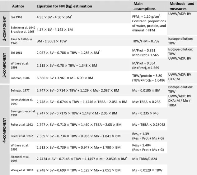
Table 2.2. Examples of body composition molecular models to estimate fat mass (kg)
Author Equation for FM (kg) estimation Main assumptions Methods and measures 2 -C O M P O NENT Siri 1961 4.95 BV - 4.50 BM* FFM D = 1.10 g/cm3 Constant proportions of water, protein, and mineral in FFM
UWW/ADP: BV
Behnke et al. 1942
Brozek et al. 1963 4.57 BV - 4.142 BM Pace & Rathbun
1945 BM - 1.3661 TBW TBW/FFM = 0.732 Isotope dilution: TBW 3 -C O M P O NENT Siri 1961 2.057 BV – 0.786 TBW – 1.286 BM* M/Prot = 0.351 M to Prot = 1.565 Isotope dilution: TBW UWW/ADP: BV Withers et al. 1998 2.115 BV – 0.78 TBW – 1.348 BM M/Prot = 0.354 (M+Prot)D = 1.569 Lohman, 1986 6.386 BV + 3.961 M – 6.09 BM TBW/protein = 3.80 (TBW+Prot)D = 1.0486 UWW/ADP: BV DXA: M 4 -C O M P O NENT
Selinger, 1977 2.747 BV - 0.714 TBW + 1.129 Mo - 2.037 BM Ms = 0.0105 BM Isotope dilution: TBW UWW/ADP: BV DXA: M / Mo / TBBA Heymsfield et al. 1990 2.748 BV – 0.6744 TBW + 1.4746 TBBA – 2.051 BM Ms= TBBA 0.235 Baumgartner et al. 1991 2.747 BV - 0.7175 TBW + 1.148 M - 2.05 BM Ms = 0.235 Mo Fuller at al. 1992 2.747 BV – 0.710 TBW + 1.460 TBBA – 2.05 BM Ms = TBBA 0.23048 Friedl et al. 1992 2.559 BV – 0.734 TBW + 0.983 Mo – 1.841 BM ResD = 1.39
(Res = Prot + Ms + G) Withers et al. 1992 2.513 BV – 0.739 TBW + 0.947 Mo – 1.790 BM ResD = 1.404 (Res = Prot + Ms + G) Siconolfi et al. 1995 2.7474 BV – 0.7145 TBW + 1.1457 M – 2.0503 BM # M = TBBA/0.824 Wang et al. 2002 2.748 BV – 0.699 TBW + 1.129 Mo – 2.051 BM Ms = 0.0129 TBW
Abbreviations: FM, fat-mass (kg); BV, body volume; BM, body mass; TBW, total body water; M, total mineral; Mo, bone mineral; TBBA, total body bone mineral; FFMD, fat-free mass density; FFM, fat-free mass; Prot, protein;
M/ProtD, total mineral + protein density; TBW/ProtD, total body water + protein density; Ms, soft mineral; ResD,
residual density; UWW, underwater weighting; ADP, air-displacement plethysmography; DXA, dual-energy X-ray absorptiometry.
*
This model was obtained considering the density of fat and fat-free mass at 37ºC; #This model was developed considering 3.037 as the density of total mineral.
The basic 2-component models lie on the premise that the body can be divided into two chemically distinct compartments, FM and FFM, with FFM corresponding to all the remaining tissues together [46]. In these models it is assumed that the density of FM and FFM are 0.9007 g/cm3 and 1.100 g/cm3 [17], and also that the FFM/TBW = 0.732 [28]. The majority of the errors associated with 2-component models falls not in the technical accuracy of the measurements but in the validity of the assumptions on composition and density of FFM, which are based on analyses of just three male cadavers [17]. Generally two component models involve the determination of body
progressed and models to estimate body composition that partition body mass up to six components are now available (Table 2.2). By including more and different measured properties or other components than 2-component models, these methods typically account for more biological variability [46, 47].
Densitometric models
The Ancient Greek civilization contribution to the study of body composition occurred when Archimedes (c.287-212 B.C.) observed that the buoyant force on a submerged object equals the body mass of the water it displaces, enabling the calculation of its specific gravity. He thus pioneered densitometry correctly observing that King Hiero’s crown was in fact an alloy which included cheaper and less dense metals and was not pure gold [48]. These findings would be of extreme usefulness in 1942 when Albert Behnke [44], refined underwater weighting to estimate body density.
The density of an object is defined as its mass per unit volume; therefore if we are able to determine a person’s body mass and volume we are able to calculate its density (body mass / volume). The body density (Db) is then usually transformed into FM using the Brozek et al. equation [17] (Table 2.1). At this regard the Siri equation [45] is also used to estimate FM (Table 2.1). However this last equation uses a value of 0.9000 g/cm3 for the density of fat as the author considered a body temperature of 37ºC. Although body core temperature approximates 37°C, the average body temperature under basal conditions in a comfortable environment is 1-2°C lower [49]. Accordingly, as Brozek et al. [17] used 0.9007 g/cm3 for the density of fat at 36ºC it seems more accurate to use this authors equation [50]. The two-component densitometric model will yield incorrect values for %FM if the overall density of the FFM components is different than 1.100 g/cm3 [50].
There are currently two methods to estimate body density: underwater weighting (UWW) and air displacement plethysmography (ADP).
The most traditional method for determining body density is UWW. The method requires the subject to be completely submerged in water [44]. The volume of water displaced and/or the subject’s body mass underwater, combined with the subject’s laboratory body mass, are used to calculate the whole body density. The main
limitations and restrictions of this method are associated with the estimates of body volume (BV) and the residual lung volume [45, 51-53].
More recently the UWW technique started to be replaced by ADP, where the subject is immersed not in water but in a close air-filled chamber. Air displacement plethysmography systems consist of a single structure that contains two chambers: the front chamber is where the participant is tested while the rear chamber is where the instrumentation is housed and serves a reference volume. The system determines body density through an air displacement method. A volume perturbing element (movable diaphragm) is mounted on the common wall separating the front and the rear chambers. When this diaphragm is oscillated under computed control, it produces complementary volume perturbations in the two chambers (equal in magnitude but opposite in sign). These volume perturbations produce very small pressure fluctuations that are analyzed to yield chamber air volume. The classic relationship of pressure versus volume, at a fixed temperature, is used to solve for the volume of the subject chamber [54].
Densitometric methods allow estimation of FM using 2-component models [17, 44, 45] but estimations of body volume are also necessary in multi-component models [47].
Hydrometric models
Water is the most abundant constituent of the body [24, 37, 55]. No other method applied in vivo can provide FM estimates in such a wide range of mammals, from the mouse to the elephant, which differ in body mass by a factor of 105 [2, 56, 57]. Unlike the other molecular body components, the water compartment consists of a single molecular species (H2O), which simplifies the task of its measurement.
Therefore, TBW is a common method for the assessment of body composition at the molecular level. The principle behind hydrometric models is that lipids are hydrophobic and thus free of water, which is therefore restricted to the FFM compartment. The calculation of FFM from TBW depends on the assumption of a constant hydration of FFM [58]. Pace and Rathbun [15] have reviewed chemical analytical data from several mammal species and observed that the FFM/TBW = 0.732. By considering that BM equals the sum of FM and FFM it is possible to derivate that FM = BM – (TBW/0.732)
occurs in infancy, with higher TBW/FFM comparing to adult values, which implicates that fat-free mass density is lower in pediatric ages [59].
Total body water can be measured by isotope dilution [58]. The basic principle of dilution techniques is that the volume of a compartment can be defined as the ratio of the dose of a tracer, administered orally or intravenously, to its concentration in the water space within a short time after the dose is administered. Usually, two samples of the same fluid (blood, saliva or urine) are collected, one before the administration of the dose as a baseline sample and other after waiting a sufficient amount of time for penetration of the tracer within the compartment of interest, as an enrichment sample. Inherent in any tracer dilution technique are four basic assumptions: 1) the tracer is distributed only in the extrachangeable pool; 2) it is equally distributed within this pool; 3) it is not metabolized during the equilibration time; and 4) tracer equilibration is achieved relatively rapid. Therefore, TBW can be measured by using a tracer dose of labelled water (tritium, deuterium, or 18-oxygen). Deuterium dilution is the most commonly used tracer to estimate TBW, as it is a stable isotope, simple to obtain and with small costs than tritium or 18-oxygen. Isotope enrichment analysis can be performed using infrared spectrometry, nuclear magnetic resonance, mass spectrometry, and isotope ratio mass spectrometry [43, 58].
Multi-component models
Traditionally body composition at the molecular level of analysis was studied as the sum of two components (Table 2.2), where the body mass equals the sum of FM and FFM [17, 44, 45]. However, at the molecular level FFM can be partitioned into several molecular components, including water, mineral, and protein [4].
Multi-component models share in common their developments from simultaneous equations, which may include two or more unknown components. As a general rule, for each unknown component estimated there must be one independent equation that includes the unknown component, the known component, and/or the measurable property. At the molecular level of analysis, measurable components include TBW by isotope dilution and mineral by Dual Energy X-ray Absorptiometry (DXA). Measurable properties used in developing molecular level multi-component models include body mass and body volume by UWW or ADP [11].
Body volume estimates are used in one term of the classical densitometric 2-component model [17, 44, 45] that serves as the basis for multi-2-component models. The addition of an estimate of TBW by isotope dilution [45, 46] or mineral by DXA [60] allows the development of 3-component models. Later investigators extended the Siri’s [45] classic 3-component model to a 4-component model by adding the bone mineral content of the FFM [61-68].
The formula for the 4-component model, which controls for biological variability in TBW, bone mineral mass, and residual can be generated using the same concept as for the two- and three-component models [49]:
(3) Where Db is body density, FM is fat mass, TBW is total-body water, Mo is bone mineral, res, is residual, and D
is density.
By assuming the densities of the molecular components it is possible to derivate the following equation (equation 4)
(4)
Where BV is body volume, FM is fat mass, TBW is total-body water, Mo is bone mineral, and res, is residual. Although multi-component models share assumed constant densities for FM, TBW, and Mo, two main strategies are applied in developing these models. In one approach the residual BM (Res) after subtracting FM, water, and bone mineral is assumed to be protein and soft tissue minerals of known densities. The other approach is to assume a combined residual mass (i.e. protein, soft tissue mineral, and other) of known density [47]. In fact, residual mass includes protein, soft tissue minerals, and glycogen. In equation 4 a value 1.404 g/cm3 is assumed for the residual mass density [69]. At this point it is important to remember that the largest components of residual mass are protein (density = 1.34 g/cm3) and glycogen (density = 1.52 g/cm3), in addition the residual mass also includes soft tissue minerals (density = 3.317 g/cm3).
In 2002 Wang at al. [67] has stated that the available 4-component models did not include an accurate estimation of soft tissue mineral, which is a small but important
electrolytes found in the extracellular and intracellular compartments of soft tissue. Although the mass of soft tissue minerals (about 400 g) is relatively small in adults, its contribution to body density should be considered because soft tissue minerals collectively have a higher density (3.317 g/cm3) than do each of the other components, including fat (0.9007 g/cm3), water (0.99371 g/cm3), protein (1.34 g/cm3), and bone mineral (2.982 g/cm3). At this regard Wang. et al [67] as developed a new 5-component model for FM which was simplified to a 4-component model (table 2.2) by assuming that Ms can be estimated from TBW (Ms = 0.0129 TBW).
More sophisticated –five and –six component models have also emerged [70, 71]. Besides estimations of molecular components these equations also incorporate measurements at the atomic level of body composition.
Dual-energy X-ray absorptiometry
Single photon absorptiometry was introduced in the early nineteen sixties as a way of quantifying appendicular bone mass. Dual photon absorptiometry methods first became clinically available in the early eighties, with the most recent advanced referred to as dual-energy X-ray absorptiometry (DXA) [72]. DXA provides whole body and regional assessment of FM, FFM and, also the estimation of bone mineral that can be used in multi-component models.
The fundamental principle of DXA is the measurement of the transmission of X-rays through the body at two different energy levels, low and high (typically 40 and 70 keV), which passes through tissues and is attenuate at rates related to its elemental composition (density and thickness of the human tissues through which they pass) (Figure 2.5). The extent to which photon energy is attenuated is a function of the initial photon energy of the X-ray beam, the mass per unit area of the absorber material, and the mass attenuation coefficient (m) of the absorber. When photons at two different
energies (e.g. 40 and 70 keV) are passed through an absorber, attenuation at the lower energy can be expressed as a ratio (R) to attenuation observed at the higher energy. For a homogeneous absorber, R is a function of mass attenuation coefficient and mass fraction of each component [72]. Therefore each element has a characteristic mass attenuation coefficient and an R value at a given energy. For instances, bone is rich in
highly attenuating minerals (Ca and P), and is readily distinguished from soft tissues [72].
Bone health assessment Body composition
assessment Major outcome:
Bone mineral density
Major outcome: Fat mass Lean soft tissue
Figure 2.5. Fundamental principle of dual-energy X-ray absorptiometry (DXA): the DXA measures the transmission of X-rays through the body at high and low energies. The X-ray beam energy is attenuated with the passage through tissue. The DXA body composition approach assumes that humans consist of three components that are distinguishable by their X-ray attenuation properties: bone mineral, fat tissue, and lean soft tissue (LST). [73]
The DXA body composition approach assumes that human consist of three components that are distinguishable by their X-ray attenuation properties: FM, bone mineral, and LST. In theory, solving for three unknown components requires measurement at three different photon energies. However, in practice, DXA can only resolve the fractional masses of a two-component mixture. Thus, DXA first separates pixels into those with soft tissue only (FM and LST) and those with soft-tissue plus bone mineral, based on the two different photon energies. This means that in pixels with bone mineral, soft tissue is not separately analyzed and the equipment assumes the FM
Detector
Low energy High energy
scan contains bone in addition to soft tissue thus, a systematic individual error is introduced as there might be variations in body composition between measured and non measured areas [74]. For example, the influence of arm and thorax on body composition estimation can be underrepresented due to the relatively large areas of bone in those regions [75]. This source of systematic error can be increased when tracking body composition compartments [76].
For athletes, DXA measurement presents several advantages over other laboratory methods due to its good precision, large availability, and low radiation dose [41, 73]. The progressive replacement of the original pencil-beam densitometers by fan-beam devices in the early 1990s allowed for better resolution and faster scan times, without compromising accuracy and without increasing radiation dose substantially, thus easing the burden of use for both patient and clinicians [73, 77]. However caution must be taken when using DXA on multiple occasions (perhaps no more than four times during a sports season), not only due to the cumulative radiation dose [41], but also due to the error of measurement [78], which limits the ability to detect small body composition changes over time, leading to misinterpreting data [41]
Despite DXA’s accuracy, precision, reliability, high speed, and non-invasive estimates with minimal radiation exposure [73, 79, 80], DXA is not without limitations. The main limitations pointed to this method are: algorithms calculations differ between manufacturers and are not published; pencil and fan-beam densitometers differ in accuracy; and limited active scan area [41]. This last limitation particularly affects athletes involved in sports where height is a major factor of performance, such as basketball and volleyball. Considering that it may be critical to measure people taller than the DXA scan area, alternative procedures are required to allow complete whole body scans (Evans, Prior, & Modlesky, 2005).
2.3.3.
C
ELLULARL
EVEL OFB
ODYC
OMPOSITIONWhole-Body Counting
Potassium is found mainly within the intracellular fluid compartment (ICF) and there is a stable intracellular potassium concentration. In addition, there is also a relatively stable relationship between ICF and BCM. The measurement of TBK by
![Figure 2.2. The five-levels of human body composition. ECS and ECF, extracellular solids and fluids, respectively [4]](https://thumb-eu.123doks.com/thumbv2/123dok_br/18851643.929591/33.892.128.773.358.761/figure-levels-human-composition-extracellular-solids-fluids-respectively.webp)
![Table 2.1. Assumed constants of composition and density (at 36ºC) of fat, fat-free mass, and body mass [17]](https://thumb-eu.123doks.com/thumbv2/123dok_br/18851643.929591/35.892.120.775.177.437/table-assumed-constants-composition-density-free-mass-body.webp)

![Figure 2.4. Classification of in vivo body composition methods [18]](https://thumb-eu.123doks.com/thumbv2/123dok_br/18851643.929591/41.892.115.775.203.500/figure-classification-vivo-body-composition-methods.webp)

![Figure 2.6. Doubly labelled water technique [134]](https://thumb-eu.123doks.com/thumbv2/123dok_br/18851643.929591/59.892.130.766.230.524/figure-doubly-labelled-water-technique.webp)
![Table 2.3. Fat mass (%) in samples of male athletes in several sports [adapted from [126]]](https://thumb-eu.123doks.com/thumbv2/123dok_br/18851643.929591/62.892.125.759.152.1124/table-fat-mass-samples-male-athletes-sports-adapted.webp)
![Table 2.4. Fat mass (%) in samples of female athletes in several sports [adapted from [126]]](https://thumb-eu.123doks.com/thumbv2/123dok_br/18851643.929591/63.892.133.759.370.987/table-fat-mass-samples-female-athletes-sports-adapted.webp)

