UNIVERSIDADE DO ALGARVE
Faculdade de Ciências e Tecnologia
SELF-ASSEMBLED NANOPARTICLES AS NEW SMART
CONTRAST AGENTS FOR MAGNETIC RESONANCE
IMAGING
Teresa Fragoso Rocheta Simão
Master’s Degree in Biomedical Sciences
Faro, Portugal
2010
UNIVERSIDADE DO ALGARVE
Faculdade de Ciências e Tecnologia
SELF-ASSEMBLED NANOPARTICLES AS NEW SMART
CONTRAST AGENTS FOR MAGNETIC RESONANCE
IMAGING
Teresa Fragoso Rocheta Simão
Master’s Degree in Biomedical Sciences
Project and thesis supervised by:
Fouzi Mouffouk, Ph.D (CBME, IBB)
Nuno Rodrigues dos Santos, Ph.D (CBME, IBB)
Faro, Portugal 2010
“The secret of getting ahead is getting started. The secret of getting started is breaking your complex, overwhelming tasks into small manageable tasks, and then starting on the first one.” Mark Twain
Dissertação de Candidatura ao Grau de Mestre em Ciências Biomédicas
Área de Bionanotecnologiapela Universidade do Algarve
MSc Thesis in Biomedical Scienses
As opiniões expressas nesta publicação são da exclusive responsabilidade do seu autor
The contents of this dissertation are of the exclusive responsibility of the author
ACKNOWLEDGEMENTS
The work described in this thesis would have been almost impossible without the support and help of many people.
First, I would like to express my gratitude to my supervisors, Dr. Fouzi Moffouk and Dr. Nuno Rodrigues dos Santos. Thanks to Dr. Fouzi Mouffouk for the opportunity to work in this great project, for teaching me and trust in my capabilities to perform this task. Thanks to Dr. Nuno Rodrigues dos Santos for receiving me in his laboratory, for being such a dedicated professor, with who I learned so much, thanks to patiently listen to my ideas and doubts, to advice, assist and support me all over this work.
A special thank you to Dr. Ana Costa, for being present whenever I needed, for gently teaching and conducting me during this work.
Thanks to Dr. Ana Grenha, Dr. Gabriela Silva and Dr. Álvaro Tavares for being always there and to supply me so many precious informations.
To my lab colleagues, Mónica Fernandes, Marinella Ghezzo and Ricardo Silva, thanks so much for sharing their knowledge, for their friendship, company and for always supporting me. Thanks to everyone at Dr. Guilherme Ferreira’s, Dr. José Belo’s and Dr. Ana Grenha’s laboratories for their suggestions and for helping me every time I needed.
Thanks to my work colleagues for several schedule shifts, support and friendship. To all my friends, special tanks for their encouragement to continue and also for their companionship. I salute them for understanding and forgiving my lack of time to be with them.
Lastly, I am very grateful to my parents and sister for being always a constant and unconditional source of support, affection, patience and understanding.
Thank you very much to everyone!
ABSTRACT
The aim of this thesis is to develop smart and targeted nanoparticle contrast agents for Magnetic Resonance Imaging. These nanoparticles were designed to improve the sensitivity of this high resolution imaging technique and thereby improve early cancer detection, which is a major factor for the reduction of cancer mortality. The designed nanoparticles are expected to accumulate in the tumor through passive and active targeting. In addition, when exposed to the characteristic low pH of the cancer microenvironment, the nanoparticles will release the contrast agent, which will turn on its imaging capability. This contrast agent consists of pH-sensitive polymeric micelles formed by self-assembly, loaded with Gadolinium (III) complexes and bioconjugated with the C595 monoclonal antibody against the human MUC1 protein, which is overexpressed during tumorigenesis since its early stages.
The capability of micelles to disintegrate and release the encapsulated contrast in acidic conditions was proved by 1.5T MRI experiments. The MRI study showed no image signal from the sample with intact micelles whereas a signal enhancement was observed from the sample at low pH. After one mouse has been intramuscularly injected with Gadolinium(III)-loaded micelles in both hind legs, the MR image demonstrated a stronger signal from the right hind leg, which was previously injected with an acid solution. To assess in vitro cytotoxicity of free and encapsulated Gadolinium (III) complexes in polymeric micelles, MTT assays were performed on different cancer cell lines. Encapsulated Gadolinium (III) complexes showed significantly lower cytotoxicity than free Gadolinium (III) complexes, even at the highest concentration. To verify the target capability of nanoparticles, fluorescent dye-loaded polymeric micelles were incubated with breast cancer cells expressing MUC1 (verified by semi-quantitative RT-PCR, Western blotting and flow cytometry) and mouse bone marrow stroma cells. Antibody-conjugated micelles had superior affinity for MUC1-expressing human breast cancer cells than for mouse bone marrow cells.
Key words
Smart contrast agent; Magnetic Resonance Imaging; Nanoparticles; Amphiphilic diblock copolymers; Mucin-1
RESUMO
A presente Tese de Mestrado aborda a criação e síntese de um agente de contraste ―inteligente‖ para imagiologia por Ressonância Magnética.
A Ressonância Magnética (RM) é uma técnica de imagem médica que apresenta excelente resolução espacial e contraste entre tecidos diferentes. No entanto, por vezes a resolução de contraste não é suficiente para demonstrar alterações patológicas no mesmo tecido, especialmente quando as alterações ainda só estão presentes a nível celular e molecular. Para aumentar a sensibilidade desta técnica de imagem e potenciar a sua aplicação ao nível da imagem molecular desenvolveram-se nanopartículas com agente de contraste, específicas para detectar cancro.
Este agente de contraste ―inteligente‖ é constituído por polímeros anfifílicos, sensíveis ao pH, que por self-assembly se organizam formando micelas. Estas, devido à característica particular do polímero que as forma, irão responder, desintegrando-se e libertando o seu conteúdo que consiste em quelatos de gadolínio (III) hidrofóbicos, exclusivamente perante ambientes com valores de pH relativamente ácidos, permanecendo intactas noutros meios. Enquanto o meio de contraste estiver encapsulado no centro hidrofóbico das micelas, elas restringem o acesso das moléculas de água ao quelato de gadolínio, o que impede a interacção dos protões da água com o ião metálico. Assim, não há diminuição do tempo de relaxação longitudinal (T1) das moléculas de água circundantes. Pretende-se, por isso, utilizar o pH extracelular relativamente ácido dos tecidos tumorais como despoletador específico da desintegração destas micelas e assim permitir que os quelatos de gadolínio aumentem o contraste da imagem de RM unicamente nestas situações. Esta diminuição localizada e específica do pH verifica-se desde as fases mais precoces do desenvolvimento tumoral, principalmente devido ao aumento da taxa de glicólise aeróbica das células cancerígenas.
As micelas foram bioconjugadas com um anticorpo monoclonal, C595, contra a proteína Mucina-1 (MUC1) humana. Esta proteína tem um papel importante no desenvolvimento de certos tipos de cancro, demonstrando alterações relativamente aos seus níveis de expressão e localização na superfície celular, assim como no seu grau de glicosilação, desde as primeiras alterações tumorais verificadas ao nível da célula. Com esta abordagem, pretende-se tornar mais específica e intensa a acumulação deste tipo de
partículas em tecidos cancerígenos, que ocorre devido à arquitectura vascular anómala, caracterizada por amplos espaços entre as células endoteliais. A referida acumulação de nanopartículas em tecidos tumorais é devida à sua dimensão reduzida que lhes permite extravasar pelos espaços endoteliais aumentados, à drenagem linfática ineficaz dos tecidos tumorais e ao longo tempo de permanência na circulação sanguínea que aumenta a probabilidade e a frequência do extravasamento.
Utilizaram-se quelatos de gadolínio como agente de contraste, pois o contraste positivo produzido por este ião paramagnético em imagens de RM ponderadas em T1 é mais fácil de assinalar que o contraste negativo induzido pelos contrastes superparamagnéticos em imagens ponderadas em T2. A quantidade relativamente elevada de quelatos de gadolínio encapsulada nas micelas, a retenção e acumulação destes transportadores na lesão tumoral vão aumentar a concentração local do meio de contraste. Este fenómeno resultará numa amplificação da intensidade de sinal emitida pela lesão tumoral, o que aumenta a sensibilidade para a sua detecção, mesmo quando apenas existe um número limitado de células alteradas. Enquanto se aumenta a biodisponibilidade local do meio de contraste, diminui-se a concentração total de contraste necessária para a obtenção de imagens com acuidade diagnóstica.
Mediante o descrito, este trabalho foi desenvolvido com o intuito de testar o mecanismo de resposta das micelas à diminuição do pH, de perceber se os quelatos de gadolínio hidrofóbicos produziam um aumento de contraste em imagens de RM compatíveis com aplicação clínica, de testar a toxicidade in vitro dos quelatos de gadolínio livres ou encapsulados nas micelas e de avaliar a especificidade deste novo sistema para as células-alvo. Para este fim sintetizaram-se micelas com complexos de gadolínio, micelas com o fluoróforo 1-metilpireno encapsulado e micelas bioconjugadas com o anticorpo anti-MUC1 e com fluoróforo encapsulado. As nanopartículas foram caracterizadas por Dispersão Dinâmica de Luz (DLS). A distribuição de tamanho mostrou que as micelas com 1-metilpireno encapsulado têm cerca de 24 nm de diâmetro. Obtiveram-se curvas de calibração para os quelatos de gadolínio e para o fluoróforo para se estimar a concentração total de cada um dos compostos numa dada suspensão de micelas. Demonstrou-se a capacidade destas micelas para se desintegrarem em pH ácido e libertarem o agente de contraste através da aquisição de imagens de duas amostras com micelas com meio de contraste encapsulado e com diferente pH, num equipamento de RM de 1.5 T. Outra experiência semelhante foi realizada, através da injecção intramuscular de micelas com meio de contraste
encapsulado em ambos os membros posteriores de um ratinho. Sendo que, previamente havia sido injectado uma solução ácida no membro posterior direito. As imagens da primeira experiência demonstraram um aumento da intensidade do sinal proveniente da amostra com pH baixo, enquanto a amostra com pH neutro não demonstrou qualquer alteração de sinal relativamente ao meio envolvente. Relativamente à segunda experiência, verificou-se que a intensidade do sinal proveniente do membro direito era superior à do no membro esquerdo. A citotoxicidade dos complexos de gadolínio foi observada in vitro através de ensaios de MTT em diferentes linhas celulares de cancro. Verificou-se que a encapsulação dos complexos de gadolínio nas micelas diminuiu significativamente a sua toxicidade comparativamente com o complexo livre. O valor de IC50 para estes quelatos de gadolínio variou entre 10.2 e 12.8 μM consoante a linha celular em estudo. As experiências de verificação da especificidade das micelas bioconjugadas com C595 para linhas celulares de carcinoma mamário que expressavam MUC1 foram realizadas através de citometria de fluxo e microscopia de fluorescência. As micelas tinham 1-metilpireno encapsulado para facilitar a sua observação por ambos os métodos. Utilizaram-se como controlo células estromais da medula óssea de ratinho. Observou-se que as partículas conjugadas ao anticorpo mostraram maior afinidade para as células que expressavam MUC1 do que as partículas sem anticorpo. A expressão de MUC1 nas linhas celulares utilizadas foi comprovada através de RT-PCR semi-quantitativo, PCR em tempo real, Western blotting e citometria de fluxo.
Os resultados obtidos demonstraram que estas micelas têm potencialidade para ser utilizadas em RM como agente de contraste, pois, relativamente aos agentes de contraste actualmente utilizados, combina a capacidade de acumular passivamente (devido à dimensão) e activamente (devido à bioconjugação com o anticorpo) em tumores que expressem a proteína MUC1. Além disso, possibilita a amplificação do sinal da imagem de RM em microambientes com pH relativamente ácido, que são habitualmente característicos de lesões cancerígenas.
Palavras-chave
Agente de contraste inteligente; Imagiologia médica; Nanopartículas; Polímeros dibloco anfifílicos; Mucina-1
TABLE OF CONTENTS
Acknowledgements ... iv
Abstract... v
Resumo ... vi
List of figures ... xi
List of tables ... xii
List of abbreviations ... xiii
Chapter I: Introduction ... 1
1. Nanotechnology ... 2
1.1. Nanoparticles in medicine ... 2
1.1.1. Micelles ... 5
1.1.1.1. pH-sensitive micelles for early cancer detection ... 7
2. Magnetic resonance imaging ... 11
2.1. Basic physical principles ... 11
2.2. Relaxation times ... 13
2.3. Main types of image contrast ... 14
3. Magnetic resonance imaging with contrast agents ... 16
3.1. Mechanism of shortening T1 and T2 times ... 17
3.2. Contrast agents for T1WI ... 18
3.2.1. Nonspecific Gd(III)-based contrast agents ... 18
3.2.2. Targeted Gd(III)-based contrast agents ... 21
3.2.3. Smart Gd(III)-based contrast agents... 21
4. Cancer microenvironment ... 23
5. MUC1 protein ... 25
5.1. A Target Molecule for cancer imaging ... 26
6. Breast cancer ... 29
6.1. Tumorigenesis ... 29
6.2. Classification ... 30
6.2.1. Histopathological subtypes... 30
6.2.2. Genetic alterations ... 31
7. Aims of the project ... 33
Chapter II: Materials and Methods ... 34
1. Bioconjugation and Polymeric micelle preparation ... 35
2. Detection of Nanoparticles-based contrast agent ―on‖ and ―off‖ states ... 36
3. Measurement of Gd(III) complexes and fluorophore concentrations ... 37
4. Cell lines and culture conditions ... 37
5. In vitro cytotoxicity tests ... 38
6. Analysis of MUC1 expression ... 40
6.1. SDS-PAGE and Western Blotting ... 40
6.2. RT-PCR ... 42
6.3. Real-time PCR ... 44
6.4. Flow cytometry ... 44
7. Assessment of specific targeting of bioconjugated Micelles ... 45
7.1. Flow cytometry ... 45
7.2. Fluorescence microscopy ... 45
Chapter III: Results ... 47
1. Demonstration of micelle pH-sensitive mechanism of content release ... 48
2. In vitro cytotoxicity tests ... 50
3. Analysis of MUC1 gene and protein expression ... 52
4. Assessment of specific targeting of bioconjugated nanoparticles ... 56
5. Gd(III) complexes and fluorophore concentrations in micelle solutions ... 61
Chapter IV: Discussion ... 63
1. Nanoparticle size and pH stability ... 64
2. Gd(III) complex and micelle cytotoxicity ... 65
3. MUC1 gene and protein differential expression between cell lines ... 67
4. Bioconjugation of nanoparticles potentiates targeting to MUC1-expressing cells 70 5. Gd(III) complexes and fluorophore concentrations in micelle solutions ... 71
Chapter V: Future perspectives ... 73
References ... 76
Appendix A ... 91
LIST OF FIGURES
Figure 1 - Micelle structure……… 5
Figure 2 - Schematic representation of amphiphilic diblock copolymer synthesis and micelle pH-sensitive disaggregation………... 7 Figure 3 - Micelle size characterization……….. 8 Figure 4 - Spectroscopic proof of polymeric micelles formation and
disassembled with parallel release of 1-methylpyrene due to low pH... 9 Figure 5 - Schematic representation of switch off/on mechanism……….. 10 Figure 6 - Schematic representation of hydrogen nuclei behavior under the
influence of an magnetic field (B0)……… 12 Figure 7 - Schematic representation of the behavior of the net magnetization
vector, M, after the system received energy from a RF pulse………… 13
Figure 8 - Sagittal images of the knee……….………... 15 Figure 9 - Negative contrast provided by uptake of SPIO by Kupffer cells
(RES)……….………. 16
Figure 10 - Nonspecific image signal enhancement following administration of
LMWCA……… 20
Figure 11 - MUC1 expression on normal and cancer cells………….…...………... 26 Figure 12 - MUC1 glycosylation pattern at a cancer cell surface membrane……... 27 Figure 13 - Demonstration of pH-sensitive nanoparticle content release.………… 48 Figure 14 - Demonstration of pH-sensitive nanoparticle content release in in vivo. 49 Figure 15 - Cytotoxicity of free Gd(III) complexes by cell count…………....…… 50 Figure 16 - Cytotoxicity of free Gd(III) complexes by MTT assay………. 51 Figure 17 - Linear regression line for cytotoxicity of free Gd(III) complexes.…… 51 Figure 18 - Cytotoxicity comparison of free and nanoparticle-encapsulated
Gd(III) complexes……….. 52
Figure 19 - Detection of MUC1 expression by RT-PCR……….. 53 Figure 20 - MUC1 relative expression, in different cell lines, by real-time PCR
analysis………... 54
Figure 21 - Electrophoresis and detection of MUC1 glycoprotein in homogenates of MCF-7 breast cancer cells and DND41 acute lymphoblastic leukemia cells by Western Blotting………... 55
Figure 22 - Analysis by fluorescent flow cytometry to detect MUC1 cell surface expression on MCF-7 and MDA-MB-468 cell lines………. 55 Figure 23 - Fluorescent flow cytometry to assess specific targeting of Anti-
MUC1 bioconjugated 1-methylpyrene loaded nanoparticles…………. 57 Figure 24 - Specific binding of anti-MUC1 1-methylpyrene-loaded micelles to
MCF-7 breast cancer cells……….. 59
Figure 25 - Specific binding of anti-MUC1 1-methylpyrene-loaded micelles to MCF-7 breast cancer cells, after washes……… 59 Figure 26 - Increased targeting of anti-MUC1 1-methylpyrene-loaded micelles to
MDA-MB-468 breast cancer cells………. 60 Figure 27 - Calibration curve for Gd(III) complexes………... 61 Figure 28 - Calibration curve for 1-methylpyrene……… 62
LIST OF TABLES
Table 1 - Delta (∆) values……… 57
Table 2 - Absorbance measurements of 1-methylpyrene-loaded micelle
LIST OF ABBREVIATIONS
CA contrast agent
CT cytoplasmic tail
CTA chain transfer agent
DCE dynamic contrast enhancement
DCIS ductal carcinoma in situ
DMF N,N-Dimethylformamide
DLS dynamic light scattering
DOTA 1,4,7,10-tetraazacyclododecane-1,4,7,10-tetraacetic acid
DTPA diethylenetriaminepentaacetic acid
DTPA-BMA {bis-[2-(carboxymethylmethylcarbamoylmethylamino)ethyl]amino} acetic acid
EGFR epidermal growth factor receptor
EPR enhanced permeability and retention
ER estrogen receptors
Gd (III) gadolinium ion
HER2 human epidermal growth factor receptor 2
HP-DO3A 1,4,7,10-tetraazacyclododecane-1-hydroxypropyl-4,7,10-trisacetic acid
HPMA N-(2-hydroxypropyl)methacrylamide
IC50 inhibitory concentration that reduces cell viability by 50%
IDC invasive ductal carcinoma
LIN lobular intra-epithelial neoplasia
LMWCA low molecular weight contrast agent
MAb monoclonal antibody
MMWCA macromolecular weight contrast agent
MR magnetic resonance
MRI magnetic resonance imaging
MTT 3-(4, 5-dimethylthiazol-2-yl)-2, 5-diphenyl tetrazolium bromide
NMR 1H Magnetic Resonance
PBS phosphate-buffered saline
PBST phosphate-buffered saline/0.1% Tween 20
PD proton density
PEG poly (ethylene glycol)
PR progesterone receptors
RAFT reversible addition-fragmentation chain transfer
RES reticuloendothelial system
RF radiofrequency
SPIO superparamagnetic particles of iron oxide
T Tesla
TE echo time
TEM transmission electron microscopy
THF tetrahydrofuran TR repetition time T1WI T1-weight image T2WI T2-weighted image UV ultraviolet Symbol Definition A UV-Vis absorption
B0 external magnetic field M net magnetization vector
T1 longitudinal magnetization time
1. NANOTECHNOLOGY
Nanotechnology has emerged in the last few years as a new multidisciplinary scientific field, which not only benefits from but also has an impact on such diverse domains as chemistry, physics, electronics, optics, energy, materials science, space technology and biomedicine (Porter & Youtie, 2009; Choi & Baker, 2007). Nanoscience offers the exceptional possibility to produce nanoscale devices with approximately 1–100 nm and containing multiple integrated properties (Liu, Kiessling, & Gatjens, 2010; Yezhelyev, Gao, Xing, Al-Hajj, Nie, & O’Regan, 2006).
1.1.NANOPARTICLES IN MEDICINE
Great progress has been made regarding nanomaterial applications to medicine. These compounds have not only been tested in in vitro experiments but also been applied clinically (Tiefenauer, 2007). The development of this research area will provide extraordinary opportunities for future individualized diagnostic strategies and therapeutic approaches for humans (Liu et al., 2010).
Nanoparticles are complex compounds made of multiple molecules and polymers that may present single or multiple functions (Bulte & Modo, 2008). They have been developed as contrast agents for medical imaging, for therapeutic applications such as target carriers for drug controlled release and for gene delivery (Kairemo, Erba, Bergström, & Pauwels, 2008; Peer et al., 2007).
An area under intensive research is the application of nanoparticles for molecular imaging (Young-wook, Jae-Hyun, & Jinwoo, 2007). Molecular imaging is an interdisciplinary research field, which covers chemistry, biology, pharmacology, and medicine, aiming to detect early, to visualize and to characterize physiological and disease processes in vitro and in vivo (Weissleder & Mahmood, 2001). This application provides valuable clinical information to help guiding therapy against a variety of diseases, such as cancer (Mankoff, 2007). The issue of early cancer diagnostics has been the object of much research. Several types of nanoparticles with variable characteristics have been engineered to optimize cancer detection through noninvasive imaging modalities, such as magnetic resonance imaging (MRI), computed tomography, ultrasound, positron emission tomography, single photon emission computed tomography and near-infra-red fluorescence imaging (Weissleder, 2002).
Critical features for successful preparation and application of nanoparticles to biological environments are reproducibility of synthesis method, creation of well-defined nanoparticles with monodispersity populations, low immune system reactions and reduced cytotoxicity and elimination from the biological system (Choi & Baker, 2007). Novel challenges have also emerged, such as the elimination of nonspecific uptake of nanoparticles by macrophages and other cells of the reticuloendothelial system (RES); the development of more specific nanoparticles that target just molecules of interest, and the design of particles small enough to penetrate endothelial barriers (blood-brain barrier and dermal tight junctions). At the same time, nanoparticles should be able to maintain long-circulating blood half-lives, allowing accumulation at the target location and release of their contents to get a localized high drug/contrast agent (CA) concentration with far less systemic effects. Another new approach is the use of diagnostic ―smart‖ agents that behave as sensors and act in response to several biological phenomena (Bulte & Modo, 2008; Kairemo et al., 2008).
Depending on the application intended, the surface of nanoparticles can be decorated with various molecules to avoid the recognition by the immune system and can be functionalized with targeting ligands to create specificity for a receptor of interest. Several molecule-specific ligands have been used, including antibodies and their fragments, aptamers, peptides, peptidomimetics, vitamins and carbohydrates ( Mulder, Strijkers, van Tilborg, Griffioen, & Nicolay, 2006; Allen, 2002). To target exclusively cancer tissues, several nanoparticles have been functionalized to bind tumor biomarkers such as transferrin receptors (Choi, Alabi, Webster, & Davis, 2010), folate receptors (Hong et al., 2008), ανβ3 integrin (Winter et al., 2003), epidermal growth factor receptor (EGFR) (Yan et al., 2009), human epidermal growth factor receptor 2 (HER2) (T. Chen et al., 2009), prostate specific membrane antigen (Wang et al., 2007) and somatostatin receptors (Li et al., 2009).
Despite the increasing diversity of nanoparticles, most can be classified into two major types: inorganic or organic. Inorganic nanoparticles have an inorganic core that confers specific fluorescence, optical, electronic and magnetic properties. The surface core is coated with a protective biocompatible organic layer that stabilizes the structure, avoids uncontrolled growth, aggregation and delays blood elimination of the nanoparticles (Yezhelyev et al., 2006). Some examples are quantum dots, superparamagnetic, silver, gold, silica and calcium nanoparticles (Tiefenauer, 2007; Xing, Chang, & Kang, 2010). These nanoparticles are disadvantageous for systemic
cancer cell targeting, because they are neither biodegradable nor small enough to be efficiently cleared from the blood through the kidneys, and can thus accumulate in the body and cause long-term toxicity (Peer et al., 2007). Organic nanoparticles such as liposomes, micelles, dendrimers, carbon nanotubes and other polymers are composed of organic materials (Lee, MacKay, Fréchet, & Szoka, 2005; Yezhelyev et al., 2006). These nanoparticles have been extensively used as drug and CA carriers. They present an increased hydrophilic and hydrophobic content, bio-availability, a high surface-area-to-volume ratio, which allows attachment of several targeting ligands to the surface, and have shown an efficient accumulation at pathological locations (Kairemo et al., 2008; Peer et al., 2007).
Polymers are the most commonly explored materials for the design of organic nanoparticles. These biomaterials are long-chain molecules composed of a large number of small repeating units (monomers), and can be derived from natural sources or from organic synthesis (Cooper, Visser, Hergenrother, & Lamba, 2004).The methods of synthetic polymer preparation can be classified into two groups: addition polymerization and condensation polymerization. In addition polymerization, monomers have to be activated by an initiator (free radicals, cations, anions or catalysts) to react through the stages of initiation, propagation, and termination to form the final polymer. For condensation polymerization, the monomers contain functional groups, so the initiator is not required. Two monomers react to form a covalent bond, usually with elimination of a small molecule such as water, hydrochloric acid, methanol, or carbon dioxide. The reaction is continuous until one of the reactants finish (Park & Lakes, 2007, pp.173-177). The choice of polymerization method strongly affects the polymer obtained. For example, considering addition polymerization of free radicals, the molecular weights of the polymer chains are difficult to control while in anionic polymerization the molecular architecture can be easily controlled. Recent methods of living free radical polymerizations called atom transfer radical polymerization and Reversible Addition-Fragmentation chain Transfer (RAFT) polymerization have been used to create amphiphilic block copolymers with well-defined structures. These methods proved to achieve low polydispersity indexes, which enable well-controlled composition, size and morphology of polymeric micelles ( Zhang, Zhang, Wen, Li, & Li, 2008; Stenzel, 2008).
Polymers can be classified as homopolymers, which are composed by one type of repeat unit, or as copolymers, which are made by two or more types of repeat units.
Regarding copolymers, the structure of polymer chains can be alternating (monomers alternate along the chain), random (monomers are randomly distributed along the chain), graft (main chain is made up by one type of monomer to which are bounded small chains of another monomer), or block copolymers (the polymer chain is formed by blocks of each monomer). As block copolymers have separate segments they display the individual features of each homopolymer (Cooper et al., 2004).
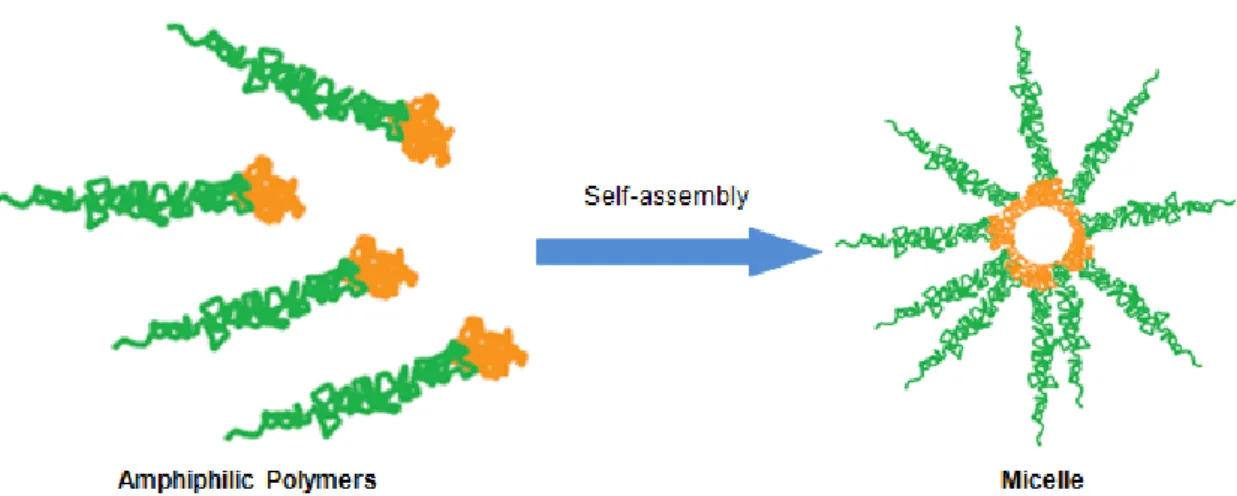
1.1.1.MICELLES
Micelles are self-assembling nanosized colloidal particles with a hydrophobic core and hydrophilic corona (Figure 1) (Torchilin, 2007). There are several composites able to form micelles. However, amphiphilic block copolymers have received a great deal of attention. These polymers are composed by hydrophilic and hydrophobic monomer units. These molecules form micelles spontaneously when they are dissolved in selective solvents, under a narrow concentration interval (critical micelle concentration) and temperature (critical micellization temperature) (Palivan, Vebert, Axthelm, & Meier, 2006; Jones & Leroux, 1999). Polymeric micelle formation involves a decrease of free energy in the system due to elimination of hydrophobic blocks from the aqueous environment. The hydrophobic region of the amphiphilic polymer forms the micelle core, while the hydrophilic region composes the micelle corona that stays in contact with water (Kwon & Okano, 1999).
Figure 1 - Micelle structure. Polymeric micelles are composed by a hydrophobic core (orange) and a hydrophilic corona (green). Adapted from (Peer et al., 2007).
Polymeric micelles have been successfully used as drug carriers due to their high stability in vitro and in vivo and improved bioavailability of the content. Micelle content can be released by surface erosion, by diffusion through progressive degradation of the hydrophobic core, or in response to environment (pH, temperature, or salt concentration sensitivity) (Rutkaite, Swanson, Li, & Armes, 2008). In addition, their smaller size compared to other particulate carriers allowed a superior permeability across physiological barriers and improved biodistribution (Nakamura, Makino, Okano, Yamamoto, & Yokoyama, 2006; Hong et al., 2008; Maeda, Wu, Sawa, Matsumura, & Hori, 2000). The use of copolymers having a poly (ethylene glycol) (PEG) extends the micelle half-life in blood. A direct correlation between the longevity of a particulate drug carrier in the circulation and its ability to reach its target site has been observed on multiple occasions ( Maeda, Sawa, & Konno, 2001; Torchilin, 2001). Micelles have also a reasonably narrow size distribution, and are very easy to prepare and to load drug or CA (Heller & Hoffman, 2004). By changing the copolymer chain size and chemical nature, the type of solvent used to dissolve the copolymer, and the critical micelle concentration of the copolymer, it is simple to modify the shape and size of polymeric micelles. Several formulations of drug-loaded micelles are currently at different stages of preclinical and clinical trials (Torchilin, 2007).
The transport of CA by micelles is a relatively new approach, which have been experimented just for diagnostic/imaging or for monitoring the drug delivery by
micelles (Liu, Zeng, & Allen, 2007; Zhang et al., 2008).
A promising approach on medical imaging is the use of stimuli-sensitive micelles whose degradation and resultant content release is due to pH or specific temperature values (Nakamura et al., 2006; Torchilin, 2007). Stimulus-sensitive
micelles are made of ―smart‖ polymers, which respond to slight alterations in physical, chemical or biochemical environmental conditions with significant changes in their physical properties (Hoffman, 2004). Consequently, micelles composed by these special polymers are able to dissociate the core and release their content only under certain pathological features (Palivan et al., 2006). Several approaches using pH-sensitive micelles, which release their content in conditions of low tumor cell pH, have been studied and used successfully to improve the efficiency of chemotherapy and to decrease the side effects and toxicity of free chemotherapeutics (Lee, Na, & Bae, 2003; Lee, Na, & Bae, 2005; Gillies & Fréchet, 2005; Hrubý, Konák, & Ulbrich, 2005).
1.1.1.1. PH-SENSITIVE MICELLES FOR EARLY CANCER DETECTION
This new kind of pH-sensitive nanoparticles seemed to be a very promising approach for early cancer detection because they disassemble exclusively in a specific pH value, have proven to be stable and are efficient carriers for hydrophobic drug molecules. Therefore a new class of nanoparticle-based CA has been developed.
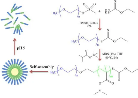
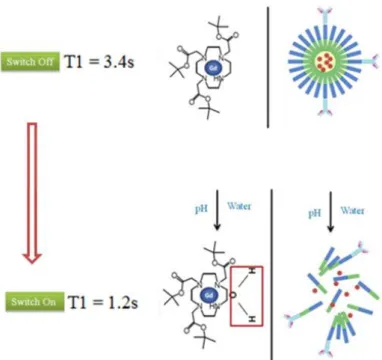
The proposed pH-sensitive polymeric micelles are able to encapsulate a large number of CA molecules (hydrophobic Gadolinium ion [Gd(III)] chelates) during its self-assembly from simple building blocks (Poly(ethylene glycol-b-trimethylsilyl methacrylate). The pH-sensitive mechanism of the designed polymeric micelles is generated by amphiphilic polymer silicon moieties that can be cleaved in a slightly acidic environment. This cleavage turns the polymer hydrophilic, which triggers micelle disassembly and release of its content (Figure 2). This pH-sensitive amphiphilic diblock copolymer was synthesized by RAFT polymerization from trimethylsilyl methacrylate using α-(O-ethylxanthate)-ώ-methylPEG 2’000 as a macro-chain transfer agent (macro-CTA), and a monomer to macro-CTA ratio of 45:1. Azobis-isobutyronitrile (AIBN) (1 mol% of the monomer), was used as radical initiator (Chiefari et al., 1998). The CA was synthesized and characterized via well known synthesis routes to generate hydrophobic Gd(III) complexes with ligands such as bipyridine (Bechara, Leygue, Galaup, Mestre, & Picard, 2009).
Figure 2 – Schematic representation of amphiphilic diblock copolymer synthesis and micelle pH-sensitive disaggregation. Synthetic approach for the synthesis of poly(ethylene glycol-b-trimethylsilyl methacrylate) and schematic representation of its self-assembly into micelles and their disaggregation due to pH decrease.
These polymeric micelles were designed to remain intact during circulation and if the target is absent, the CA remains switched-off until their removal from the system. However if micelles reach the target site (cancer tissue), they disassemble due to low pH. When micelles are intact, they restrict water access to the hydrophobic Gd(III) chelates that are located in the micelle core. Consequently, the relaxivity of nearby water molecules remain unchanged, however when these Gd(III) complexes are released from the micelle core, they will have access to water molecule protons and thus reduce the longitudinal magnetization time (T1) of the water surrounding the targeted tissue. This new approach will therefore provide the medicine with better tools to understand the causes and mechanisms of disease and the associated structural and functional alterations.
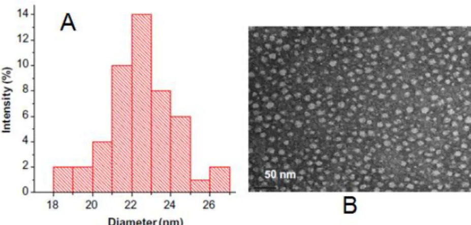
The size distribution profile of the novel micelles (with encapsulated 1-methylpyrene) was found to be around 24 nm (Figure 3), as measured by dynamic light scattering (DLS) (Figure 3-A). These micelles were also visualized under transmission electron microscopy (TEM) (Figure 3-B) and the size matches the values obtained from DLS.
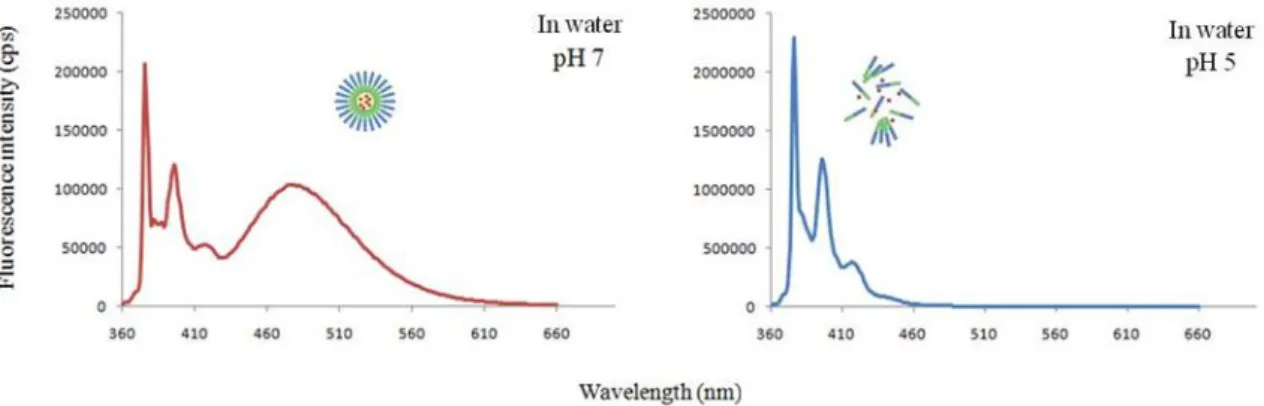
To establish the amphiphilic aggregation of the copolymer and its disaggregation, with parallel tracer release under pH change, it was used the 1-methylpyrene fluorescence spectrum. When 1-1-methylpyrene is dissolved and encapsulated inside micelles, it reaches a threshold concentration (104 M) that induces the formation of excimers, which have a different emission wavelength than
1-Figure 3 – Micelle size characterization. (A) Dynamic Light Scattering results showing the size distribution profile of 1-methylpyrene loaded polymeric micelles. (B) Transmission electron microscopy of 1-methylpyrene loaded polymeric micelles after the self assembly of poly(ethylene glycol-b-trimethylsilyl methacrylate), bar scale 50nm.
methypyrene monomers. The monomer emission occurs in less concentrated solutions and in aqueous solutions due to the low solubility of 1-methylpyrene. The emission spectrum of 1-methylpyrene-loaded micelles in a pH=7 aqueous solution revealed both monomer (at 375 nm) and excimer (at 480 nm) emission bands, indicating that micelles existed in the aqueous solution. On the other hand, the emission spectrum of 1-methylpyrene-loaded micelles in a pH=5 aqueous solution showed only the monomer emission band, indicating that nanoparticles were decomposed and had release their content. The release causes contact of excimer with water and consequent decrease of the threshold concentration required to form them (Figure 4).
After proving the ability of these nanoparticles to release their content in acidic conditions, we demonstrated by 1H Magnetic Resonance (NMR) Spectroscopy that the CA (tetraaquodichloro(4,4'-ditBu-2,2'-bipyridine)gadolinium(III) chloride) encapsulated within the micelle core was shield from water molecules in solution (Figure 5). This conclusion stems from the observation that the T1 measured in a pH=7 aqueous solution with Gd(III) complex-loaded micelles was 3.4 seconds, while at pH=5 the T1 time obtained was 1.7 seconds. At pH=7, intact polymeric micelles restrict the water access to the Gd(III) complexes and the T1 value is the same as the T1 measured in pure water (switched-off state). In contrast, micelles disassembled due to low pH (similar to those found in cancer tissue) and the water molecules had accessed and exchanged with Gd(III) complexes, thus significantly decreasing the T1 of the aqueous solution surrounding these nanoparticles (switched-on state).
Figure 4 - Spectroscopic proof of polymeric micelles formation and disassembled with parallel release of 1-methylpyrene due to low pH. At pH=7 the polymeric micelles are stable as the excimer band (480 nm) holds. However at pH=5.5 only the monomer emission (375 nm) stands due to complete release of 1-methypyrene.
Because the existing imaging techniques are generally unsatisfactory for many molecular imaging applications, it is important to develop high-performance imaging systems capable of identifying detailed biological processes at the molecular and subcellular levels. Currently, MRI is one of the most powerful medical diagnostic tools available mainly due to its high spatial resolution and by the fact that physiological and anatomical information can be acquired simultaneously (Li, Fraser, & Meade, 1999; Medarova, 2009). However, in terms of sensitivity MRI lags behind other tools (Massoud & Gambhir, 2003). To overcome this hurdle, several attempts have been made to improve the sensitivity of MRI using CAs. With the increasing ability of nanotechnology to create devices at the cellular and molecular scale, more powerful imaging CAs for MRI such as superparamagnetic particles of iron oxide (SPIO) and magnetodendrimers nanoparticles have developed. SPIO have gained great attention for molecular MRI and has been experimentally and clinically used to detect infarction, inflammation, angiogenesis, primary malignant lesions, and lymph node metastasis (Priest et al., 2006; Russell & Anzai, 2007; Gambarota et al., 2006; Corot, Robert, Idée, & Port, 2006). Although much progress has been made to develop these nanoparticles during the past few years, their successful use has been limited to in vitro systems,
Figure 5 - Schematic representation of switch off/on mechanism. Relaxometric experiment that measures the T1time before and after the
except for a few in vivo cases. These difficulties lie on two factors: poor MRI contrast effects and limited stability and biocompatibility under in vivo conditions.
The new micelles have therefore a great potential to be used as CA for MRI. They present the advantage over other CAs of imaging cancer tissues only and not normal cells, because they expose Gd(III) chelates to the aqueous surroundings and enhance the relaxivity of the paramagnetic metal only under characteristic low pH cancer environments. These micelles should increase the half-life in blood of the Gd(III) complexes, by providing a protection from the outside elements via their encapsulation within their the core. Furthermore, this CA system will provide an increase in sensitivity through signal amplification of the targeted tissue. The signal is amplified via the massive amount of the CA that has been encapsulated within these micelles. This last feature allows detecting cancer in very early stage when cancer related molecular and cellular changes are minor, and reduces the amount of CA required for imaging.
2. MAGNETIC RESONANCE IMAGING
MRI is a non-invasive medical imaging technique with several advantages over others, such as the ability to obtain direct multiplanar images and physiological data and to avoid ionizing radiation. In addition, MRI allows submillimeter spatial resolution, which is the capacity to identify an object as a separate and different element from another object. It has an excellent contrast resolution, which is the ability to differentiate tissues with low contrast, and has a good sensitivity, which reflects how well an imaging system can detect slight differences in anatomy (Bushong, 2003, pp.3-15).
2.1.BASIC PHYSICAL PRINCIPLES
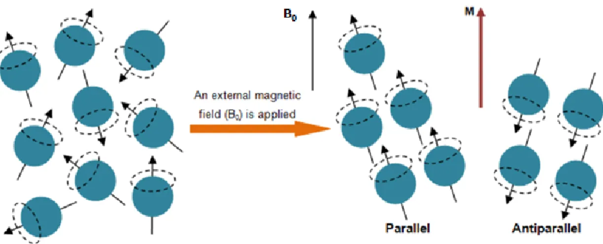
The human body is constituted by approximately 80% of hydrogen atoms. MRI collects the signal from the nuclei of hydrogen atoms to produce images (Bushong, 2003, pp.3-15). The proton of the hydrogen atom has a spin movement, which means that it turns around an axis. The orientation of its rotational axis is normally random during the rotation movement. Since the proton is a mass with an electrical charge in movement, it has a magnetic moment and acts as a bar magnet (Weishaupt, Kochli, & Marincek, 2006, pp.1-5). When the protons are exposed to a strong external magnetic field (B0)
parallel or anti-parallel (Figure 6). These alignments imply distinct levels of energy, so the one that takes less energy, the parallel alignment, is the one which has a slight larger number of protons (Westbrook & Kaut, 2000, pp.1-11). The protons have now a wobble movement, called precession. This precession movement has a characteristic speed, which is positively correlated with the strength of the applied B0 and is named
precession frequency or Larmor frequency (McRobbie, Moore, Graves, & Prince, 2006, pp.137-144). When the protons are in precession movement, their individual magnetic moments add together and cancel each other as some protons align parallel and others antiparallel. However, there are more protons aligned parallel than antiparallel. Consequently, some magnetic moments are not cancelled and the resultant magnetization is represented by a vector in the z-direction, aligned with B0 direction,
called net magnetization vector (M) (Brown & Semelka, 2003, pp.1-9). This vector originates the signal for the MR image but while it has the same direction of B0 it is
impossible to measure it directly. For this purpose M has to be perpendicular to B0. To
obtain this, the system needs to have an energy supply, so an electromagnetic wave with the same frequency as the Larmor frequency will be applied to the system (McRobbie et al., 2006, pp.137-144).
The radiofrequency (RF) pulse, emitted from a radio antenna, called coil, will perturb the stable aligned precession movement of the protons and make them obtain energy, a process named resonance (Bushong, 2003, pp.3-15). The energy transfer has two consequences on the protons: changes the energy state of each proton and some will
Figure 6 – Schematic representation of hydrogen nuclei behavior under the influence of a magnetic field (B0).
Protons usually spin with a random direction, however when applied B0, they aligned in two ways: parallel and antiparallel. As little more protons align parallel to B0, they create a longitudinal magnetization (M). Adapted from (Weishaupt et al., 2006, pp. 1-5).
raise to a higher level of energy, being into the negative z-direction (antiparallel to B0)
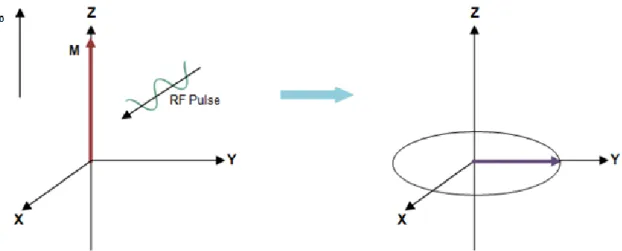
and also makes the protons to precess in phase coherence, which means that they all have an equal position on the precession movement (McRobbie et al., 2006, pp.137-144). These changes will traduce in a decrease of the longitudinal magnetization and in an appearance of a new magnetization in the xy-plane that is perpendicular to the direction of B0, called transversal magnetization (Figure 7). The transverse
magnetization precesses around the z-axis, being a constant oscillating magnetic field, which induces a voltage varying at the Larmor frequency. This signal, known free induction decay (FID) will be collected as a radio signal emitted from the human body by a receiver coil and processed by computers, giving rise to the MR image (Nitz & Reimer, 1999). The MR signal is fluctuating and decreases with time due to the proton spin relaxation in order to get the original state and is represented by time constants called relaxation times (Westbrook & Kaut, 2000, pp.1-11).
Figure 7 – Schematic representation of the behavior of the net magnetization vector, M, after the system received energy from a RF pulse. The RF pulse tips some protons to a higher level of energy and make the protons
to precess is phase. As a consequence there is a decrease in longitudinal magnetization and appear a new transverse magnetization vector in xy plane, which rotate around z-axis. Adapted from (McRobbie et al., 2006).
2.2.RELAXATION TIMES
When the RF pulse is turned off, the system relaxes and returns to its lower state of energy, consequently the transversal magnetization diminishes until extinction (transversal relaxation) and the longitudinal magnetization increases until its original value (longitudinal relaxation) (McRobbie et al., 2006, pp.148-153).
Longitudinal or spin-lattice relaxation happens due to energy transfer for the environment or lattice and the time constant that represents how quickly is the recovery of longitudinal magnetization is named longitudinal magnetization time or T1. This time constant is dependent on the strength of B0 and the inner movement of the molecules
(Brownian motion). Transversal or spin-spin relaxation is caused by internal magnetic field variations such as different proton precession frequencies because of inhomogeneity in B0 and the influence of the magnetic field of each proton in the nearby
nuclei. The different precession frequencies cause the protons to be out of phase. This will cause the decrease of transversal magnetization and the constant time which describes this process is the transversal relaxation time or T2 (Brown & Semelka, 2003, pp.21-31). These two relaxations times T1 and T2 represent two independent processes which occur at the same time (Bushong, 2003, pp.64-71).
2.3.MAIN TYPES OF IMAGE CONTRAST
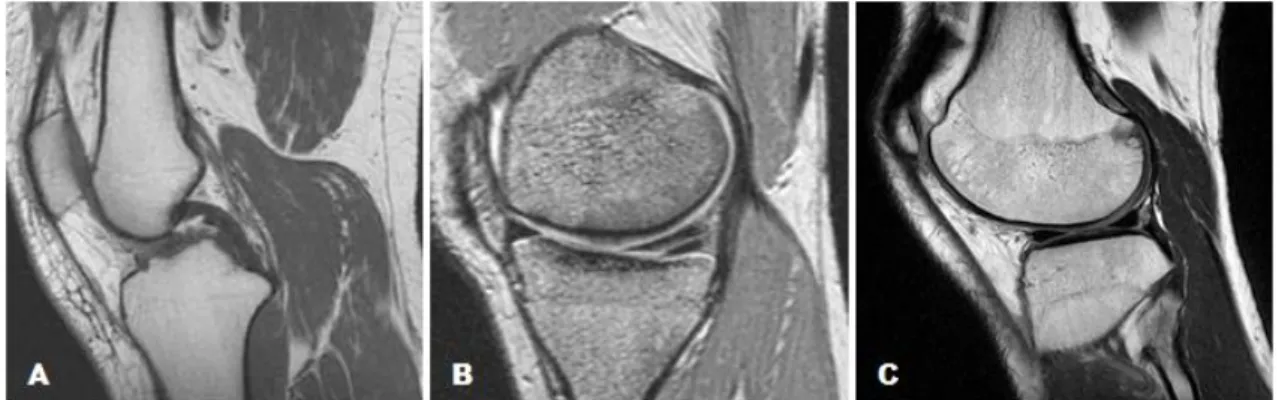
Contrast in MR images is reached based on tissue differences in T1, T2, and proton density (PD). These parameters are intrinsic features of biological tissues which using different RF pulse sequences can produce an image intensity that can be weighted with respect to T1, T2 or PD (Figure 8) (Nitz & Reimer, 1999). As these parameters are very different from one tissue to another, this allows soft-tissue discrimination and diagnostic potential of MRI. To create an MR image, a slice of the body has to be excited with more than one RF pulse (a succession of RF pulses is a pulse sequence) and the emitted signal recorded many times. The repetition time (TR) is the interval between two successive excitations of the same slice and consequently the duration of the relaxation period between two excitation pulses. The echo time (TE) is the time-period between application of the RF pulse and the collection of the MR signal (Weishaupt et al., 2006, pp. 11-20). The generation of T1-, T2-, or PD-weighted images depends on the TR and TE values (Nitz & Reimer, 1999).
The time constant T1 is short when the change of energy is efficient (the lattice has precession frequencies near the Larmor frequency). When the molecules in the environment do not move at the Larmor frequency, the protons are not able to send its energy fast to the surroundings, so they will be slower to return to their lower energy level. Consequently the longitudinal magnetization will take a long time to recover, and the T1 time will be long (Westbrook & Kaut, 2000, pp.12-20). To get advantage of this
biological characteristic one can use a short TR to create a difference in signal intensity between different tissues. This difference will be possible because the tissues with short T1 will recover faster than others with long T1. However, if a long TR is used, all tissues recover its longitudinal magnetization and the signal differences disappear. So the resulting MR image of a short TR is classified as T1-weight image (T1WI) (Figure 8-A), since tissue contrast is mostly created by their difference in T1 times. Tissues with short T1 appear bright and others with a long T1 present a weak signal (Nitz & Reimer, 1999). The T2 time constant represents the velocity of decrease in transversal magnetization vector after the RF pulse excitation. When the image contrast is almost dependent of the T2 times of the tissues, the image is classified as T2-weighted image (T2WI) (Figure 8-B). If a long TE is used, the tissues show different signal intensities and a good contrast on the MR image. The tissues with short T2 lose their transversal magnetization and appear dark, while tissues with a long T2 maintain the magnetization longer and produce a stronger signal appearing bright (Nitz & Reimer, 1999).
PD is the number of hydrogen atoms in a particular volume. To get a PD-weighted image (Figure 8-C) the influence of the other two parameters, T1 and T2, has to be suppressed. To this end, a long TR and a short TE is used, so that the collected signal is neither T1WI nor T2WI, but mostly influenced by differences in proton density. Thus, tissues with large content in hydrogen emit a stronger signal (McRobbie et al., 2006, pp.32-36).
Figure 8 - Sagittal images of the knee. (A) T1-weight image. (B) T2-weight image. (C) PD-weight image. Adapted
3. MAGNETIC RESONANCE IMAGING WITH CONTRAST AGENTS
MRI easily creates a distinction between different tissues based on differences in T1, T2 and PD. However, healthy and pathological tissues as well as distinct diseases show similar magnetic moments producing a poor image contrast. To get a better anatomical differentiation and to improve sensitivity, CAs are used. Due to their physico-chemical properties, CAs induce different effects on image signal intensities by modifying the intrinsic contrast properties of biological samples (Weishaupt et al., 2006, pp.103-123). CAs can also increase image quality, allow higher resolution, and provide kinetic information about an enhanced lesion (McRobbie et al.,2006, pp.42-44).
CAs act by shortening T1 and T2 relaxation times, however, some tracers decrease specially T1 time. Consequently, tissues with a short T1 appear bright on T1WIs. These CAs are called positive because they enhance the image signal. They are also classified as T1 CAs because effects of lower concentrations are easily observed on T1WIs. On the other hand the contrasts which have a greater change in T2 time of tissues are called negative, because they cause a decrease in image signal (Figure 9-B). They are T2 CAs, because their effect is clearly evident on T2WI (Gandhi, Brown, Wong, Aguirre, & Sirlin, 2006)
The present thesis is about a T1 CA based on Gd(III) chelates. Therefore the mechanism of action and classification will be detail describe for Gd(III)-based CAs.
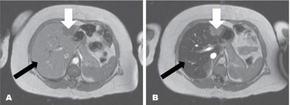
Figure 9 – Negative contrast provided by uptake of SPIO by Kupffer cells (RES). (A) Axial T2WI obtained
before the administration of SPIO. (B) Axial T2WI after injection of SPIO demonstrates signal image decrease
on normal liver parenchyma due to uptake of iron oxide nanoparticles (black arrow) and maintenance of the enhancement by metastatic liver lesion (white arrow), which tumor cells replaced kupffer cells. Adapted from (McRobbie et al., 2006, pp.42-44).
3.1.MECHANISM OF SHORTENING T1 AND T2 TIMES
In the human body, the water molecules possess a rotation movement faster than Larmor frequency, consequently their relaxation is inefficient and they have a long T1. The spin molecules used as CA have a high magnetic moment and in close contact with the protons of water they will induce a fluctuation in the neighboring magnetic fields near the Larmor frequency. This influence will facilitate the water molecules to decrease the energy that have previously gained from the RF pulse. Therefore the relaxation time T1 of the neighbor water protons is reduced and water will appear bright in a T1WI. There is also a decrease of T2 due to the magnetic moments of unpaired electrons, which alter the local magnetic field strength, causing a faster dephasing of the protons (Westbrook & Kaut, 2000, pp.193-197). The magnetic field inhomogeneity created by Gd(III) has a very small distance of action, however the newly affected protons will also exchange with other protons further away from Gd(III). So there is an overall reduction of T1 and T2 (Gandhi et al., 2006).
The interference in the local magnetic field strength is due to dipole-dipole interactions between the unpaired electron spins or protons of the CA and the neighboring excited hydrogen nuclei of the water, fat, or protein molecules which compose the tissue.Therefore, a fraction of the action mechanism of the CA is based on the electron shell and not simply due to nuclear interactions. The magnetic moments of the electrons are much higher than those of protons, thus the electron shell contains powerful paramagnetic properties. The relaxation produced by contrast on surrounding tissues is a result of inner-sphere and outer-sphere effects. The first one is caused by the relaxation of the hydrogen nuclei of water molecules directly bound to the paramagnetic ion of the CA, and the second is caused by interactions between paramagnetic ions and closely diffusing water molecules (McRobbie et al., 2006, pp.162-166).
The interaction efficiency of CA with nearby water molecules is translated by the measure of relaxivity or relaxation rate (R1 and R2). This parameter is the inverse of the relaxation time, so it is determined by measuring T1 or T2, respectively, in a one molar solution. A higher relaxivity reflects an efficient interaction between the CA and the water protons, thus a faster relaxation of the protons and an increase in signal on T1WI (Weishaupt et al., 2006, pp.109-112).
3.2.CONTRAST AGENTS FOR T1WI
Paramagnetic substances are atoms or molecules which have a strong magnetic moment due to unpaired electrons in their outer electron shells or unpaired nucleons in their atomic nuclei (Westbrook & Kaut, 2000, pp.193-197). When these paramagnetic materials are under an external magnetic field, their magnetic moments align, add up and create a positive and strong net magnetization, while most body tissues, which are diamagnetic, become only weakly magnetized. Some metal ions that can be used as CA are Gd(III) and Manganese (II) and Manganese (III) (Kozlowska et al., 2009).
Gadolinium belongs to the lanthanide series of rare earth elements. It has seven unpaired electrons, and therefore very strong paramagnetic properties (Que & Chang, 2006). Free Gd(III) is toxic because its diameter is close to that of calcium ions. Indeed, gadolinium ions bind to calcium channels, preventing binding of calcium ions. For this reason Gd(III) cannot be used in their elemental state but have to be chelated to a ligand. Some examples of ligands used for complexing the Gd(III) are DTPA, DOTA, DTPA-BMA, HP-DO3A. These ligands reduce significantly Gd(III) toxicity and influence the pharmacokinetics of the complex. Several Gd(III) formulations are available for commercial use and others are under experimental scrutiny (Weishaupt et al., 2006, pp.107-109).
3.2.1.NONSPECIFIC Gd(III)-BASED CONTRAST AGENTS
The most frequent CAs used in clinical MRI are small molecule Gd(III) chelates that can distribute uniformly to all perfused tissues throughout the vasculature, and can diffuse across endothelial wall vessels into the extracellular spaces (Gandhi et al., 2006; Gillies, Raghunand, Karczmar, & Bhujwalla, 2002). These molecules are however too large to cross the blood-brain barrier, except when this is disrupted by pathological conditions (e.g. primary tumors and metastasis). In these settings, low molecular weight contrast agents (LMWCA)s can enter and accumulate in the affected brain tissue, resulting in increased visibility of the lesion. After systemic body distribution, these tracers are rapidly eliminated by kidneys (Padhani, 2002).
LMWCAs have been extensively used to improve tumor localization and characterization through dynamic contrast enhancement (DCE) MRI. Some examples are cancers of the breast (Turnbulla, 2009), liver (Goshima et al., 2009), bone (Reddick
et al., 2001), lung (Hunter et al., 1998), pancreas ( Murakami, Nawano, Moriyama, & Onuma, 1998), prostate (Ocak et al., 2007), and brain (Padhani, 2002).
DCE MRI consists in the acquisition of data after intravenous administration of CA. This method provides anatomical images and physiologic data such as a time-enhancement curve, which is used to assess the time-enhancement of the lesion during the CA uptake and washout. Analysis of the curve shape aids the physician in the diagnosis of the tumor and in the distinction between a benign or malignant lesion (Rausch & Hendrick, 2006). A large portion of invasive breast cancers usually exhibit a quick and strong enhancement, and this is followed by either stabilization or fast loss of the signal intensity. On the other hand, benign lesions show a weaker but continuous enhancement. Contrast enhancement analyses are an excellent diagnostic tool for breast cancer in that more than 90% of breast cancers lesions show a strong enhancement (Kuhl, 2007). However, contrast enhancement is nonspecific because of considerable common contrast enhancement features between benign and malignant lesions (Rausch & Hendrick, 2006). For this reason, LMWCAs showed a high sensivity for tumor detection (e.g. breast tumors) but lack of specificity (Mattrey & Aguirre, 2003).
LMWCAs are not taken up by particular organs, do not target specific tissues or pathological areas, and do not respond to the cellular microenvironment. They enhance all vessels and high vascular tissues (Figure 10-B). Thus only lesions with significant blood flow volumes can be distinguished by MRI, making it almost impossible the detection of primary cancer or metastasis when they measure just a few millimeters. Also, since these contrast agents leak through both normal and neoplastic vessels, it
may be difficult to distinguish between normal and angiogenic vessels (Hartman et al., 2008; McDonald & Choyke, 2003). These CAs have also a short half-life in blood (Mattrey & Aguirre, 2003) and a large part of administered CA is eliminated before images are taken. Therefore, in order to get informative images, a substantial quantity of CA needs to be injected in the patient, which may constitute at times a health risk for some patients (Nakamura et al., 2006).
To overcome the drawbacks showed by LMWCAs, macromolecular weight contrast agents (MMWCA)s are in development. Some examples of MMWCAs are iron oxide particles and Gd(III) bound to larger molecules such as albumin, polylysine, dendrimers, micelles and liposomes (Padhani, 2002).
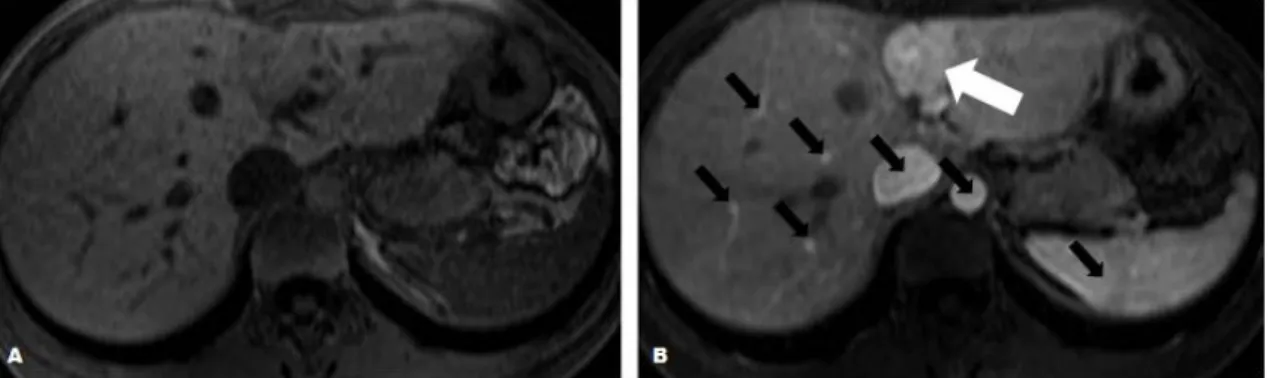
Figure 10 - Nonspecific image signal enhancement following administration of LMWCA. (A) Axial T1WI
obtained before the administration of Gd(III) chelates, image with no apparent lesion. (B) Axial T1WI after injection
of CA demonstrates enhancement of a liver lesion (white arrow), blood vessels and high perfused organs (black arrows). Adapted from (Gandhi et al., 2006).
These MMWCAs diffuse poorly or not at all through normal vessels, leaking only when the vessel wall is abnormal, as is the case for neoangiogenic vessels (Bhujwalla, Artemov, Natarajan, Ackerstaff, & Solaiyappan, 2001). MMWCAs are frequently used to blood pool imaging because of their long circulation times and slower diffusion as well as clearance from interstitial spaces (Gillies et al., 2002). This slower kinetics allows a better quantification of vascular leakage, thus helping to differentiate benign from malignant tumors. MMWCAs have therefore been reported to be better suited for assessment of microvasculature permeability of tumors lesions (Daldrup et al., 1998; Padhani, 2002). However, hyperpermeability to MMWCAs is not exclusive of cancer microvasculature, since it has also been observed in inflammatory, ischemic and transplant rejection tissue models (Mattrey & Aguirre, 2003).
Chen and colleagues created a MMWCA for hepatocellular carcinoma detection in rats. They used self-assembled micelles made by poly lactic acid–PEG and commercial Gd(III)–DTPA. The Gd(III)–DTPA was absorbed onto the surface of the nanoparticles. This MMWCA provided better and prolonged image contrast effects than commercial LMWCA in liver, even with a lower dose of gadolinium per kilogram of body weight (Z. Chen et al., 2009). Zhang and colleagues reported a successful production of micelles based on biodegradable poly(L-glutamic acid)-bpolylactide block copolymer with DTPA-Gd(III) chelated to the micelle shell. This complex showed two-fold higher relaxivity than LMWCA (Zhang et al., 2008). Bertini and coworkers developed a MMWCA for detection of solid tumors. This probe consisted of PEG-stabilized paramagnetic liposomes, with Gd(III)–DTPA on their surface. This
MMWCA showed increased relaxivity compared to conventional LMWCA and achieved a prolonged visualization of neoplastic lesions in mice (Bertini et al., 2004).
3.2.2.TARGETED Gd(III)-BASED CONTRAST AGENTS
Since current MRI CAs have low specificity and since MRI has proven to be a valuable molecular imaging tool, efforts have been taken to develop targeted and specifically activated CAs. These tracers aim to target distinct molecules related with different pathologies, accumulate selectively in a exact biological site providing an increased local concentration of CAs, allow more specific diagnosis, and have the potential to characterize diseases at the molecular level in vivo (Aime et al., 2002; Kozlowska et al., 2009). Targeted CAs identify specific cell types by internalization or interaction with proteins expressed on the cell surface (Lyons, 2005). Most MRI CAs used for molecular imaging are linear polymers and dendrimers conjugated to metal chelates as well as liposomes and micelles containing paramagnetic ions (Liu et al., 2010).
An approach of target Gd(III)-based CA consisted of perfluorocarbon nanoparticles able to detect integrins expressed on neovasculature in nascent Vx-2 rabbit tumors. In this experiment, a small arginine-glycine-aspartic acid-peptidomimetic was covalently attached to the nanoparticle. These nanoparticles showed specific signal enhancement in sites of tumor angiogenesis. Furthermore, the leakage in tumor vessels was greater than in muscle (Winter et al., 2003). Another research group successfully combined a long-circulating liposome with membrane-incorporated Gd(III)-chelates and a cancer specific antibody, 2C5, attached to the liposome surface (Erdogan, Medarova, Roby, Moore, & Torchilin, 2008). To detect tumor cell death after chemotherapy, Krishnan and colleagues produced specific CA based on Gd(III)-chelates conjugated to the C2A domain of synaptotagmin I, which targets the plasma membrane phospholipid phosphatidylserine expressed by apoptotic cells. This probe was able to identify tumor cell death in vivo (Krishnan et al., 2008).
3.2.3.SMART Gd(III)-BASED CONTRAST AGENTS
Targeted CAs may greatly improve the accuracy and extent of diagnostic imaging. However, another approach based on physiological activatable or stimulus-sensitive CAs, seems to offer a significant improvement of the MRI potential for disease detection. Unlike standard targeted agents, which enhance MR image constantly, smart
CAs change between two conformational states dependent on a certain stimulus. The change is detected as an alteration in signal, because one of the conformational states has low or no enhancement, the ―off state‖, and other has high enhancement, the ―on state‖. These stimulus-sensitive agents can be detected when switched from one state to another after exposure to a metabolic or physiological event in a specific molecular target (Lyons, 2005). Smart CAs have been built on Gd(III) systems because their relaxivity can be dictated by their environment. The numbers of water molecules in the first coordination sphere, the water exchange rate and the rotational correlation time have a strong effect on the relaxivity of the compounds and can be influenced by many factors. There are CAs responsive to metal ions such as calcium (Li et al., 1999), copper (Que & Chang, 2006) and zinc (Hanaoka et al., 2002), while others are sensitive to oxygenated hemoglobin (Aime et al., 1999b), the presence of radicals (Glogard, Stensrud, & Aime, 2003), or are enzimatically activated to provide a means of measuring enzyme activity and enzyme localization (Moats, Fraser, & Meade, 1997).
The pH-sensitive CAs are of interest to tumor MRI imaging, because a common feature of cancerous tissues is a significantly lower extracellular pH as compared to healthy tissues. Thus, pH seems to be a promising parameter for detecting early stage cancer (Hartman et al., 2008; Gillies, Raghunand, Garcia-Martin, & Gatenby, 2004). To this aim, Zhang and colleagues created a pH-sensitive Gd(III) complex of a DOTA tetramide derivative. They observed an interesting behavior of the complex relaxivity versus pH. Indeed, starting from pH 4, the relaxivity first increased until pH 6 and then decreased until reaching a minimum at pH 8.5. It remained at this minimum between pH 8.5 and 10.5 and then increased again (Zhang, Wu, & Sherry, 1999). Aime and coworkers reported a pH-sensitive CA with Gd(III)-chelates and ornithin residues. The chelates were conjugated to the amino acid chain via squaric esters, which readily reacts with amines. At low pH, the amines are protonated and do not interact with the squaric ester residues. When the pH increases in a range of 4.5 to 8.5, the amine side chains become deprotonated and interact with squaric ester linkers. This interaction rigidifies the polymer creating and increasing relaxivity (Aime et al., 1999a). Also Hartman and colleagues synthetised ultrashort singe-walled carbon nanotube capsules that provided extremely high and pH-dependent relaxivity. These so-called gadonanotubes showed an increased relaxivity upon a decrease in pH from 7.4 to 7.0 (Hartman et al., 2008).
These smart contrast agents have proven to be excellent MRI tools for very specific targets in vitro, but failed to show the same performance in vivo, due to the