art fnafreire structural properties
Texto
Imagem


Documentos relacionados
The resulting powders were characterized by thermogravimetric analysis, Fourier-transform infrared spectroscopy (FTIR), X-ray diffraction (XRD), scanning electron microscopy
samples, prior to and after functionalization, were characterized by chemical analyses, thermogravimetric analysis (TGA), x ray diffraction (XRD), diffuse reflectance infrared
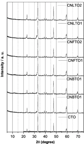
The structural features, electrical, thermal and optical properties of the ceria nanoparticles were determined in depth with X-ray powder diffraction (XRD), high resolution
The structural properties of particles were investigated by X-ray diffraction (XRD), atomic force microscopy (AFM), differential thermal analysis (DTA), and N 2
The results obtained through X-ray structural analysis, magnetic properties and elemental analysis and the critical temperature of the compounds indicate the interlocked structure to
Using the optical and electron microscopy, X-ray powder diffraction and Fourier transform infrared spectroscopy we attempted to correlate the results obtained on 8
Its composition was determined by X-ray diffractometry (XRD), Fourier transform infrared spectroscopy (FTIR) and Raman micro-spectroscopy, and its topography by atomic
Detailed optical properties were evaluated using UV-Vis spectroscopy, and photoluminescence (PL) emission spectra of doped and undoped CT were employed to examine the
