Braz Dent J 20(1) 2009
74 A.C.M.N. Rosales et al.
Correspondence: Prof. Dr. Márcio Ajudarte Lopes, Avenida Limeira, 901, Areão, 13414-903 Piracicaba, SP, Brasil. Tel: +55-19-2106-5319. Fax: +55-19-2106-5218. e-mail: [email protected]
ISSN 0103-6440
Dental Needs in Brazilian Patients Subjected
to Head and Neck Radiotherapy
Ana Carolina de Mesquita Netto ROSALES1 Sérgio Carlos Barros ESTEVES2
Jacks JORGE1 Oslei Paes de ALMEIDA1
Márcio Ajudarte LOPES1
1Department of Oral Diagnosis, Piracicaba Dental School, University of Campinas, Piracicaba, SP, Brazil
2Center of Oncology, Piracicaba, SP, Brazil
In spite of its recognized beneits in the treatment of malignant tumors, radiation therapy have several side effects in the head and neck region. The evaluation of oral conditions by a dentist is important to prevent or minimize these problems. The aim of this retrospec
-tive review was to analyze the dental needs in 357 patients who received radiotherapy in the head and neck region and were treated at Orocentro/FOP/UNICAMP, between January 1990 and December 2004. Review of patient iles showed that dental examination before radiotherapy was not performed in 148 patients (41.5%) and was done in 209 patients (58.5%). From the total of examined patients, 94 (45%) did not require dental procedures at the moment of examination, while 115 (55%) presented some sort of dental need. Fol
-lowing the patients after the radiotherapy, it was observed that the group of patients that was evaluated before radiation presented less need of restorations, root canal illing and dental extractions than those who were not evaluated. The results of this study conirm that the evaluation of oral conditions prior to radiotherapy is essential to minimize the dental needs, emphasizing the importance of the dentist in the multidisciplinary team that treats cancer patients.
Key Words: oral cavity, cancer, radiotherapy, oral complications.
INTRODUCTION
Radiation therapy may be either an effective alternative to surgery or a valuable adjuvant therapy to surgery and/or chemotherapy in the treatment and loco-regional control of malignant head and neck tumors (1).
However, in addition to acting in tumor cells, ionizing radiation causes damage in normal tissues located in
the radiation ield. Direct cell damage combined with
regional loss of vascular perfusion result in xerostomia
and decrease in the healing capabilities of some tissues (1). In addition, irradiated patients are at signiicant risk of potentially debilitating dental complications, such as aggressive caries activity (2,3). Oral alterations can be prevented or at least more properly managed if dental and medical health care providers work together. Dental evaluation and treatment with a long-term oral
care regimen is recognized as an important aspect to
be considered before, during and after radiotherapy (4,5). Pre-radiation and post-radiation dental treatment in patients with head and neck cancer is often a clinical challenge (6). However, for these treatments to be ef
-fective, good patient compliance with close professional supervision is required for an indeinite period after ra
-diation. Therefore, the aim of this study was to evaluate the dental needs in patients subjected to radiotherapy in the head and neck region.
MATERIAL AND METHODS
The basis of this study was a retrospective review of the iles of the Department of Oral Diagnosis of the Dental School of Piracicaba, University of Campinas, Brazil, where 357 patients with history of radiation therapy in the head and neck region were treated be
Braz Dent J 20(1) 2009
Dental needs in irradiated patients 75
age, gender, tobacco smoking and alcohol consump
-tion history, tumor loca-tion, histological type, clinical stage, treatment were obtained from the patient’s charts. Indications of dental treatment, such as restorations, root canal illing, dental extractions and prosthesis, before and after radiotherapy were assessed. The study was conducted after approval of the Human Research Ethics Committee of the Dental School of Piracicaba, University of Campinas, Brazil.
RESULTS
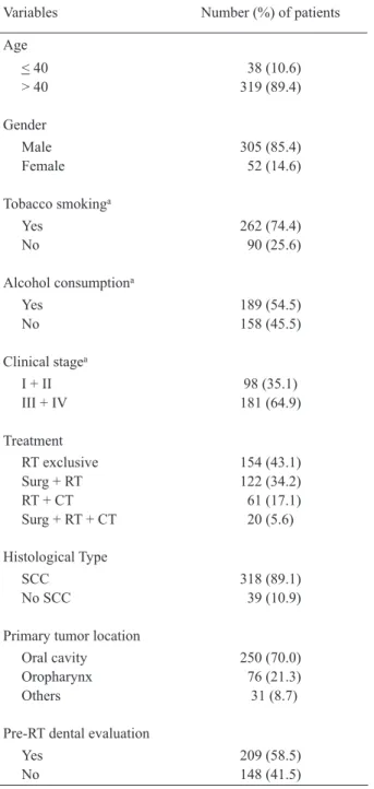
Table 1 shows the distribution of the 357 irradi
-ated patients with head and neck cancer according to demographic, lifestyle, clinical and treatment variables.
The age of the patients ranged from 12 to 87 years (mean age = 54.9 years) and most individuals were male (85.4%). Tobacco smoking and alcohol consumption were reported by 74.4% and 54.5% of the patients, respectively.
The most common site of the primary tumors was the oral cavity corresponding to 70% of the cases. Squamous cell carcinoma was the histopathological diagnosis of 89.1% of the tumors. Advanced clinical
stages were more frequent, stages III and IV being
de-tected in 64.9% of the patients. Consequently, exclusive radiotherapy was the treatement option for 43.1% of the patients followed by surgery plus radiotherapy in 34.2%.
Dental examination before radiotherapy was performed in 209 patients (58.5%), while 148 patients (41.5%) were not examined prior to radiation and sought dental treatment only afterwards. From the total of examined patients, 94 (45%) did not require dental procedures at the moment of examination, while 115 (55%) presented some sort of dental need.
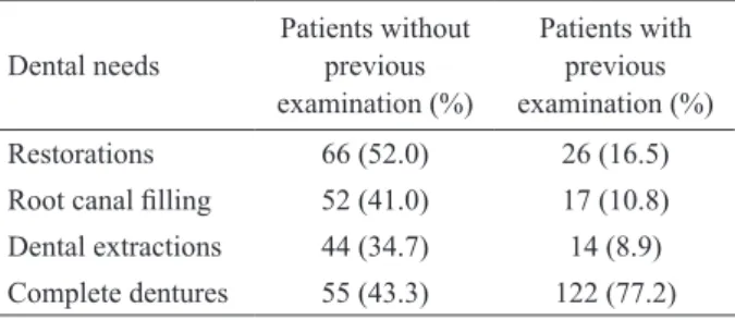
Patients that were evaluated before radiotherapy required less dental procedures after radiation than those that were not previously evaluated. Needs of restora
-tions were observed in 52% of the patients that were not previously evaluated versus 16.5% of the patients
that were examined by a dentist before head and neck radiation. Root canal illing was required in 41% of the non-examined patients and in only 10.8% of the patients
examined for dental conditions. Dental extractions were
also more indicated in patients that were not examined (34.7%) than in those that were seem by a dentist be
-fore radiotherapy (8.9%). On the other hand, the need
of complete dentures was more frequent in patients
that were examined before radiotherapy (77.2%) com
-pared to the patients that were not previously examined (43.3%) (Table 2).
Table 1. Distribution of 357 cases of head and neck irradiated patients according to demographic, lifestyle, clinical and treatment
variables.
Variables Number (%) of patients
Age
< 40 > 40
38 (10.6) 319 (89.4)
Gender Male Female
305 (85.4) 52 (14.6)
Tobacco smokinga
Yes No
262 (74.4) 90 (25.6)
Alcohol consumptiona
Yes No
189 (54.5) 158 (45.5)
Clinical stagea
I + II III + IV
98 (35.1) 181 (64.9)
Treatment RT exclusive Surg + RT RT + CT Surg + RT + CT
154 (43.1) 122 (34.2) 61 (17.1) 20 (5.6)
Histological Type
SCC No SCC
318 (89.1) 39 (10.9)
Primary tumor location Oral cavity Oropharynx Others
250 (70.0) 76 (21.3)
31 (8.7)
Pre-RT dental evaluation Yes
No
209 (58.5) 148 (41.5)
a:Excludes cases with missing information; RT: radiotherapy; Surg.:
Braz Dent J 20(1) 2009
76 A.C.M.N. Rosales et al.
DISCUSSION
Head and neck cancer is a serious, debilitating,
and potentially life-threatening disease. However, ad
-vances in its management have resulted in signiicant improvements in survival and functional outcome (1,7). The use of radiotherapy has proven to be an effective technique in the control and cure of the malignant tumors (1,7). Unfortunately, radiotherapy also has a signiicant negative impact on the oral health increasing morbidity (8). In this study, the majority of patients that underwent radiotherapy were male, over 40 years of age, reported tobacco and alcohol consumption and had advanced oral squamous cell carcinoma, conirming the indings in the literature (1,3,5,9-11).
Oral complications following radiation therapy for head and neck cancer are common and affect quality of life (8). The importance of adequate pre-radiotherapy dental screening is well documented (6,12,13). In the present study, dental examination was performed in 58.5% of the sample while 41.5% were not previously evaluated. It was observed that fewer dental procedures
were needed in patients evaluated prior to radiation exposure.
The initial dental evaluation should include
assessment and elimination of all existing oral
condi-tions that are likely to precipitate complicacondi-tions, such as extensive carious lesions, periapical pathology and advanced periodontal disease (3,6,14,15). Poor dental condition frequently requires multiple extractions prior to initiation of radiotherapy (3,5). An adequate time for healing of extraction sites before radiation exposure is considered mandatory (5). A minimum period of 2 weeks between extraction and the onset of radiation therapy is recommended in order to avoid the occurrence of osteoradionecrosis (14). In the present study, 89.5% of
the patients evaluated before radiotherapy needed dental extractions and this was the most common performed
procedure.
It is important for the patients to understand that therapeutic irradiation of head and neck can lead to an
increased rate of dental caries and its management can
prove to be extremely dificult and frustrating (3). In the present case series, 52% of the patients who were not evaluated prior to radiotherapy needed some type of restorative dental procedure at the post-radiation period, in contrast to only 16.5% of the individuals that were examined by a dentist before radiation of the head and neck region. This inding emphasizes the importance of oral hygiene and dietary counseling to prevent potential complications (12).
As much as 41% of the patients who were not evaluated for dental conditions before radiation therapy needed endodontic treatment. In contrast, only 10.8% of the evaluated ones needed root canal treatment at the post-radiation period. This inding may be explained by large number of prophylactic dental extractions performed in this group of individuals allied to the im
-provement, to some extent, of the patients’ oral hygiene. Whenever possible, endodontic treatment should be the
option for pain control, preservation of function and
mainly prevention of osteoradionecrosis by avoiding dental extractions (16). However, endodontic treatment in irradiated patients may be time-consuming and re -quires optimal patient compliance to be concluded. In
a signiicant number of cases, cervical caries may lead to amputation of dental crown, which, in turn, makes endodontic therapy more complicated. Teeth that are partial or totally destroyed by deep carious lesion can be treated endodontically to preserve bone and periodontal ligament integrity to support a removable complete denture that will be more comfortable and will improve the life quality of these patients. Trismus is a potential complication of radiation therapy, impairing the proper mouth opening and hence limiting adequate restorative and endodontic dental care (17).
This study revealed that 34.7% of patients who were not evaluated for the dental conditions before ra
-diation therapy required dental extractions against only 8.9% in the evaluated group. These results are in agree
-ment with the indings of previous studies, conirming that dental extractions represent a common dental need in post-irradiated patients who did not have previous access to appropriate dental care (11).
Table 2. Comparative analysis between patients without and with dental examination before radiotherapy regarding dental needs.
Dental needs
Patients without
previous
examination (%)
Patients with
previous
examination (%)
Restorations 66 (52.0) 26 (16.5)
Root canal illing 52 (41.0) 17 (10.8)
Dental extractions 44 (34.7) 14 (8.9)
Braz Dent J 20(1) 2009
Dental needs in irradiated patients 77
Studies regarding quality of life demonstrate that patients unable to have appropriate function in swallowing and speech frequently become depressed. Consequently, most patients can obtain signiicant ben
-eit from oral rehabilitation, which, in some instances, may be achieved by constructing functional removal dentures (18-20). In the present study, a large number of patients needed complete dentures (77.2%), which may be explained in part by the multiple prophylactic
pre-radiation dental extractions.
In conclusion, the indings of this study reinforce that: 1. a multidisciplinary approach is ideal for the management of patients scheduled to receive radiation therapy, particularly in the head and neck region; 2. dental examination before radiation therapy may prevent or minimize complications in the post-radiation period and provide better oral health conditions to the patients.
RESUMO
Apesar dos benefícios da radioterapia no tratamento de tumores malignos, vários são os seus efeitos colaterais na região de cabeça e pescoço. Sendo assim, a avaliação das condições bucais pelo cirurgião dentista é fundamental para prevenir e/ou minimizar
estes danos. Este estudo retrospectivo teve como objetivo veriicar
as condições dentárias e as necessidades de tratamento odon-tológico dos 357 pacientes que receberam radioterapia na região de cabeça e pescoço, atendidos pelo Orocentro/ FOP/UNICAMP, no período de janeiro de 1990 a dezembro de 2004. Em 148
(41,5%) do total dos pacientes a avaliação odontológica não foi
realizada previamente à radioterapia. A avaliação odontológica
pré-radioterápica foi realizada em 209 pacientes (58,5%) dos quais 94 (45%) não tinham necessidades de tratamento odontológico no momento da avaliação, enquanto 115 (55%) apresentavam
algum tipo de necessidade odontológica. O grupo de pacientes avaliados antes da radioterapia apresentou menores necessidades de restaurações, endodontias e exodontias que os pacientes não avaliados. Conclui-se que a avaliação das condições bucais previamente à radioterapia é essencial para diminuir as neces-sidades de tratamento odontológico enfatizando a importância da participação do cirurgião-dentista na equipe multidisciplinar que trata pacientes com câncer.
REFERENCES
1. Boyle J, Patel S, Shah JP. Management of oral and oropharyngeal cancers. Oral Dis 2003;9:109-111.
2. Andrews N, Grifiths C. Dental complications of head and neck radiotherapy: Part 1. Aust Dent J 2001;46:88-94.
3. Bonan PR, Lopes MA, Pires FR, Almeida OP. Dental
manage-ment of low socioeconomic level patients before radiotherapy of the head and neck with special emphasis on the prevention of osteoradionecrosis. Braz Dent J 2006;17:336-342.
4. Cacchillo D, Barker GJ, Barker BF. Late effects of head and neck radiation therapy and patient/dentist compliance with recom
-mended dental care. Spec Care Dentist 1993;13:159-162.
5. Doerr TD, Marunick MT. Timing of edentulation and extraction in
the management of oral cavity and oropharyngeal malignancies. Head Neck 1997;19:426-430.
6. Bruins HH, Jolly DE, Koole R. Preradiation dental extraction deci
-sions in patients with head and neck cancer. Oral Surg Oral Med Oral Pathol Oral Radiol Endod 1999;88:406-412.
7. Sygula M, Skladowski K, Pilecki B, Wygoda A, Hutnik M, Sasi
-adek W. Eficacy of primary and combined radiotherapy in locally advanced cancer of oropharynx and nasopharynx in III and IV stage. Otolaryngol Pol 2005;59:229-234.
8. Epstein JB, Robertson M, Emerton S, Phillips N, Stevenson-Moore P. Quality of life and oral function in patients treated with radiation therapy for head and neck cancer. Head Neck 2001;23:389-398.
9. Brugere J, Guenel P, Leclerc A, Rodrigues J. Differential effects of
tobacco and alcohol in cancer of the larynx, pharynx and mouth. Cancer 1986;57:391-395.
10. Kotz T, Costello R, Li Y, Posner MR. Swallowing dysfunction after chemoradiation for advanced squamous cell carcinoma of the head and neck. Head Neck 2004;26:365-372.
11. Sulaiman F, Huryn JM, Zlotolow IM. Dental extractions in the irra
-diated head and neck patient: a retrospective analysis of Memorial
Sloan-Kettering Cancer Center protocols, criteria, and end results.
J Oral Maxillofac Surg 2003;61:1123-1131.
12. Andrews N, Grifiths C. Dental complications of head and neck radiotherapy: Part 2. Aust Dent J 2001b;46:174-182.
13. Koga DH, Salvajoli JV, Alves FA. Dental extractions and
radio-therapy in head and neck oncology: review of the literature. Oral Dis 2008;14:40-44.
14. Beumer J 3rd, Curtis T, Harrison RE. Radiation therapy of the oral cavity: sequelae and management, part 1. Head Neck Surg 1979;1:301-312.
15. Hancock PJ, Epstein JB, Sadler GR. Oral and dental management
related to radiation therapy for head and neck cancer. J Can Dent Assoc 2003;69:585-590.
16. Seto BG, Beumer J 3rd, Kagawa T, Klokkevold P, Wolinsky L. Analysis of endodontic therapy in patients irradiated for head and neck cancer. Oral Surg Oral Med Oral Pathol 1985;60:540-545. 17. Scully C, Epstein JB. Oral health care for the cancer patient. Eur
J Cancer B Oral Oncol 1996;32B:281-292.
18. Rogers SN, Lowe D, Fisher SE, Brown JS, Vaughan ED. Health-related quality of life and clinical function after primary surgery for oral cancer. Br J Oral Maxillofac Surg 2002;40:11-18. 19. Rogers SN, McNally D, Mahmoud M, Chan MF, Humphris GM.
Psychologic response of the edentulous patient after primary surgery for oral cancer: a cross-sectional study. J Prosthet Dent 1999;82:317-321.
20. Shaw RJ, Sutton AF, Cawood JI, Howell RA, Lowe D, Brown JS, et al.. Oral rehabilitation after treatment for head and neck malignancy. Head Neck 2005;27:459-470.